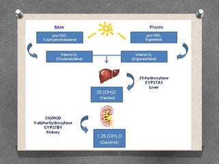
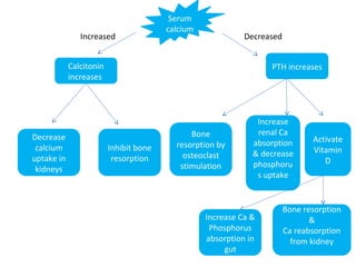
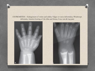
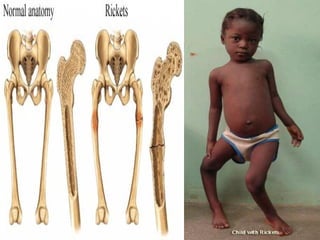
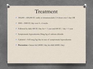
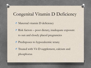
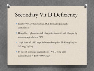
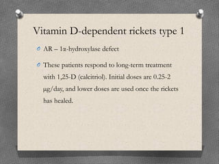
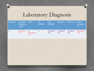
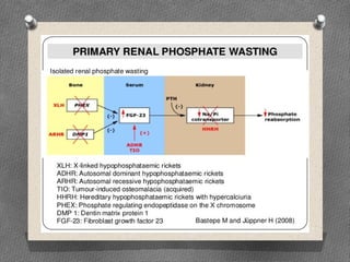
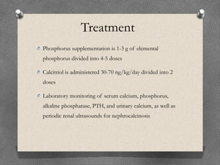
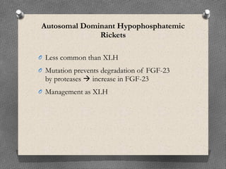
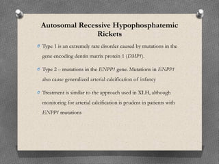
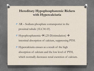
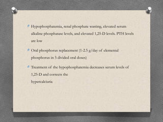
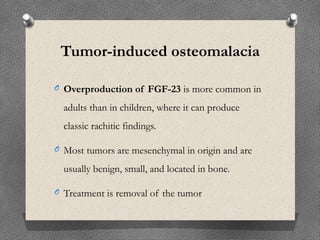
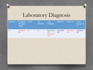
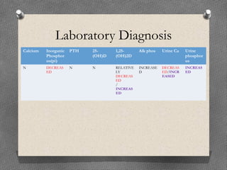
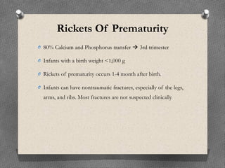
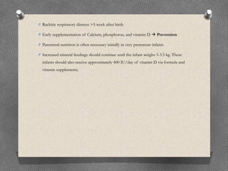
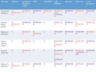
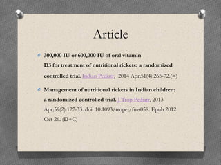
Calcium Homeostasis
Rickets – Types & Management
Dr Raaghul c
Calcium
O 98% – Bone
O Normal ionized calcium level – 1.12-
1.23mmol/L
Serum
calcium
Decreased
PTH increases
Inhibit bone
resorption
Calcitonin
increases
Increase
renal Ca
absorption
& decrease
phosphoru
s uptake
Bone
resorption by
osteoclast
stimulation
Activate
Vitamin
D
Increase Ca &
Phosphorus
absorption in
gut
Decrease
calcium
uptake in
kidneys
Increased
Bone resorption
&
Ca reabsorption
from kidney
Rickets
O It is a disease of growing bone, unmineralized matrix at the growth plates
and occurs in children only before fusion of the
epiphyses.
O An increase in the circumference of the growth plate and the metaphysis,
increasing bone width at the location of the growth plates.
Causes
O VITAMIN D DISORDERS
Nutritional vitamin D deficiency
Congenital vitamin D deficiency
Secondary vitamin D deficiency
Malabsorption
Increased degradation
Decreased liver 25-hydroxylase
Vitamin D–dependent rickets type 1 A and B
Vitamin D–dependent rickets type 2 A and B
Chronic kidney disease
O CALCIUM DEFICIENCY
Low intake
Diet
Premature infants (rickets of prematurity)
Malabsorption
Primary disease
Dietary inhibitors of calcium absorption
O PHOSPHORUS DEFICIENCY
Inadequate intake
Premature infants (rickets of prematurity)
Aluminum-containing antacids
O RENAL LOSSES
X-linked hypophosphatemic rickets
Autosomal dominant hypophosphatemic rickets
Autosomal recessive hypophosphatemic rickets (1 and 2)
Hereditary hypophosphatemic rickets with hypercalciuria
Overproduction of fibroblast growth factor-23
Tumor-induced rickets
McCune-Albright syndrome
Epidermal nevus syndrome
Neurofibromatosis
Fanconi syndrome
Dent disease
Distal renal tubular acidosis
Clinical features
O GENERAL – Failure to thrive, Listlessness, Protruding
abdomen, Muscle weakness (especially proximal) &
Fractures
O BACK – Scoliosis, Kyphosis & Lordosis
O HYPOCALCEMIC SYMPTOMS – Tetany, Seizures &
Stridor due to laryngeal spasm
HEAD – Craniotabes, Frontal bossing, Delayed fontanel closure
Delayed dentition; Caries & Craniosynostosis
CHEST – Rachitic rosary, Harrison groove &
Respiratory infections and atelectasis
EXTREMITIES – Enlargement of wrists and ankles, Valgus or varus deformities, Windswept
deformity, Anterior bowing of the tibia and femur, Coxa vara & Leg pain
Vitamin D Deficiency
Nutritional
O 1st two months of life no deficiency
O Sun – melanin
O These children have increased risk of
pneumonia and muscle weakness leading to
delayed motor development
Laboratory Diagnosis
Calcium Inorganic
Phosphor
us(pi)
PTH 25-
(OH)D
1,25-
(OH)2D
Alk phos Urine Ca Urine
phosphor
us
N,
DECREAS
ED
DECREAS
ED
INCREAS
ED
DECREAS
ED
DECREAS
ED, N,
INCREAS
ED
INCREAS
ED
DECREAS
ED
INCREAS
ED
Treatment
O 300,000 – 600,000 IU orally or intramuscularly 2-4 doses over 1 day OR
O 2000 – 5000 IU/day over 4 – 6 weeks
O Followed by daily 400 IU/day for < 1 year and 600 IU / day > 1 year
O Symptomatic hypocalcemia 20mg/kg of calcium chloride
O Calcitriol – 0.05 mcg/kg/day in case of symptomatic hypocalcemia
O Prevention – breast fed (400IU/day) & child (600IU/day)
Congenital Vitamin D Deficiency
O Maternal vitamin D deficiency
O Risk factors – poor dietary, inadequate exposure
to sun and closely placed pregnancies
O Predisposes to hypocalcemic tetany
O Treated with Vit D supplement, calcium and
phosphorus
Secondary Vit D Deficiency
O Liver (>90% dysfunction) and GI disorders (pancreatic
dysfunction)
O Drugs like - phenobarbital, phenytoin, isoniazid and rifampin by
activating cytochrome P450
O High dose of 25 D helps in better absorption 25-50mcg/day or
5-7 mcg/kg/day
O In case of increased degradation of Vit D long term
administration = 1000-4000IU/day
Vitamin D-dependent rickets type 1
O AR – 1α-hydroxylase defect
O These patients respond to long-term treatment
with 1,25-D (calcitriol). Initial doses are 0.25-2
µg/day, and lower doses are used once the rickets
has healed.
O Targeting a low-normal calcium concentration
and a high-normal PTH level avoids excessive
dosing of calcitriol, which can cause
hypercalciuria and nephrocalcinosis.
O Periodic assessment of urinary calcium
excretion, with a target of <4 mg/kg/day.
Vitamin D-dependent rickets type 2
O AR mutation in Vit D receptor
O 1,25-D increased
O Less likely to have rickets due to partially functional D
receptors
O 50-70%  have alopecia
O Managed initially with high does of 1,25-D 2mcg/day ---- 50-
60mcg/day and calcium 1000 to 3000mg/day
Chronic Kidney Disease
Therapy requires Calcitriol
Dietary phosphorus restriction and the use of
oral phosphate binders is as important as the
use of activated vitamin D
Calcium Deficiency
O Excellent sources of calcium – breast milk and formula
feed
O Intravenous nutrition without adequate calcium or
malabsorption in celiac disease, intestinal
abetalipoproteinemia, and after small bowel resection.
There may be concurrent malabsorption of vitamin D.
Laboratory Diagnosis
Calcium Inorganic
Phosphor
us(pi)
PTH 25-
(OH)D
1,25-
(OH)2D
Alk phos Urine Ca Urine
phosphor
us
N,
DECREAS
ED
DECREAS
ED
INCREAS
ED
N INCREAS
ED
INCREAS
ED
DECREAS
ED
INCREAS
ED
Treatment
O Elemental calcium 700mg/day – 1 to 3 years of
age
O 1000mg/day – 4 to 8 years of age
O 1300mg/day – 9 to 18 years
Phosphorus Deficiency
O Malabsorption (celiac disease, cystic fibrosis,
cholestatic liver disease), but if rickets develops,
the primary problem is usually malabsorption of
vitamin D and/or calcium.
O Isolated malabsorption of phosphorus  long-
term use of aluminum-containing antacids.
Laboratory Diagnosis
Calcium Inorganic
Phosphor
us(pi)
PTH 25-
(OH)D
1,25-
(OH)2D
Alk phos Urine Ca Urine
phosphor
us
N DECREAS
ED
N,
DECREAS
ED
N INCREAS
ED
INCREAS
ED
INCREAS
ED
DECREAS
ED
Fibroblast Growth Factor-23
O Fibroblast growth factor-23 (FGF-23) is a
humoral mediator that decreases renal tubular
reabsorption of phosphate and therefore
decreases serum phosphorus.
O It is synthesized by osteocytes, also decreases the
activity of renal 1α-hydroxylase, resulting in a
decrease in the production of 1,25-D.
X-Linked Hypophosphatemic Rickets
O AD – Defective gene is on the X chromosome
O PHEX – PHosphate-regulating gene with homology to
Endopeptidases on the X chromosome
O Indirect role in inactivating FGF-23, defect in this gene
causes over production of FGF-23
O Rickets with involving lower extremities and poor growth
are the dominant features
Treatment
O Phosphorus supplementation is 1-3 g of elemental
phosphorus divided into 4-5 doses
O Calcitriol is administered 30-70 ng/kg/day divided into 2
doses
O Laboratory monitoring of serum calcium, phosphorus,
alkaline phosphatase, PTH, and urinary calcium, as well as
periodic renal ultrasounds for nephrocalcinosis
Autosomal Dominant Hypophosphatemic
Rickets
O Less common than XLH
O Mutation prevents degradation of FGF-23
by proteases  increase in FGF-23
O Management as XLH
Autosomal Recessive Hypophosphatemic
Rickets
O Type 1 is an extremely rare disorder caused by mutations in the
gene encoding dentin matrix protein 1 (DMP1).
O Type 2 – mutations in the ENPP1 gene. Mutations in ENPP1
also cause generalized arterial calcification of infancy
O Treatment is similar to the approach used in XLH, although
monitoring for arterial calcification is prudent in patients with
ENPP1 mutations
Hereditary Hypophosphatemic Rickets
with Hypercalciuria
O AR – Sodium-phosphate cotransporter in the
proximal tubule (SLC34A3).
O Hypophosphatemia 1,25-D(stimulation) 
intestinal absorption of calcium, suppressing PTH.
O Hypercalciuria ensues as a result of the high
absorption of calcium and the low level of PTH,
which normally decreases renal excretion of calcium.
O Hypophosphatemia, renal phosphate wasting, elevated serum
alkaline phosphatase levels, and elevated 1,25-D levels. PTH levels
are low
O Oral phosphorus replacement (1-2.5 g/day of elemental
phosphorus in 5 divided oral doses)
O Treatment of the hypophosphatemia decreases serum levels of
1,25-D and corrects the
hypercalciuria
Tumor-induced osteomalacia
O Overproduction of FGF-23 is more common in
adults than in children, where it can produce
classic rachitic findings.
O Most tumors are mesenchymal in origin and are
usually benign, small, and located in bone.
O Treatment is removal of the tumor
Laboratory Diagnosis
Calcium Inorganic
Phosphor
us(pi)
PTH 25-
(OH)D
1,25-
(OH)2D
Alk phos Urine Ca Urine
phosphor
us
N DECREAS
ED
N N RELATIVE
LY
DECREAS
ED
INCREAS
ED
DECREAS
ED
INCREAS
ED
Fanconi Syndrome
O It is secondary to generalized dysfunction of the
renal proximal tubule.
O Hypophosphatemia caused by phosphate losses
and proximal renal tubular acidosis caused by
bicarbonate losses. Failure to thrive is a
consequence of both rickets and renal tubular
acidosis.
Laboratory Diagnosis
Calcium Inorganic
Phosphor
us(pi)
PTH 25-
(OH)D
1,25-
(OH)2D
Alk phos Urine Ca Urine
phosphor
us
N DECREAS
ED
N N RELATIVE
LY
DECREAS
ED
/
INCREAS
ED
INCREASE
D
DECREAS
ED/INCR
EASED
INCREAS
ED
Rickets Of Prematurity
O 80% Calcium and Phosphorus transfer  3rd trimester
O Infants with a birth weight <1,000 g
O Rickets of prematurity occurs 1-4 month after birth.
O Infants can have nontraumatic fractures, especially of the legs,
arms, and ribs. Most fractures are not suspected clinically
O Rachitic respiratory distress >5 week after birth
O Early supplementation of Calcium, phosphorus, and vitamin D  Prevention
O Parenteral nutrition is often necessary initially in very premature infants
O Increased mineral feedings should continue until the infant weighs 3-3.5 kg. These
infants should also receive approximately 400 IU/day of vitamin D via formula and
vitamin supplements.
Disorder Calcium Inorganic
Phosphor
us(pi)
PTH 25-(OH)D 1,25-
(OH)2D
Alk phos Urine Ca Urine
phosphoru
s
Vitamin D
deficiency
N,
DECREASE
D
DECREASE
D
INCREASE
D
DECREASE
D
DECREASE
D, N,
INCREASE
D
INCREASE
D
DECREASE
D
INCREASE
D
Chronic
kidney
disease
N,
DECREASE
D
INCREASE
D
INCREASE
D
N DECREASE
D
INCREASE
D
N,
DECREASE
D
DECREASE
D
Dietary pi
deficiency
N DECREASE
D
N,
DECREASE
D
N INCREASE
D
INCREASE
D
INCREASE
D
DECREASE
D
Tumor-
induced
rickets
N DECREASE
D
N N RELATIVEL
Y
DECREASE
D
INCREASE
D
DECREASE
D
INCREASE
D
Fanconi
syndrome
N DECREASE
D
N N RELATIVEL
Y
DECREASE
D
/
INCREASE
D
INCREASE
D
DECREASE
D/INCREA
SED
INCREASE
D
Dietary Ca
deficiency
N,
DECREASE
D
DECREASE
D
INCREASE
D
N INCREASE
D
INCREASE
D
DECREASE
D
INCREASE
D
Article
O 300,000 IU or 600,000 IU of oral vitamin
D3 for treatment of nutritional rickets: a randomized
controlled trial. Indian Pediatr.  2014 Apr;51(4):265-72.(=)
O Management of nutritional rickets in Indian children:
a randomized controlled trial. J Trop Pediatr. 2013
Apr;59(2):127-33. doi: 10.1093/tropej/fms058. Epub 2012
Oct 26. (D+C)
O A Randomized controlled trial on safety and efficacy of
single intramuscular versus staggered oral dose
of 600000IU Vitamin D in treatment of nutritional rickets.
J Trop Pediatr. 2014 Jun;60(3):203-10. doi:
10.1093/tropej/fmt105. Epub 2014 Jan 8.
O Comparing the Effects of Two Feeding Methods on
Metabolic Bone Disease in Newborns with Very Low Birth
Weights. Glob J Health Sci. 2015 May 29;8(1):249-54. doi:
10.5539/gjhs.v8n1p249.

Calcium homeostasis &amp; rickets

  • 1.
    Calcium Homeostasis Rickets –Types & Management Dr Raaghul c
  • 2.
    Calcium O 98% –Bone O Normal ionized calcium level – 1.12- 1.23mmol/L
  • 5.
    Serum calcium Decreased PTH increases Inhibit bone resorption Calcitonin increases Increase renalCa absorption & decrease phosphoru s uptake Bone resorption by osteoclast stimulation Activate Vitamin D Increase Ca & Phosphorus absorption in gut Decrease calcium uptake in kidneys Increased Bone resorption & Ca reabsorption from kidney
  • 6.
    Rickets O It isa disease of growing bone, unmineralized matrix at the growth plates and occurs in children only before fusion of the epiphyses. O An increase in the circumference of the growth plate and the metaphysis, increasing bone width at the location of the growth plates.
  • 7.
    Causes O VITAMIN DDISORDERS Nutritional vitamin D deficiency Congenital vitamin D deficiency Secondary vitamin D deficiency Malabsorption Increased degradation Decreased liver 25-hydroxylase Vitamin D–dependent rickets type 1 A and B Vitamin D–dependent rickets type 2 A and B Chronic kidney disease
  • 8.
    O CALCIUM DEFICIENCY Lowintake Diet Premature infants (rickets of prematurity) Malabsorption Primary disease Dietary inhibitors of calcium absorption O PHOSPHORUS DEFICIENCY Inadequate intake Premature infants (rickets of prematurity) Aluminum-containing antacids
  • 9.
    O RENAL LOSSES X-linkedhypophosphatemic rickets Autosomal dominant hypophosphatemic rickets Autosomal recessive hypophosphatemic rickets (1 and 2) Hereditary hypophosphatemic rickets with hypercalciuria Overproduction of fibroblast growth factor-23 Tumor-induced rickets McCune-Albright syndrome Epidermal nevus syndrome Neurofibromatosis Fanconi syndrome Dent disease Distal renal tubular acidosis
  • 10.
    Clinical features O GENERAL– Failure to thrive, Listlessness, Protruding abdomen, Muscle weakness (especially proximal) & Fractures O BACK – Scoliosis, Kyphosis & Lordosis O HYPOCALCEMIC SYMPTOMS – Tetany, Seizures & Stridor due to laryngeal spasm
  • 11.
    HEAD – Craniotabes,Frontal bossing, Delayed fontanel closure Delayed dentition; Caries & Craniosynostosis
  • 12.
    CHEST – Rachiticrosary, Harrison groove & Respiratory infections and atelectasis
  • 13.
    EXTREMITIES – Enlargementof wrists and ankles, Valgus or varus deformities, Windswept deformity, Anterior bowing of the tibia and femur, Coxa vara & Leg pain
  • 15.
    Vitamin D Deficiency Nutritional O1st two months of life no deficiency O Sun – melanin O These children have increased risk of pneumonia and muscle weakness leading to delayed motor development
  • 16.
    Laboratory Diagnosis Calcium Inorganic Phosphor us(pi) PTH25- (OH)D 1,25- (OH)2D Alk phos Urine Ca Urine phosphor us N, DECREAS ED DECREAS ED INCREAS ED DECREAS ED DECREAS ED, N, INCREAS ED INCREAS ED DECREAS ED INCREAS ED
  • 17.
    Treatment O 300,000 –600,000 IU orally or intramuscularly 2-4 doses over 1 day OR O 2000 – 5000 IU/day over 4 – 6 weeks O Followed by daily 400 IU/day for < 1 year and 600 IU / day > 1 year O Symptomatic hypocalcemia 20mg/kg of calcium chloride O Calcitriol – 0.05 mcg/kg/day in case of symptomatic hypocalcemia O Prevention – breast fed (400IU/day) & child (600IU/day)
  • 18.
    Congenital Vitamin DDeficiency O Maternal vitamin D deficiency O Risk factors – poor dietary, inadequate exposure to sun and closely placed pregnancies O Predisposes to hypocalcemic tetany O Treated with Vit D supplement, calcium and phosphorus
  • 19.
    Secondary Vit DDeficiency O Liver (>90% dysfunction) and GI disorders (pancreatic dysfunction) O Drugs like - phenobarbital, phenytoin, isoniazid and rifampin by activating cytochrome P450 O High dose of 25 D helps in better absorption 25-50mcg/day or 5-7 mcg/kg/day O In case of increased degradation of Vit D long term administration = 1000-4000IU/day
  • 20.
    Vitamin D-dependent ricketstype 1 O AR – 1α-hydroxylase defect O These patients respond to long-term treatment with 1,25-D (calcitriol). Initial doses are 0.25-2 µg/day, and lower doses are used once the rickets has healed.
  • 21.
    O Targeting alow-normal calcium concentration and a high-normal PTH level avoids excessive dosing of calcitriol, which can cause hypercalciuria and nephrocalcinosis. O Periodic assessment of urinary calcium excretion, with a target of <4 mg/kg/day.
  • 22.
    Vitamin D-dependent ricketstype 2 O AR mutation in Vit D receptor O 1,25-D increased O Less likely to have rickets due to partially functional D receptors O 50-70%  have alopecia O Managed initially with high does of 1,25-D 2mcg/day ---- 50- 60mcg/day and calcium 1000 to 3000mg/day
  • 24.
    Chronic Kidney Disease Therapyrequires Calcitriol Dietary phosphorus restriction and the use of oral phosphate binders is as important as the use of activated vitamin D
  • 25.
    Calcium Deficiency O Excellentsources of calcium – breast milk and formula feed O Intravenous nutrition without adequate calcium or malabsorption in celiac disease, intestinal abetalipoproteinemia, and after small bowel resection. There may be concurrent malabsorption of vitamin D.
  • 26.
    Laboratory Diagnosis Calcium Inorganic Phosphor us(pi) PTH25- (OH)D 1,25- (OH)2D Alk phos Urine Ca Urine phosphor us N, DECREAS ED DECREAS ED INCREAS ED N INCREAS ED INCREAS ED DECREAS ED INCREAS ED
  • 27.
    Treatment O Elemental calcium700mg/day – 1 to 3 years of age O 1000mg/day – 4 to 8 years of age O 1300mg/day – 9 to 18 years
  • 28.
    Phosphorus Deficiency O Malabsorption(celiac disease, cystic fibrosis, cholestatic liver disease), but if rickets develops, the primary problem is usually malabsorption of vitamin D and/or calcium. O Isolated malabsorption of phosphorus  long- term use of aluminum-containing antacids.
  • 29.
    Laboratory Diagnosis Calcium Inorganic Phosphor us(pi) PTH25- (OH)D 1,25- (OH)2D Alk phos Urine Ca Urine phosphor us N DECREAS ED N, DECREAS ED N INCREAS ED INCREAS ED INCREAS ED DECREAS ED
  • 31.
    Fibroblast Growth Factor-23 OFibroblast growth factor-23 (FGF-23) is a humoral mediator that decreases renal tubular reabsorption of phosphate and therefore decreases serum phosphorus. O It is synthesized by osteocytes, also decreases the activity of renal 1α-hydroxylase, resulting in a decrease in the production of 1,25-D.
  • 32.
    X-Linked Hypophosphatemic Rickets OAD – Defective gene is on the X chromosome O PHEX – PHosphate-regulating gene with homology to Endopeptidases on the X chromosome O Indirect role in inactivating FGF-23, defect in this gene causes over production of FGF-23 O Rickets with involving lower extremities and poor growth are the dominant features
  • 33.
    Treatment O Phosphorus supplementationis 1-3 g of elemental phosphorus divided into 4-5 doses O Calcitriol is administered 30-70 ng/kg/day divided into 2 doses O Laboratory monitoring of serum calcium, phosphorus, alkaline phosphatase, PTH, and urinary calcium, as well as periodic renal ultrasounds for nephrocalcinosis
  • 34.
    Autosomal Dominant Hypophosphatemic Rickets OLess common than XLH O Mutation prevents degradation of FGF-23 by proteases  increase in FGF-23 O Management as XLH
  • 35.
    Autosomal Recessive Hypophosphatemic Rickets OType 1 is an extremely rare disorder caused by mutations in the gene encoding dentin matrix protein 1 (DMP1). O Type 2 – mutations in the ENPP1 gene. Mutations in ENPP1 also cause generalized arterial calcification of infancy O Treatment is similar to the approach used in XLH, although monitoring for arterial calcification is prudent in patients with ENPP1 mutations
  • 36.
    Hereditary Hypophosphatemic Rickets withHypercalciuria O AR – Sodium-phosphate cotransporter in the proximal tubule (SLC34A3). O Hypophosphatemia 1,25-D(stimulation)  intestinal absorption of calcium, suppressing PTH. O Hypercalciuria ensues as a result of the high absorption of calcium and the low level of PTH, which normally decreases renal excretion of calcium.
  • 37.
    O Hypophosphatemia, renalphosphate wasting, elevated serum alkaline phosphatase levels, and elevated 1,25-D levels. PTH levels are low O Oral phosphorus replacement (1-2.5 g/day of elemental phosphorus in 5 divided oral doses) O Treatment of the hypophosphatemia decreases serum levels of 1,25-D and corrects the hypercalciuria
  • 38.
    Tumor-induced osteomalacia O Overproductionof FGF-23 is more common in adults than in children, where it can produce classic rachitic findings. O Most tumors are mesenchymal in origin and are usually benign, small, and located in bone. O Treatment is removal of the tumor
  • 39.
    Laboratory Diagnosis Calcium Inorganic Phosphor us(pi) PTH25- (OH)D 1,25- (OH)2D Alk phos Urine Ca Urine phosphor us N DECREAS ED N N RELATIVE LY DECREAS ED INCREAS ED DECREAS ED INCREAS ED
  • 40.
    Fanconi Syndrome O Itis secondary to generalized dysfunction of the renal proximal tubule. O Hypophosphatemia caused by phosphate losses and proximal renal tubular acidosis caused by bicarbonate losses. Failure to thrive is a consequence of both rickets and renal tubular acidosis.
  • 41.
    Laboratory Diagnosis Calcium Inorganic Phosphor us(pi) PTH25- (OH)D 1,25- (OH)2D Alk phos Urine Ca Urine phosphor us N DECREAS ED N N RELATIVE LY DECREAS ED / INCREAS ED INCREASE D DECREAS ED/INCR EASED INCREAS ED
  • 42.
    Rickets Of Prematurity O80% Calcium and Phosphorus transfer  3rd trimester O Infants with a birth weight <1,000 g O Rickets of prematurity occurs 1-4 month after birth. O Infants can have nontraumatic fractures, especially of the legs, arms, and ribs. Most fractures are not suspected clinically
  • 43.
    O Rachitic respiratorydistress >5 week after birth O Early supplementation of Calcium, phosphorus, and vitamin D  Prevention O Parenteral nutrition is often necessary initially in very premature infants O Increased mineral feedings should continue until the infant weighs 3-3.5 kg. These infants should also receive approximately 400 IU/day of vitamin D via formula and vitamin supplements.
  • 44.
    Disorder Calcium Inorganic Phosphor us(pi) PTH25-(OH)D 1,25- (OH)2D Alk phos Urine Ca Urine phosphoru s Vitamin D deficiency N, DECREASE D DECREASE D INCREASE D DECREASE D DECREASE D, N, INCREASE D INCREASE D DECREASE D INCREASE D Chronic kidney disease N, DECREASE D INCREASE D INCREASE D N DECREASE D INCREASE D N, DECREASE D DECREASE D Dietary pi deficiency N DECREASE D N, DECREASE D N INCREASE D INCREASE D INCREASE D DECREASE D Tumor- induced rickets N DECREASE D N N RELATIVEL Y DECREASE D INCREASE D DECREASE D INCREASE D Fanconi syndrome N DECREASE D N N RELATIVEL Y DECREASE D / INCREASE D INCREASE D DECREASE D/INCREA SED INCREASE D Dietary Ca deficiency N, DECREASE D DECREASE D INCREASE D N INCREASE D INCREASE D DECREASE D INCREASE D
  • 45.
    Article O 300,000 IU or 600,000 IU of oral vitamin D3 for treatment of nutritional rickets: a randomized controlledtrial. Indian Pediatr.  2014 Apr;51(4):265-72.(=) O Management of nutritional rickets in Indian children: a randomized controlled trial. J Trop Pediatr. 2013 Apr;59(2):127-33. doi: 10.1093/tropej/fms058. Epub 2012 Oct 26. (D+C)
  • 46.
    O A Randomized controlledtrial on safety and efficacy of single intramuscular versus staggered oral dose of 600000IU Vitamin D in treatment of nutritional rickets. J Trop Pediatr. 2014 Jun;60(3):203-10. doi: 10.1093/tropej/fmt105. Epub 2014 Jan 8. O Comparing the Effects of Two Feeding Methods on Metabolic Bone Disease in Newborns with Very Low Birth Weights. Glob J Health Sci. 2015 May 29;8(1):249-54. doi: 10.5539/gjhs.v8n1p249.

Editor's Notes

  • #6 &amp;lt;number&amp;gt;