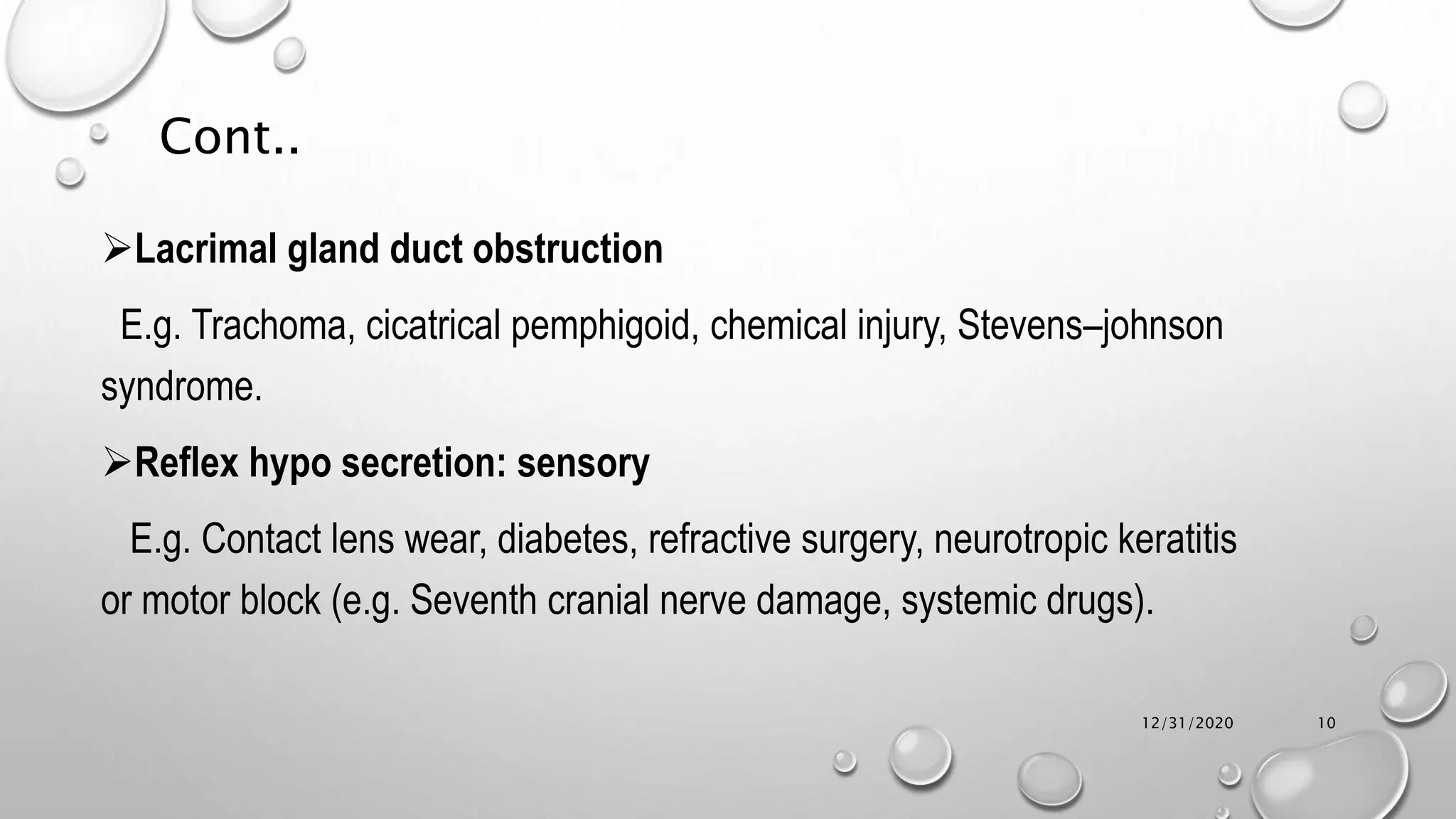
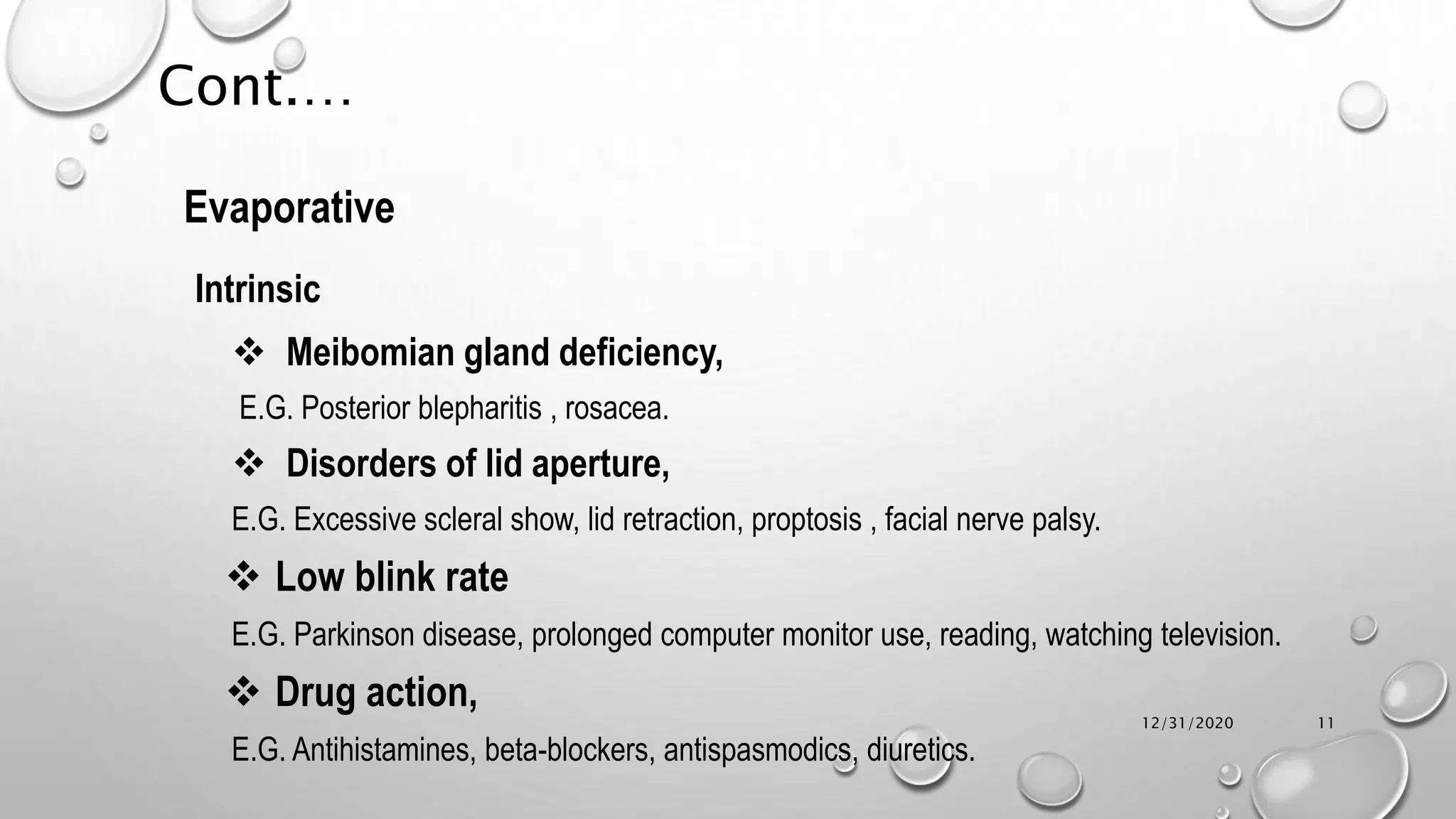
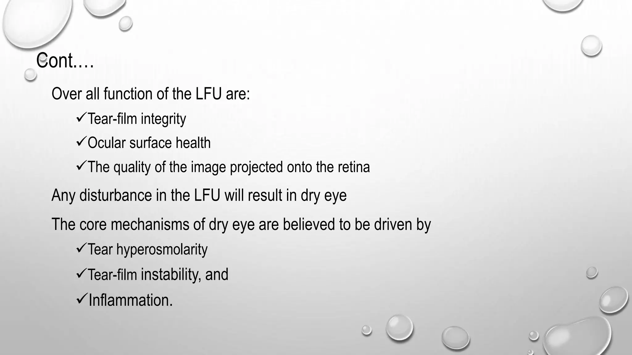
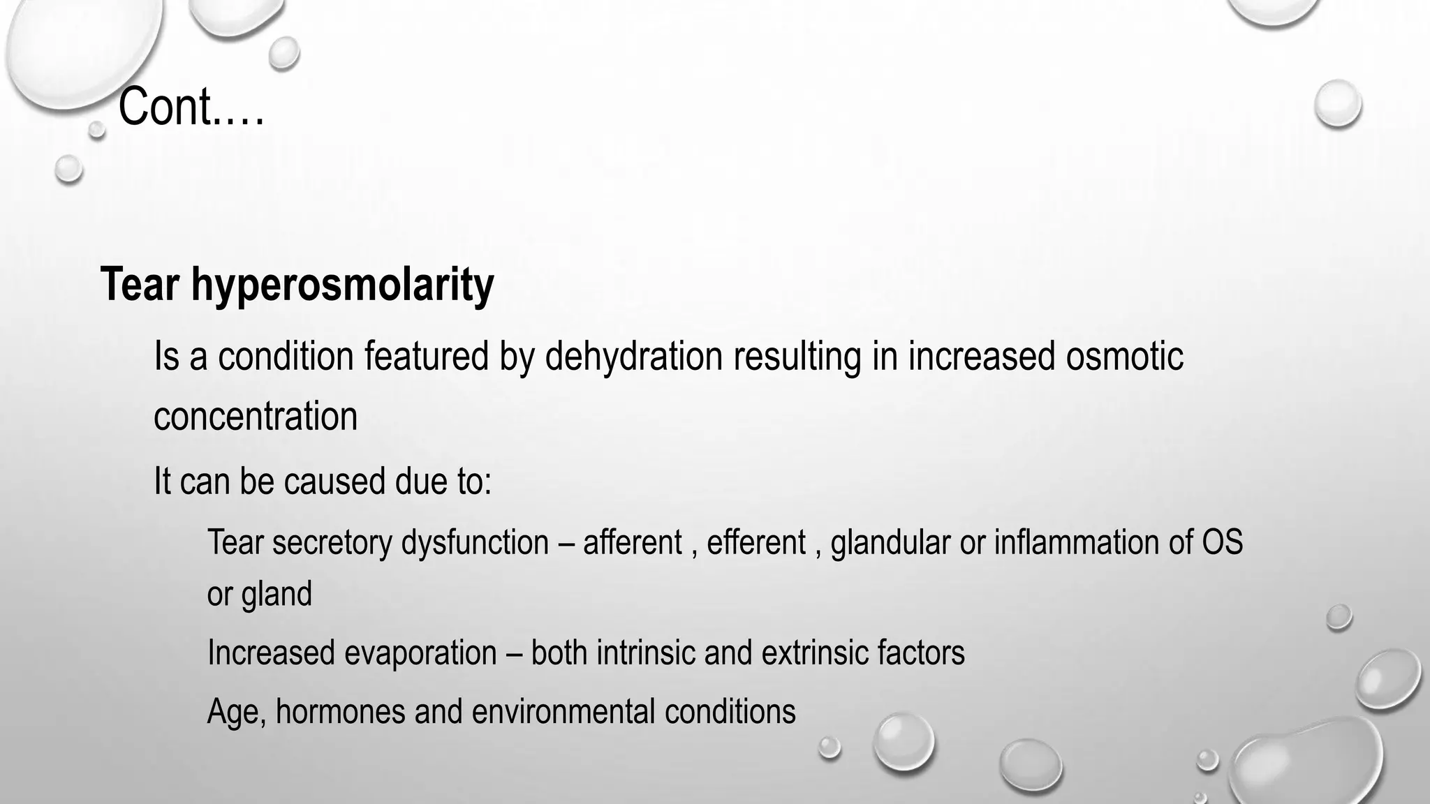
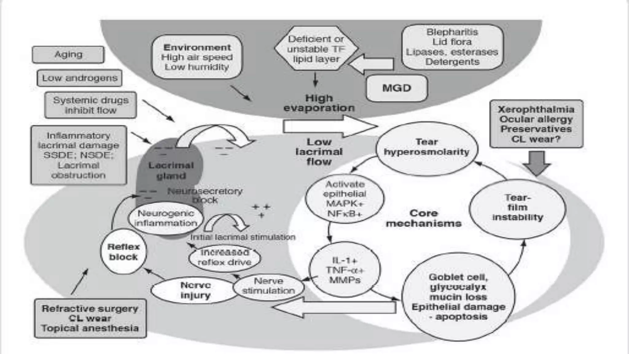
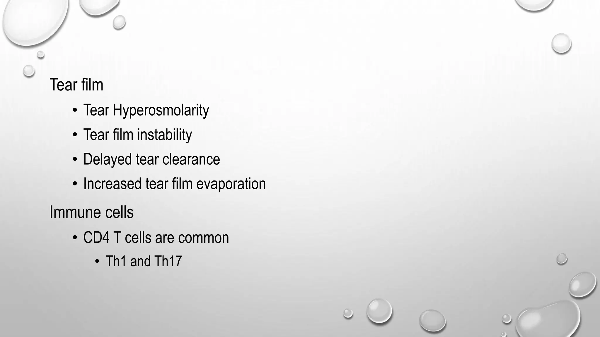
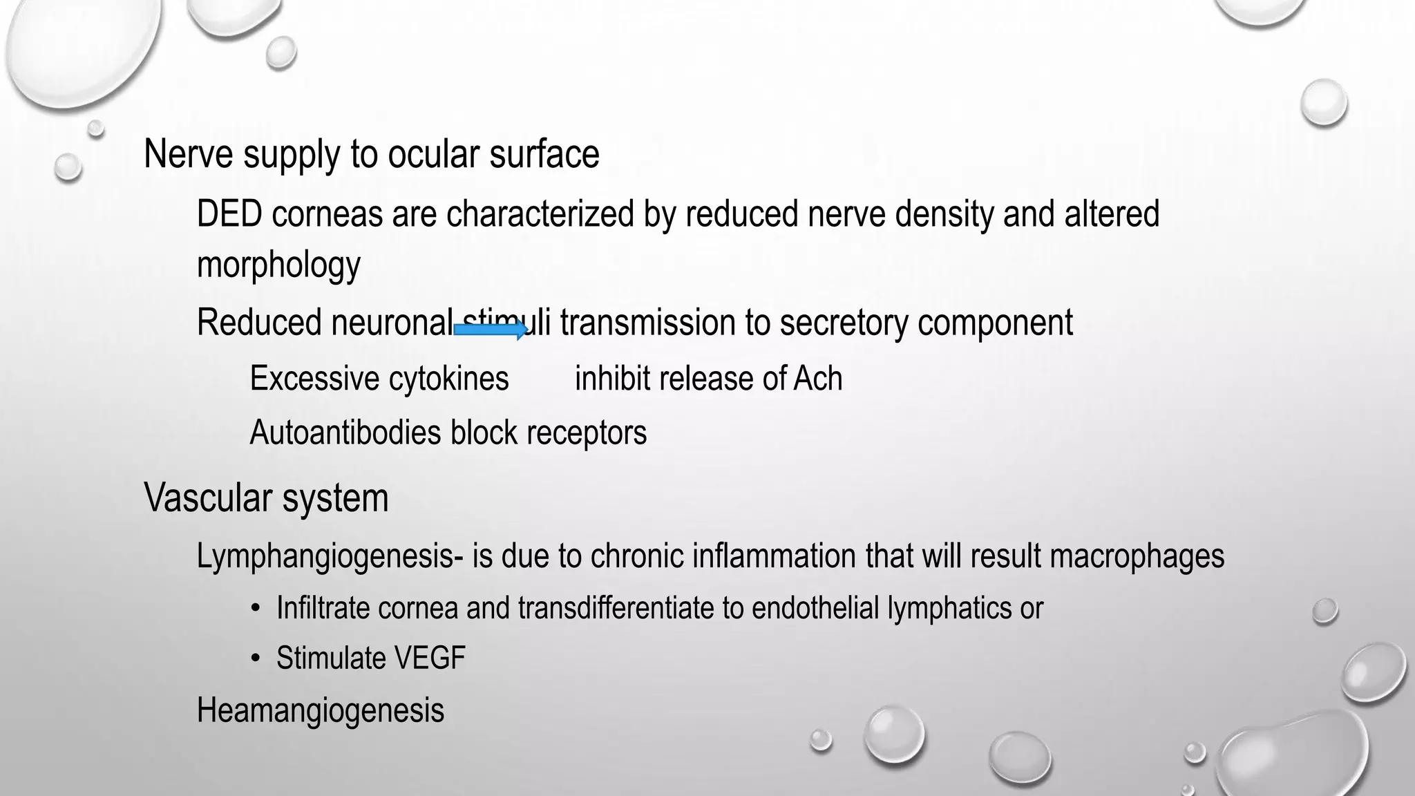
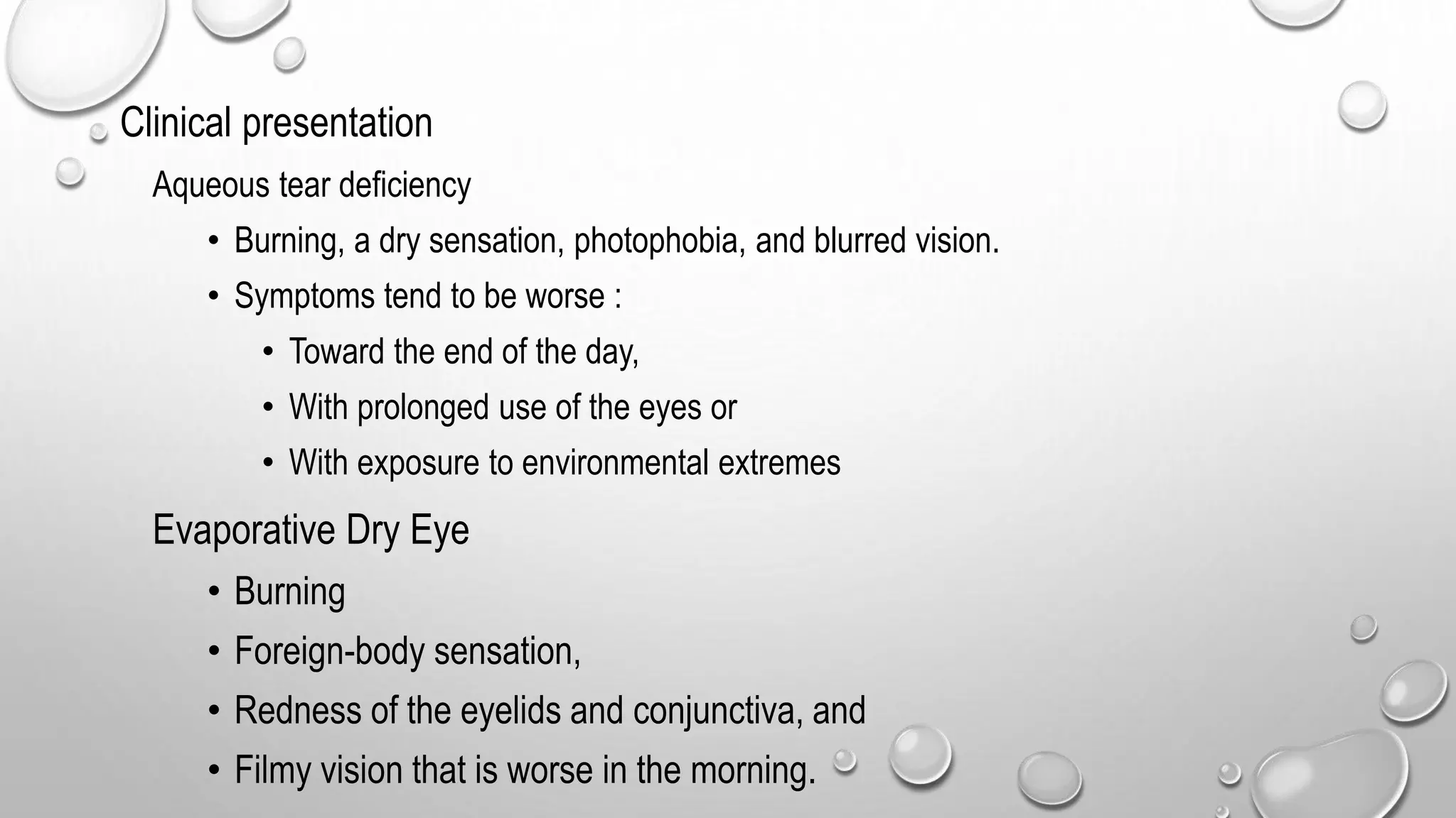
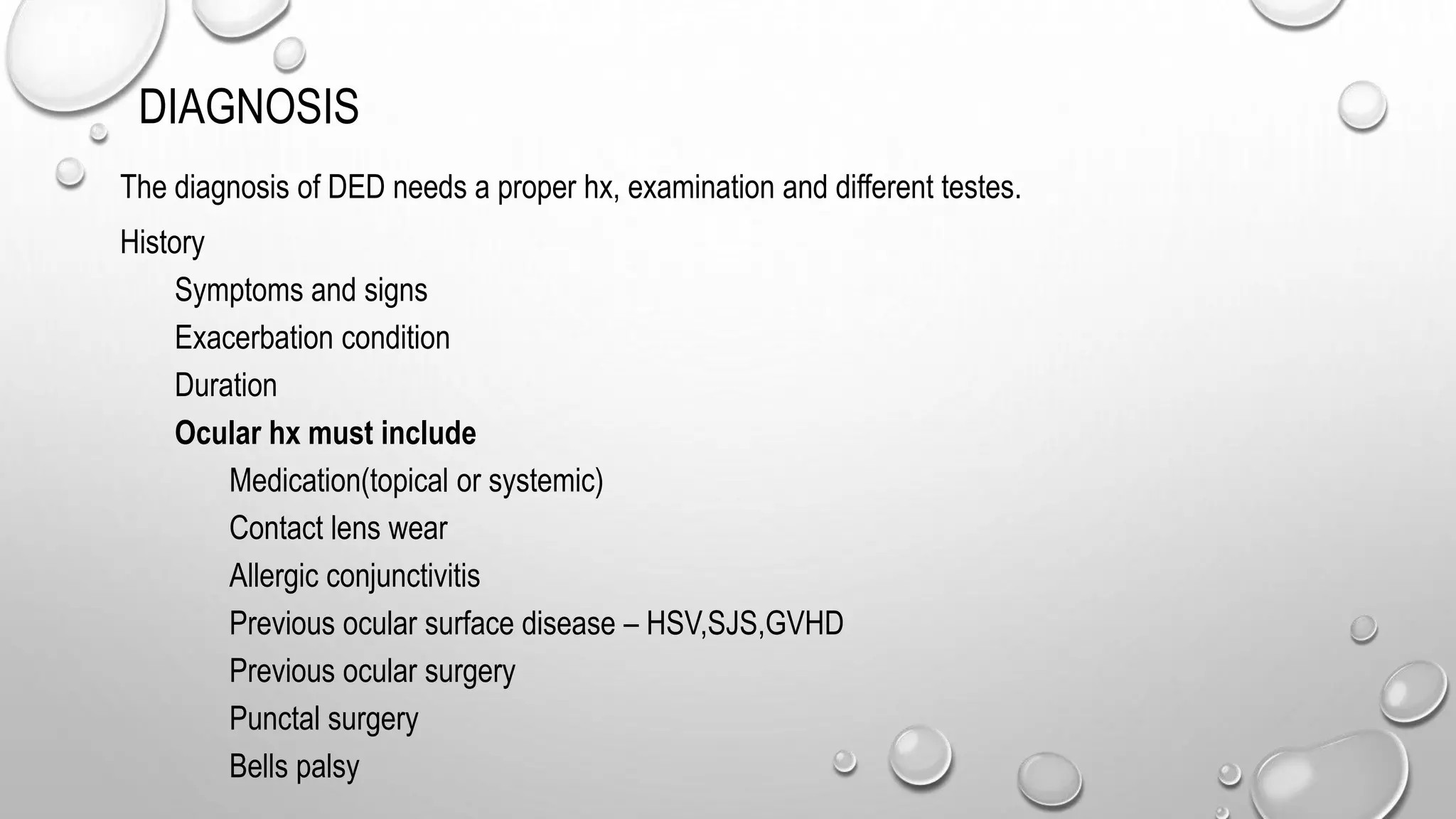
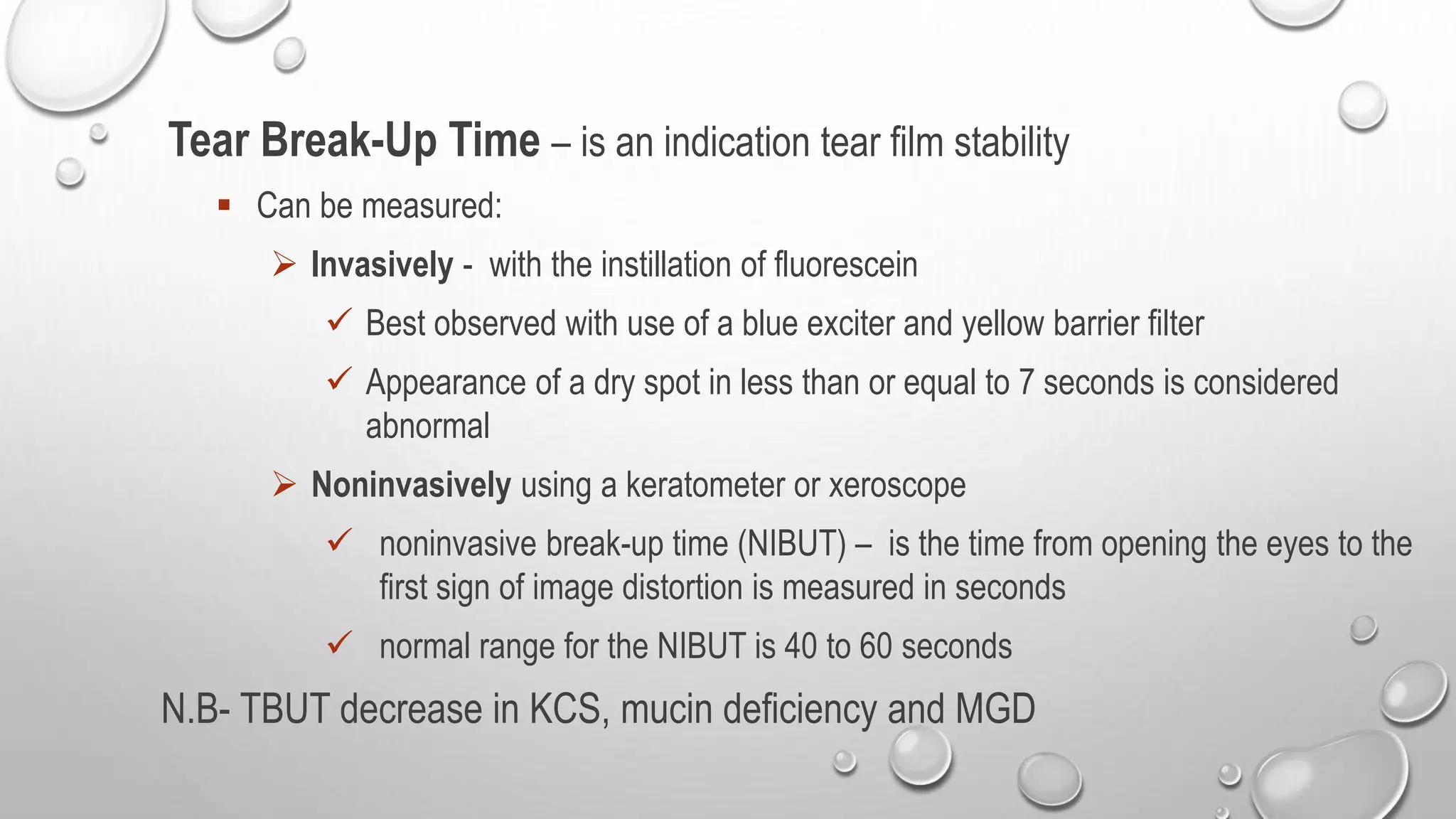
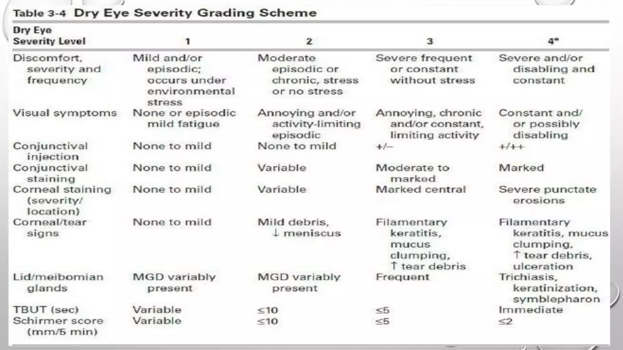
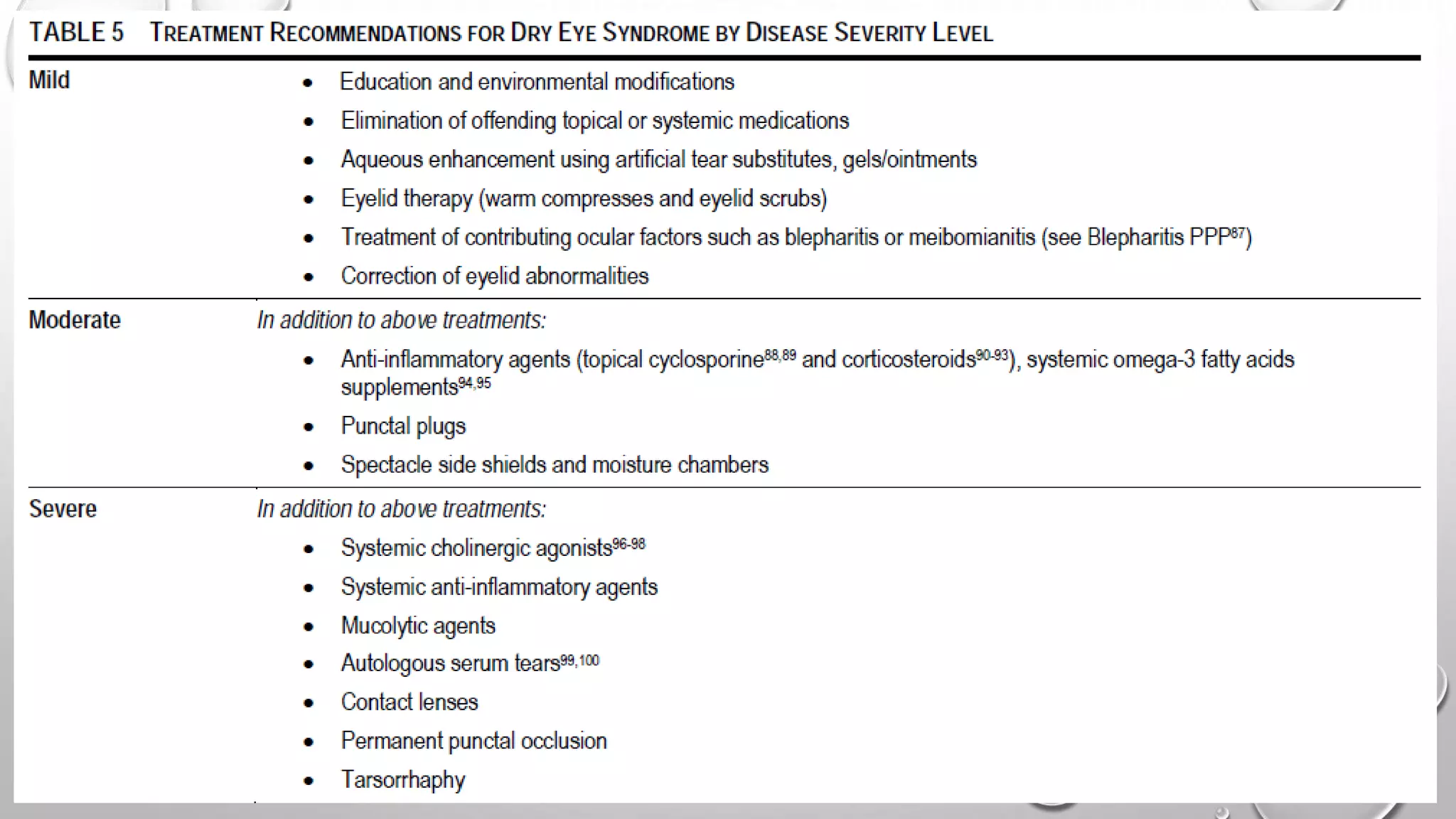
Dry eye syndrome is a highly prevalent disease characterized by loss of tear film homeostasis. It can be classified as aqueous-deficient or evaporative dry eye. Aqueous-deficient dry eye includes Sjögren's syndrome dry eye and non-Sjögren's syndrome dry eye. The pathophysiology involves tear hyperosmolarity, tear film instability, and inflammation of the ocular surface. Diagnosis is based on patient history, examination findings, tear break-up time, ocular surface staining, and tear secretion tests. Treatment depends on disease severity and may include artificial tears, anti-inflammatory medications, punctal plugs, and secretagogues.
Keratoconus is a non-inflammatory thinning of the cornea that causes it to take on a conical shape. It typically develops in adolescence and causes vision impairment due to irregular astigmatism. It is classified into four stages based on refractive error, corneal thickness and shape. While the exact cause is unknown, theories include genetic and enzymatic factors. It is often associated with eye rubbing and connective tissue disorders. Clinical features include corneal thinning, Fleischer's ring, Munson's sign, and scarring in advanced cases. Diagnosis involves topography, pachymetry and biomicroscopy to detect corneal shape changes.
Dry eye occurs when there is inadequate tear production or function, resulting in an unstable tear film and ocular surface disorder. It can be caused by conditions that reduce tear production such as Sjogren's syndrome, vitamin A deficiency, Stevens-Johnson syndrome, or medications. Other causes affect the tear film layers, like meibomian gland dysfunction reducing the outer lipid layer. Symptoms include dryness, burning, and blurred vision. Treatment focuses on replacing tears, improving ocular surface health, addressing underlying causes, and escalating care based on severity through the DEWS treatment guidelines.
Keratoplasty involves replacing diseased cornea with donor tissue. The main types are penetrating keratoplasty (PK), which replaces the full corneal thickness, and lamellar keratoplasty, which replaces only diseased layers. PK indications include scarring, infections, dystrophies and injuries. It has risks of rejection, infection, and high astigmatism. Newer techniques like deep anterior lamellar keratoplasty (DALK) and Descemet's membrane endothelial keratoplasty (DMEK) replace only diseased layers, reducing risks. Careful donor screening, surgical technique and postoperative management including steroids can reduce complications of keratoplasty.
Dry eye syndrome is a common condition characterized by discomfort, visual disturbance, and tear film instability. It results from reduced tear production or increased tear evaporation, causing tear hyperosmolarity and ocular surface inflammation. Diagnosis involves evaluating tear production via tests like Schirmer's test and tear breakup time, and examining the ocular surface for signs of damage. Treatment focuses on replacing tears and reducing inflammation with artificial tears, cyclosporine drops, and punctal plugs. More severe cases may require procedures like tarsorrhaphy.
Dry eye is a disease of the tear film and ocular surface caused by reduced tear production or increased tear evaporation. It results in eye discomfort, visual disturbance, and potential ocular surface damage. Dry eye can be caused by problems with the lacrimal functional unit such as aging, autoimmune disease like Sjogren's syndrome, or environmental factors. Diagnosis involves evaluating tear production via tests like Schirmer's test and tear breakup time, and assessing ocular surface staining. Treatment depends on dry eye severity and may include artificial tears, anti-inflammatories, punctal plugs, and management of underlying conditions. The goal is to supplement tears, reduce evaporation, stimulate natural tear production, and minimize
This document discusses various types of uveitis including classification, signs, symptoms, complications, investigations, and treatments. It covers infective causes like toxoplasmosis, tuberculosis, syphilis, and fungal infections. It also discusses immune-mediated uveitis associated with conditions like VKH syndrome, Behcet's disease, and sarcoidosis. Intermediate uveitis is characterized by inflammation of the pars plana, peripheral retina, and choroid. Posterior uveitis involves the retina and choroid and can be caused by infections, autoimmune diseases, or unknown etiologies.
Eales' disease is an idiopathic inflammatory retinal condition that primarily affects veins in the retina. It is characterized by perivascular inflammation that can lead to retinal ischemia and neovascularization. The disease progresses through stages from inflammation to ischemia to proliferation of new blood vessels. Treatment involves corticosteroids to reduce inflammation, laser photocoagulation to ablate new vessels, and vitrectomy for complications like vitreous hemorrhage or retinal detachment. While the exact cause is unknown, hypersensitivity to tuberculosis has been proposed as a potential trigger in many cases.
1. The tear film consists of three layers - an outer lipid layer, intermediate aqueous layer, and inner mucin layer. The lipid layer prevents evaporation while the aqueous layer nourishes the cornea and washes away debris.
2. Tears are produced both through basal secretion by accessory glands and reflex secretion by the main lacrimal gland in response to irritation. The tear film forms on the cornea through spreading of layers.
3. Diagnosis of dry eye involves tests like tear film break-up time (BUT), Schirmer's test, and Jones tests to evaluate tear production and drainage. Multiple factors can contribute to dry eye and tear film instability.
This document discusses surgical induced astigmatism following cataract surgery. It notes that astigmatism has a significant impact on vision and is influenced by surgical technique and incision size and type. Various factors can induce astigmatism including incision location and size, suture type and placement, and wound compression or gape. Evaluating astigmatism involves tools like retinoscopy, keratometry and corneal topography. Managing astigmatism may involve selective suture removal to reduce cylindrical error over time.
This document provides an overview of dry eye disease. It defines dry eye as a multifactorial disease resulting from tear deficiency or excess evaporation, causing symptoms like eye discomfort. Diagnostic tests evaluate tear secretion, stability, and damage to the ocular surface. Clinical features include irritation, redness, blurred vision, and staining of the cornea or conjunctiva. Dry eye exists on a spectrum of severity and has many predisposing factors like age, gender, medication use, contact lens wear, surgery, and autoimmune diseases.
Pseudoexfoliation syndrome is a systemic condition characterized by grey-white fibrillar deposits that can lead to open-angle glaucoma. It involves the trabecular meshwork, lens, ciliary body and other ocular tissues, and is a major risk factor for glaucoma. Treatment involves managing elevated intraocular pressure through medications, laser trabeculoplasty, trabeculectomy or cataract surgery due to the increased risk of complications from zonular weakness.
This document discusses corticosteroids and their use in ophthalmology. It begins by describing the basic structure and functions of steroids produced naturally in the body. It then outlines the history of corticosteroid discovery and use in medicine, including their introduction to ocular therapy in the 1950s. The document goes on to explain the mechanisms of action of corticosteroids and their effects on inflammation. It provides details on the administration, pharmacokinetics, efficacy and side effects of systemic corticosteroid use as well as topical ocular administration through eye drops, ointments and injections. Guidelines are given for dosing and monitoring patients on long-term corticosteroid therapy.
The cornea is the transparent front part of the eye that transmits and focuses light. It has 3 main layers - an outer epithelial layer, a thick middle stromal layer made of collagen, and an inner single-cell endothelial layer. The cornea derives its strength and curvature from the orderly arrangement of collagen in the stroma. It remains transparent due to its regular structure without blood vessels and the deturgescent properties maintained by the endothelial pump. The cornea has a high metabolic rate powered by glucose and oxygen and is innervated by nerves for vision and protection.
Techniques of tear film evaluation by Raju KaitiRaju Kaiti
The document summarizes techniques for evaluating the tear film, which has three layers: an outer lipid layer, intermediate aqueous layer, and inner mucous layer. Non-invasive techniques discussed include tear break-up time tests, lipid layer evaluation using interferometry, and inferior tear meniscus height measurements. Invasive techniques involve Schirmer's tests to evaluate tear secretion, fluorescein and rose bengal staining of the ocular surface, and conjunctival impression cytology to examine goblet cell density. The document provides details on procedures and normal results for each evaluation method.
This document discusses branch retinal vein occlusion (BRVO), including its pathogenesis, clinical features, complications, investigations, and management. BRVO is caused by obstruction of one of the retinal veins, usually at the arteriovenous crossing point. It can lead to macular edema, neovascularization, vitreous hemorrhage, and retinal detachment. Treatment involves anti-VEGF injections, steroids, laser photocoagulation, and occasionally surgery. Several clinical trials have evaluated therapies for BRVO, finding that anti-VEGF drugs and steroids reduce macular edema but laser provides little additional benefit when combined with anti-VEGF treatment.
The document discusses the anatomy and physiology of the lacrimal system and tear film. It describes the main structures of the lacrimal apparatus including the lacrimal gland, accessory lacrimal glands, lacrimal passages, puncta, canaliculi, lacrimal sac, and nasolacrimal duct. It discusses tear film layers, secretion, and functions. It also summarizes common lacrimal system disorders like dry eye, dacryocystitis, epiphora, dacryoadenitis and their signs, symptoms, etiologies, and treatments. Surgical procedures for these conditions like dacryocystorhinostomy are also briefly outlined.
This document discusses various types of corneal dystrophies presented by Dr. Puneet Sharma. It defines corneal dystrophies as a group of progressive, genetically determined disorders that cause non-inflammatory opacification of the cornea. It then describes several specific corneal dystrophies categorized by the layer of the cornea they affect (epithelial, stromal, endothelial). For each dystrophy, it covers inheritance pattern, histology, onset, signs/symptoms, and treatment. The document provides detailed information on Cogan dystrophy, Meesmann dystrophy, Reis-Bucklers dystrophy, granular dystrophy types 1 and 2, lattice dystrophy types 1 and 2, and Fuchs endothelial
This document discusses fungal corneal ulcers. It begins by describing the signs and symptoms of fungal ulcers, including pain, redness, defective vision, lid edema, and corneal opacity staining with fluorescein. Diagnosis involves smears, cultures and microscopy to identify causative fungi. Common fungi include Fusarium, Aspergillus, and Candida. Treatment involves topical natamycin or voriconazole drops. Prognosis includes potential complications like scar formation, astigmatism, perforation and fistula formation. Close monitoring is needed due to the difficulty treating fungal infections.
Cornea is the clear front surface of the eye. It lies directly in front of the iris and pupil, and it allows light to enter the eye.
Cornea forms the transparent and anterior 1/6th of the external fibrous coat of the globe of the eyeball.
The cornea is the eye's most powerful structure for focusing light that provides approximately 65 to 75 percent of the focusing power of the eye.
The cornea has unmyelinated nerve endings sensitive to touch, temperature and chemicals; a touch of the cornea causes an involuntary reflex to close the eyelid.
The document discusses the anatomy, embryology, and function tests of the macula lutea. It describes the macula lutea as a 5.5mm circular area at the posterior pole of the retina that subserves central vision. It notes the macula's delayed development until 8 months gestation and specialization of the fovea which contains the highest concentration of cones. The document outlines various macular function tests used to evaluate macular diseases, including visual acuity, Amsler grid, microperimetry, and electroretinography. It provides details on the anatomy and cell layers of the fovea centralis and techniques for assessing macular integrity with tests like the Maddox rod.
Chronic conjunctivitis can be caused by infection or allergy. Infectious causes include trachoma caused by Chlamydia trachomatis, which is the leading cause of preventable blindness worldwide. If left untreated, repeated trachoma infections can lead to scarring of the conjunctiva and complications like trichiasis and corneal opacity. Granulomatous conjunctivitis presents with localized conjunctival granulomas and lymphadenopathy in conditions like cat scratch disease or tuberculosis. Non-specific chronic conjunctivitis can be caused by chronic irritation or hypersensitivity reactions.
This document provides an overview of evaluating a glaucoma patient. It discusses the classification, clinical evaluation, history, clinical examination including slit lamp biomicroscopy, tonometry, gonioscopy, optic disc evaluation, visual field testing, and OCT. Classification is based on whether glaucoma is congenital or acquired, primary or secondary, open angle or angle-closure. The clinical evaluation aims to diagnose the specific form of glaucoma, determine severity, and assess disease progression. A thorough history and clinical examination are essential for appropriate glaucoma management.
- Aphakia is the absence of the crystalline lens from the eye. It can be congenital or caused by surgery or trauma.
- In aphakia, the eye becomes highly hyperopic, the anterior focal point moves forward, and the retinal image is magnified. This decreases visual acuity and field of view.
- Aphakia is treated with spectacles, contact lenses, or intraocular lenses. Spectacles cause issues like increased image size, ring scotomas, and reduced field of view. Contact lenses and IOLs provide better image quality but have risks of complications.
Lacrimation refers to watering of the eye due to excessive tear production from the lacrimal gland, while epiphora is watering caused by obstruction of the tear drainage system. Differentiating the two is important to provide proper treatment. The document discusses the anatomy and physiology of tear production, drainage, and evaluates causes of watering eye including lacrimation, dry eye, blepharitis, and epiphora due to punctal stenosis or nasolacrimal duct obstruction. Treatment depends on the underlying cause and may include punctal dilation, dacryocystorhinostomy, Jones tube insertion, or endoscopic lacrimal surgery.
Global estimates indicate that the prevalence of glaucoma is 3.54% worldwide, and the number of people affected is projected to increase from 64.3 million in 2013 to 111.8 million in 2040. Glaucoma is the leading cause of irreversible blindness, with open-angle glaucoma affecting an estimated 4.5 million people and angle-closure glaucoma affecting 3.9 million people globally. Pseudoexfoliation syndrome is the most common cause of secondary open-angle glaucoma worldwide and is characterized by the deposition of pseudoexfoliative material in various structures of the anterior eye segment.
Dry Eye Disease is a multifactorial disease that results in discomfort and visual disturbances due to tear film instability and inflammation of the ocular surface. It is caused by a disturbance in the lacrimal functional unit involving tear production, distribution, and clearance. Diagnosis involves evaluating tear secretion, volume, and stability as well as assessing ocular surface damage. Management focuses on eliminating exacerbating factors, supplementing tears, anti-inflammatory therapy, punctal plugs, and newer treatments targeting tear stimulation and mucous secretion when needed.
The document discusses dry eye, defining it as a disorder of the tear film due to reduced tear production or excessive evaporation, causing damage to the ocular surface. It describes the layers of the tear film and their functions in lubricating the eye and protecting it. Symptoms of dry eye are discussed. Diagnostic tests for dry eye including tear break-up time, ocular surface staining, and Schirmer test are summarized. Management strategies are outlined for mild, moderate, and severe dry eye, including artificial tears, anti-inflammatory therapies, punctal plugs, and systemic treatments.
Dry eye is a multifactorial disease caused by tear deficiency or excessive evaporation that damages the ocular surface. It is characterized by discomfort, visual disturbance, and tear film instability. The tear film consists of an outer lipid layer, middle aqueous layer, and inner mucin layer that together form a protective film over the cornea. Dry eye can be caused by deficiencies in tear production, meibomian gland dysfunction, or other factors. It leads to ocular surface inflammation, damage, and patient symptoms that degrade vision and cause fatigue.
Dry eye is a multifactorial disease that results in ocular discomfort and visual disturbance. It is caused by decreased tear production or increased tear evaporation, which leads to ocular surface inflammation and damage. Dry eye ranges in severity from mild intermittent symptoms to severe constant symptoms that limit daily activities. It is more common in older adults and women, and environmental factors, medications, contact lens use, and autoimmune diseases can trigger or exacerbate dry eye.
This document discusses dry eye, including its definition, causes, symptoms, and treatment approaches. It begins by defining dry eye as a multifactorial disease that results in discomfort, visual disturbance, and tear film instability. Various etiological factors that can contribute to dry eye are then outlined, including age, gender, medications, environmental conditions, and ocular surface diseases. Treatment is tailored based on the severity of dry eye and may include artificial tears, anti-inflammatory therapies, punctal plugs, and procedures to improve meibomian gland function. Modern technologies like LipiFlow are also mentioned for managing meibomian gland dysfunction.
This document provides information about dry eye disease (DED). It discusses the anatomy and function of the tear film layers. DED is defined as a multifactorial disease caused by tear film instability and inflammation. Risk factors include older age, female sex, menopause, medications, contact lens use and environmental exposures. Signs and symptoms include eye irritation, punctate keratitis, and decreased tear production. Tests to diagnose DED include tear breakup time, Schirmer test, and ocular surface staining. DED can range from mild to severe and cause complications like corneal neovascularization if left untreated.
This document provides information about keratoconus, a non-inflammatory thinning of the cornea that causes a cone-shaped bulge. It is most common in teenagers and young adults. The document discusses the definition, stages, etiology, associations, clinical features, investigations, and management of keratoconus. Keratoconus is typically managed initially with glasses or contact lenses, and more advanced cases may require collagen cross-linking, intracorneal ring segments, deep anterior lamellar keratoplasty, or penetrating keratoplasty to correct vision and stop further thinning. Differential diagnoses include keratoglobus and pellucid marginal degeneration.
This document provides an overview of dry eye syndrome. It begins by defining dry eye syndrome as a condition caused by a breakdown of the tear film layer that coats the front of the eye. It then describes the three layers that make up the tear film - lipid, aqueous, and mucus layers. The document outlines the symptoms, etiologies, diagnostic tests, and management of dry eye syndrome. Key tests include tear breakup time, ocular surface staining, Schirmer test, and tear osmolarity measurement. Psychiatric conditions and medications can also contribute to dry eye.
Dry eye, also known as dysfunctional tear syndrome, is a multifactorial disease of the tears and ocular surface. It results in symptoms of discomfort, visual disturbance, and tear film instability, and can potentially damage the ocular surface. The document discusses the prevalence, classification, signs, symptoms, and diagnostic tests and tools for dry eye. The prevalence increases significantly with age and is higher in women. Diagnosis involves evaluating symptoms, performing a slit lamp exam to check for signs of damage, and conducting tests to assess tear film stability, tear secretion, and ocular surface damage. Management involves classifying dry eye severity levels based on signs and symptoms to determine appropriate treatment.
Dry eye is a common disease caused by insufficient tear production or increased tear evaporation. It affects 10-15% of the population and prevalence is higher in females and older individuals. The tear film consists of an outer lipid layer, middle aqueous layer, and inner mucus layer which together form a smooth optical surface and nourish the cornea. Dry eye is diagnosed based on symptoms, tear film breakup time, Schirmer's test, and ocular surface staining. Treatment involves artificial tears, punctal plugs, anti-inflammatories, and managing underlying conditions. Advanced cases may require punctal occlusion, contact lenses, or autologous serum tears.
This document discusses the management of dry eye. It begins by defining dry eye as a multifactorial disease resulting in ocular discomfort and potential damage due to increased tear film osmolarity and ocular surface inflammation. Dry eye is commonly seen in clinical practice and can range from mild to severe. The document then discusses the anatomy and physiology of tear production, the tear film layers, contributing factors, classifications, signs and symptoms, diagnostic tests, and treatment strategies including artificial tears and anti-inflammatory therapies. Treatment is tailored based on dry eye severity levels with the goal of alleviating symptoms and preventing complications.
Keratoconus is a degenerative eye condition where the cornea thins and changes shape, causing vision problems. The cause is unknown but risk factors include eye rubbing and genetics. Symptoms include progressively worsening vision not fully corrected by glasses. Diagnosis involves examining the cornea shape using keratometry and topography to detect thinning, steepening, and irregular astigmatism. Mild cases may need no treatment, while progressive cases can be managed with contact lenses or corneal cross-linking depending on severity.
Epiphora, or watery eyes, is caused by a disruption in the balance between tear production and drainage. It is a common complaint that can develop at any age but is more frequent in babies under 1 and adults over 60. The causes include abnormalities that increase tearing or decrease drainage. Diagnosis involves examining for eyelid issues, dry eye, and conducting tests like syringing and probing. Treatment depends on the underlying causes but may include lid hygiene, artificial tears, lacrimal sac massage, punctal plugs, and surgery to repair drainage issues. Management involves a holistic approach targeting contributing factors and appropriate referral for surgical cases.
Dry eye is a disease of the ocular surface caused by disturbances in the tear film. The normal tear film consists of an inner mucin layer, middle aqueous layer, and outer lipid layer, which work together to form a stable tear film. Disruptions to the tear film components or their functions can lead to dry eye. Common tests to diagnose dry eye include tear break-up time, fluorescein clearance, and tear osmolarity measurement, which help identify tear film instability and inflammation associated with the condition.
This document summarizes several pediatric ocular diseases and their systemic associations. It describes the clinical features and treatments of Alport's syndrome, Alstrom syndrome, CHARGE association syndrome, Stevens-Johnson syndrome, cat scratch disease, herpes simplex virus, rubella, varicella-zoster virus, and references for further information. For each condition, it outlines ocular findings such as lenticonus, flecks, uveitis, conjunctivitis and keratitis as well as associated systemic symptoms and recommended treatment approaches.
Dry eye disease, also known as keratoconjunctivitis sicca, is a condition caused by disturbances in the tear film that leads to an unstable tear film when the eye is open. It is a common reason for visits to ophthalmologists and affects about 75% of people over age 65, with the average patient being 54 years old. The disease involves inflammation that disrupts the normal feedback loop controlling tear production. Diagnosis involves tests like tear breakup time and Schirmer tests, and treatment focuses on replacing tears through artificial drops or increasing natural tear production through drugs like cyclosporine drops.
Keratoconus is a non-inflammatory bilateral ectatic condition of the cornea that causes progressive thinning and conical protrusion. It typically starts during puberty and progresses slowly. Symptoms include irregular astigmatism and myopia. Signs include Fleischer's ring, Munson's sign, and irregular astigmatism on retinoscopy. Treatment options include glasses, rigid gas permeable contact lenses, INTACS, collagen cross-linking, and keratoplasty for later stages. Keratoglobus is a rare noninflammatory diffuse corneal ectasia present from birth. Lenticonus is a rare congenital anomaly of the crystalline lens characterized by a conical protrusion on the lens capsule.
Dry eye syndrome is characterized by ocular irritation and visual disturbance caused by alterations to the tear film and ocular surface. It can range from minor inconvenience to sight-threatening complications. While traditionally focused on inadequate tear secretion, the tear film depends on normal function of multiple components. Current treatment targets tear supplementation but often ignores multiple deficiency states, resulting in treatment failure. There is a need for improved treatment options.
Red eye, or ocular hyperemia, is caused by enlarged, dilated blood vessels leading to redness of the eye's surface. It can be caused by various conditions affecting the conjunctiva, cornea, iris, anterior chamber, eyelids, or orbit. Common causes include conjunctivitis (bacterial, viral, allergic), corneal abrasions, iritis, acute glaucoma, eyelid disorders like entropion or ectropion, and orbital cellulitis. A thorough history and examination is needed to determine the underlying cause and guide appropriate treatment.
Adhd Medication Shortage Uk - trinexpharmacy.comreignlana06
The UK is currently facing a Adhd Medication Shortage Uk, which has left many patients and their families grappling with uncertainty and frustration. ADHD, or Attention Deficit Hyperactivity Disorder, is a chronic condition that requires consistent medication to manage effectively. This shortage has highlighted the critical role these medications play in the daily lives of those affected by ADHD. Contact : +1 (747) 209 – 3649 E-mail : sales@trinexpharmacy.com
Recomendações da OMS sobre cuidados maternos e neonatais para uma experiência pós-natal positiva.
Em consonância com os ODS – Objetivos do Desenvolvimento Sustentável e a Estratégia Global para a Saúde das Mulheres, Crianças e Adolescentes, e aplicando uma abordagem baseada nos direitos humanos, os esforços de cuidados pós-natais devem expandir-se para além da cobertura e da simples sobrevivência, de modo a incluir cuidados de qualidade.
Estas diretrizes visam melhorar a qualidade dos cuidados pós-natais essenciais e de rotina prestados às mulheres e aos recém-nascidos, com o objetivo final de melhorar a saúde e o bem-estar materno e neonatal.
Uma “experiência pós-natal positiva” é um resultado importante para todas as mulheres que dão à luz e para os seus recém-nascidos, estabelecendo as bases para a melhoria da saúde e do bem-estar a curto e longo prazo. Uma experiência pós-natal positiva é definida como aquela em que as mulheres, pessoas que gestam, os recém-nascidos, os casais, os pais, os cuidadores e as famílias recebem informação consistente, garantia e apoio de profissionais de saúde motivados; e onde um sistema de saúde flexível e com recursos reconheça as necessidades das mulheres e dos bebês e respeite o seu contexto cultural.
Estas diretrizes consolidadas apresentam algumas recomendações novas e já bem fundamentadas sobre cuidados pós-natais de rotina para mulheres e neonatos que recebem cuidados no pós-parto em unidades de saúde ou na comunidade, independentemente dos recursos disponíveis.
É fornecido um conjunto abrangente de recomendações para cuidados durante o período puerperal, com ênfase nos cuidados essenciais que todas as mulheres e recém-nascidos devem receber, e com a devida atenção à qualidade dos cuidados; isto é, a entrega e a experiência do cuidado recebido. Estas diretrizes atualizam e ampliam as recomendações da OMS de 2014 sobre cuidados pós-natais da mãe e do recém-nascido e complementam as atuais diretrizes da OMS sobre a gestão de complicações pós-natais.
O estabelecimento da amamentação e o manejo das principais intercorrências é contemplada.
Recomendamos muito.
Vamos discutir essas recomendações no nosso curso de pós-graduação em Aleitamento no Instituto Ciclos.
Esta publicação só está disponível em inglês até o momento.
Prof. Marcus Renato de Carvalho
www.agostodourado.com
Rasamanikya is a excellent preparation in the field of Rasashastra, it is used in various Kushtha Roga, Shwasa, Vicharchika, Bhagandara, Vatarakta, and Phiranga Roga. In this article Preparation& Comparative analytical profile for both Formulationon i.e Rasamanikya prepared by Kushmanda swarasa & Churnodhaka Shodita Haratala. The study aims to provide insights into the comparative efficacy and analytical aspects of these formulations for enhanced therapeutic outcomes.
Here is the updated list of Top Best Ayurvedic medicine for Gas and Indigestion and those are Gas-O-Go Syp for Dyspepsia | Lavizyme Syrup for Acidity | Yumzyme Hepatoprotective Capsules etc
ABDOMINAL TRAUMA in pediatrics part one.drhasanrajab
Abdominal trauma in pediatrics refers to injuries or damage to the abdominal organs in children. It can occur due to various causes such as falls, motor vehicle accidents, sports-related injuries, and physical abuse. Children are more vulnerable to abdominal trauma due to their unique anatomical and physiological characteristics. Signs and symptoms include abdominal pain, tenderness, distension, vomiting, and signs of shock. Diagnosis involves physical examination, imaging studies, and laboratory tests. Management depends on the severity and may involve conservative treatment or surgical intervention. Prevention is crucial in reducing the incidence of abdominal trauma in children.
Integrating Ayurveda into Parkinson’s Management: A Holistic ApproachAyurveda ForAll
Explore the benefits of combining Ayurveda with conventional Parkinson's treatments. Learn how a holistic approach can manage symptoms, enhance well-being, and balance body energies. Discover the steps to safely integrate Ayurvedic practices into your Parkinson’s care plan, including expert guidance on diet, herbal remedies, and lifestyle modifications.
Vestibulocochlear Nerve by Dr. Rabia Inam Gandapore.pptx
Dry eye syndrome
1. DRY EYE SYNDROME
PRESENTER – DR.BEMNET T.
MODERATOR – DR.YARED A.(MD ,MPH,
ASSOCIATE PROFESSOR OF OPHTHALMOLOGY,
SUBSPECIALTY IN ANTERIOR SEGMENT AND CORNEAL
DISEASE)
3. INTRODUCTION
• Dry eye syndrome is a highly prevalent and multifactorial disease of the
tear film and ocular surface
• Can cause problems ranging from mildly irritating to impaired vision
• And also can affect the outcome of corneal, cataract and refractive
surgeries
• Mostly symptoms of dry eye improves with treatment but usually its not
curable
4. DEFINITION
• The definition of dry eye syndrome is still under continues revision
• According to 2017 TFOS DEWS II Dry eye is:
“A multifactorial disease of the ocular surface characterized by a loss
of homeostasis of the tear film and accompanied by ocular symptoms,
in which tear film instability and hyperosmolarity, ocular surface
inflammation and damage, and neuro sensory abnormalities play
etiological roles. ”
5. CLASSIFICATION
In 2007/2017 international dry eye workshop (DEWS),classify dry eye into:
Aqueous-deficient – is due to lacrimal gland dysfunction
Sjögren syndrome dry eye (primary or secondary).
Non- Sjögren syndrome dry eye.
Evaporative
Intrinsic
Extrinsic
6.
7. Cont.…
Aqueous deficient dry eye
Sjögren syndrome dry eye
Is an autoimmune disorder characterized by lymphocytic
inflammation
Mainly involving lacrimal and parotid gland but it also can affect any
exocrine gland.
Inflammation block release of neurotransmitters or impact the
cellular response to neuromediators.
12/31/2020 7
8. Cont..
The classic clinical triad consists of dry eyes, dry mouth and parotid
gland enlargement
It can present as:
Primary – when it present in isolation
Secondary – when it present with another autoimmune disorder.
9. Cont..
Non - Sjögren syndrome dry eye
It can be due to:
Lacrimal deficiency:
Primary (e.g. Age-related dry eye , congenital alacrima , familial
dysautonomia ) or
Secondary (e.g. Inflammatory and neoplastic lacrimal gland
infiltration, acquired immunodeficiency syndrome (AIDS), graft-
versus-host disease, lacrimal gland or nerve ablation).
12/31/2020 9
10. Cont..
Lacrimal gland duct obstruction
E.g. Trachoma, cicatrical pemphigoid, chemical injury, Stevens–johnson
syndrome.
Reflex hypo secretion: sensory
E.g. Contact lens wear, diabetes, refractive surgery, neurotropic keratitis
or motor block (e.g. Seventh cranial nerve damage, systemic drugs).
12/31/2020 10
11. Cont.…
Evaporative
Intrinsic
Meibomian gland deficiency,
E.G. Posterior blepharitis , rosacea.
Disorders of lid aperture,
E.G. Excessive scleral show, lid retraction, proptosis , facial nerve palsy.
Low blink rate
E.G. Parkinson disease, prolonged computer monitor use, reading, watching television.
Drug action,
E.G. Antihistamines, beta-blockers, antispasmodics, diuretics.
12/31/2020 11
12. Cont..
Extrinsic
○ Vitamin A deficiency.
○ Topical drugs including the effect of preservatives.
○ Contact lens wear.
○ Ocular surface disease such as allergic conjunctivitis
12/31/2020 12
13. Pathophysiology
• The well accepted theory is based on the concept of LFU
• The LFU Consist :
Ocular surface – cornea , conjunctiva and meibomian gland
Main and accessory lacrimal gland
Eye lid
Neural network – CN V AND VII
• OSM
14. Cont.…
Over all function of the LFU are:
Tear-film integrity
Ocular surface health
The quality of the image projected onto the retina
Any disturbance in the LFU will result in dry eye
The core mechanisms of dry eye are believed to be driven by
Tear hyperosmolarity
Tear-film instability, and
Inflammation.
15. Cont.…
Tear hyperosmolarity
Is a condition featured by dehydration resulting in increased osmotic
concentration
It can be caused due to:
Tear secretory dysfunction – afferent , efferent , glandular or inflammation of OS
or gland
Increased evaporation – both intrinsic and extrinsic factors
Age, hormones and environmental conditions
16. Tear film instability
Could be secondary to hyperosmolarity or prime to it
Is caused by;
• Reduced tear production
• Delayed tear clearance
• Reduced quantity and quality of lipid
• OS Irregularity and inflammation
17. Cont.…
Inflammation
The osmotic stress on ocular surface will result in the release of : IL-1B ,TNF-a ,IL-
8, MMP
This cytokines will result in localized autoimmunity
They also obstruct tearing reflex
• By stimulating release of opioids
• By directly binding to receptors
Once inflammation occur it will result ocular surface damage
18.
19. Ocular surface microenvironment change in dry eye
Cornea
Corneal scaring, opacification and degeneration
Corneal thinning
Epithelial microvilli reduction
Central endothelial density reduction
NV, pannus formation and ulcers
20. Conjunctiva
• Squamous metaplasia
• Goblet cell reduction
• Conjunctivochalasis
Lacrimal gland
• Loss of secretory acinar cells
• Fibrosis and gland atrophy
22. Tear film
• Tear Hyperosmolarity
• Tear film instability
• Delayed tear clearance
• Increased tear film evaporation
Immune cells
• CD4 T cells are common
• Th1 and Th17
23. Nerve supply to ocular surface
DED corneas are characterized by reduced nerve density and altered
morphology
Reduced neuronal stimuli transmission to secretory component
Excessive cytokines inhibit release of Ach
Autoantibodies block receptors
Vascular system
Lymphangiogenesis- is due to chronic inflammation that will result macrophages
• Infiltrate cornea and transdifferentiate to endothelial lymphatics or
• Stimulate VEGF
Heamangiogenesis
24. Clinical presentation
Aqueous tear deficiency
• Burning, a dry sensation, photophobia, and blurred vision.
• Symptoms tend to be worse :
• Toward the end of the day,
• With prolonged use of the eyes or
• With exposure to environmental extremes
Evaporative Dry Eye
• Burning
• Foreign-body sensation,
• Redness of the eyelids and conjunctiva, and
• Filmy vision that is worse in the morning.
25. DIAGNOSIS
The diagnosis of DED needs a proper hx, examination and different testes.
History
Symptoms and signs
Exacerbation condition
Duration
Ocular hx must include
Medication(topical or systemic)
Contact lens wear
Allergic conjunctivitis
Previous ocular surface disease – HSV,SJS,GVHD
Previous ocular surgery
Punctal surgery
Bells palsy
31. Tear Break-Up Time – is an indication tear film stability
Can be measured:
Invasively - with the instillation of fluorescein
Best observed with use of a blue exciter and yellow barrier filter
Appearance of a dry spot in less than or equal to 7 seconds is considered
abnormal
Noninvasively using a keratometer or xeroscope
noninvasive break-up time (NIBUT) – is the time from opening the eyes to the
first sign of image distortion is measured in seconds
normal range for the NIBUT is 40 to 60 seconds
N.B- TBUT decrease in KCS, mucin deficiency and MGD
32. Ocular surface staining
Fluorescein sodium
• Common stain in ophthalmology
• Stain areas where corneal or conjunctival surface
epithelial cells are loose or desquamated
• Fluorescein uptake graded after the cornea is
divided into 5 areas
• Each area is graded for punctate staining on a
scale of 0 to 3 (the van Bjisterveld grading system)
• Scores > 3 indicates unstable tear film
33. Rose Bengal
• Is more sensitive for conjunctival staining
• Less tolerated than fluorescein
• Stain devitalized epithelial cells
• In ATD interpalpebral area is the common staining area
34. Lissamine green
Have similar staining characteristic of Rose Bengal
Much better tolerated than Rose Bengal
In summery
35. Testes for tear secretion
SCHIRMER I
Used to evaluate aqueous-deficient dry eye
This is done by measuring reflex tear secretion in response to conjunctival
stimulation
The test is performed without anesthesia
Steps in doing the test
A. Place whatman #41 paper at the junction of the middle and lateral thirds of the lower
eyelid of each eye
B. Ask the patient to look forward and blink normally while the strip is held in place for 5
minutes
C. Record amount of wetting in millimeter
Interpretation of results:
Anything less than 10 mm is considered diagnostic of aqueous tear deficiency
Drawback - sensory and psychological stimuli
12/31/2020 35
37. CONT..
SCHIRMER II
Measures reflex tear secretion in response to nasal stimulation
Done like schirmer I test but with nasal stimulation by a cotton-tipped applicator
Interpretation of results:
Wetting of less than 15 mm after 5 minutes is associated with a defect in reflex
secretion
Drawback with test is patient discomfort
12/31/2020 37
39. CONT..
Phenol red thread test
Phenol red is a ph sensitive dye
Indicate tear volume by a schirmer-like test
Measured over a period of 15 seconds
Interpretation of results:
<6 mm is diagnostic
Meniscometry
Is a noninvasive way to assess tear volume indirectly
This is done by measuring the tear meniscus radius
Dry eye patients have significantly smaller menisci than normal 12/31/2020 39
40. Meniscometry
• Is a noninvasive way to assess tear volume indirectly
• This is done by measuring the tear meniscus radius
• Dry eye patients have significantly smaller menisci than normal
MMP-9
The normal level is 3- 40ng/mL
Is done using inflammDry test
41. Tear Film Osmolarity
1. Tear Film Osmolarity
- effective & sensitive for diagnosing dry eye
- requires expensive equipment & skilled person
- doesn’t differentiate b/n tear deficient & evaporative dry eye
2. Tear Ferning
- depends on the ratio of Na+ & K+ to Ca2+ Mg2+
- diffences in electrolyte concentration related to different ferning patterns
- dry eye patients show less ferning
42.
43. Management
Is dependent on severity of DED
Mild DED
Education – cessation of smoking, regular breaks and increase blinking during reading
Environmental modification like using humidifier
Revise both topical and systemic medication
If the severity increase
Aqueous enhancement using artificial tear, gel or ointment
Treat contributing factor
Correction of lid abnormality
44. Tear substitutes
Preservative-free tear substitutes are recommended
In mild cases we can use them with preservative
If installation of drops is frequent its better to use preservative free in mild cases
Emulsions ,gels and ointments can be used
Treat contributing factors like blepharitis and meibomianitis
Correct eye lid abnormality
45. Moderate DED
Mild DED Management plus
1.Anti- inflammatory medications
Cyclosporine a
• Inhibits the T cell activation and act as immunosuppression
• Inhibits apoptosis of lacrimal and conjunctival epithelial cells
• Dose is 0.005%
• Therapy is often initiated in combination with a short course of topical steroids
Tacrolimus – is macrolide and have an immunosuppressive effect
• Can be used in patients with GVHD,SJS and intolerance to cyclosporine
• Is prepared as 0.03%
46. Corticosteroids
Are highly effective in suppressing OS inflammation
Have to be used in low dose and infrequently for short duration(2 -3 wk)
Doxycycline and azithromycin
Dietary supplementation
Omega-3 fatty acids has been shown to increase average tear production and
tear volume
It also block the proinflammatory eicosanoids and cytokines
47. 2.Punctal plugs
• Use to retain lubricants and
inhibit the natural tear
drainage
• Are effective in patient with
ATD
• Can be temporary, reversible
prolonged and permanent
48. 3. Use of eyeglass side shields
This is to decrease tear film evaporation
Moisture retaining goggles, swimming goggles and gas permeable scleral
lenses could be used
49. Sever DED
In addition of the above treatments for mild and moderate DED we have to add
Secretagogues
Topical
Aqueous Secretagogues – Diquafosol tetrasodium 3%,lacritin
Mucin Secretagogues – rebamipide suspension increase MUC1,4 and
16experession
Lipid stimulation – IGF -1, androgen
Oral medication –to improve tear secretion by stimulation of lacrimal gland by inducing
cholinergic signaling pathway
Pilocarpine
-5mg po QID
Cevimeline - Is also a muscarinic agent acting as agonist on M1 and M3
50. • Autologous serum drop
• Can induce proliferation, migration and restore tight junction of OS epithelia
• Can be prepared from 3 to 4 red toped blood drown tubes
• Topical EGF - is also being tried to stimulate integrity of epithelium and
improve tear stability
• Human amniotic membrane
• Anti MMP-9
• Mucolytic - acetylcysteine 10% qid is used in filamentary keratopathy
• Permanent Punctal occlusion
• Limited tarsoraphy
51. Prognosis and Follow up
Most patient who keep up with their regimen could function either symptom free
or with minimal difficulty
Frequency and extent of follow up depend on
Severity
Therapeutic approach and
Response
On follow up evaluation we have to monitor for any OS damage
52.
53.
54. REFERENCE
• BCSC ,Section 8
• KANSKI 8TH EDITION
• PRACTICAL OPHTHALMOLOGY FOR BEGINNERS
• DRY EYE SYNDROME PPP BY AAO
• INTERNATIONAL JOURNAL OF MOLECULAR SCIENCE
• eyewiki.com
Thank u