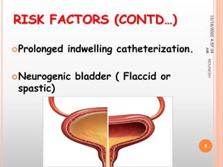
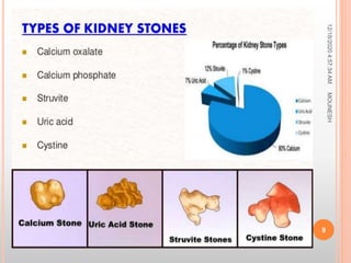
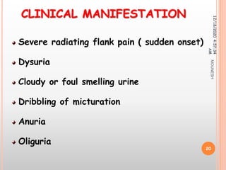
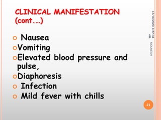
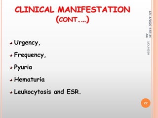
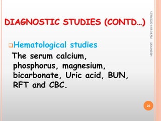
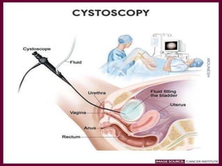
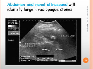
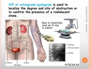
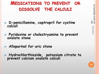
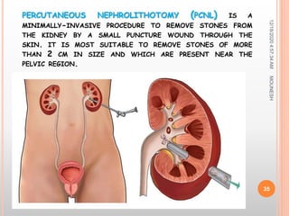
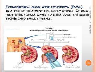
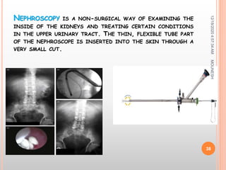
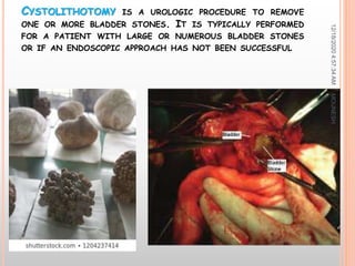
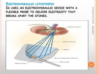
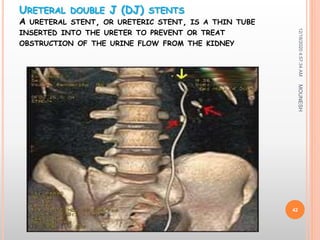
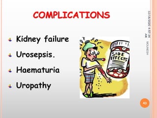
MR. MOUNESH. D. BADIGER
SENIOR TUTOR
MEDICAL SURGICAL DEPARTMENT
KAHER INSTITUTE OF NURSING SCIENCES,
BELAGAVI,
12/18/20204:57:34AM
1
MOUNESH
DEFINITION
 Renal calculi also known as urolithiasis.
It is a kidney stone disease where a
solid piece of material (kidney stone)
occurs in the urinary tract.
12/18/20204:57:34
AM
2
MOUNESH
ETIOLOGY
Idiopathic
Super saturation of urine.
Obstruction with urinary
stasis
12/18/20204:57:34
AM
3
MOUNESH
 Urinary tract infection
 Medications may induce calculus
formation,
(e.g., calcium carbonate and sodium
bicarbonate, and aluminum hydroxide).
 Massive dose of vitamin C increases
urinary oxalate levels
12/18/20204:57:34AM
4
ETIOLOGY
MOUNESH
RISK FACTORS
 Immobility and sedentary lifestyle
 Dehydration, which leads to super
saturation.
 Previous history of urinary calculi.
 Imbalance of pH in urine.
12/18/20204:57:34
AM
5
MOUNESH
RISK FACTORS (CONTD…)
Metabolic disturbances that result
in an increase in calcium or other
ions in the urine.
 Dietary intake
 Gout ( Normal Uric acid levels are
2.4-6.0 mg/dL (female) and 3.4-
7.0 mg/dL (male).)
12/18/20204:57:34
AM
6
MOUNESH
RISK FACTORS (CONTD…)
High mineral content in drinking
water.
A diet high in purines, oxalates,
calcium supplements, animal
proteins.
12/18/20204:57:34AM
7
MOUNESH
RISK FACTORS (CONTD…)
Prolonged indwelling catheterization.
Neurogenic bladder ( Flaccid or
spastic)
12/18/20204:57:34
AM
8
MOUNESH
12/18/20204:57:34AM
9
MOUNESH
CALCIUM OXALATE
Incidence (%): 35-40
Color: Black/dark brown
Sensitivity: Radio-opaque
12/18/20204:57:34
AM
10
MOUNESH
CALCIUM PHOSPHATE
Incidence (%): 8-10
Color: Dirty white
Sensitivity: Radio opaque
12/18/20204:57:34
AM
11
MOUNESH
STRUVITE (MAGNESIUM
AMMONIUM PHOSPHATE)
Incidence (%): 10-15%
Color: Dirty white
Sensitivity: Radio opaque
12/18/20204:57:34
AM
12
MOUNESH
URIC ACID
Incidence (%): 5-8
Color: Yellow/reddish brown
Sensitivity: Radiolucent
12/18/20204:57:34
AM
13
MOUNESH
CYSTINE
Incidence (%): 1-2
Color: Pink/Yellow
Sensitivity: Radio-opaque
12/18/20204:57:34
AM
14
MOUNESH
PATHOPHYSIOLOGY
12/18/20204:57:34AM
15
MOUNESH
Saturation
Super saturation
Nucleation ( liquid to solid)
Crystal growth and aggregation
(hard mass)
Crystal retention
Stone formation
12/18/20204:57:34AM
16
MOUNESH
Calcium and oxalate come
together to make the tiny
crystals.
Supersaturation promotes
their combination
Continued deposition at the
renal papillae leads to growth
of the kidney stones.
12/18/20204:57:34AM
17
MOUNESH
Kidney stones grow and
collect debris .
The kidney stones block
all routes to the renal
papillae
The complete stone forms
and retention occurs.
12/18/20204:57:34AM
18
MOUNESH
12/18/20204:57:34AM
19
MOUNESH
CLINICAL MANIFESTATION
Severe radiating flank pain ( sudden onset)
Dysuria
Cloudy or foul smelling urine
Dribbling of micturation
Anuria
Oliguria
12/18/20204:57:34
AM
20
MOUNESH
 Nausea
Vomiting
Elevated blood pressure and
pulse,
Diaphoresis
 Infection
 Mild fever with chills
12/18/20204:57:34
AM
21
CLINICAL MANIFESTATION
(cont.…)
MOUNESH
CLINICAL MANIFESTATION
(CONT.…)
Urgency,
Frequency,
Pyuria
Hematuria
Leukocytosis and ESR.
12/18/20204:57:34
AM
22
MOUNESH
12/18/20204:57:34AM
23
MOUNESH
DIAGNOSTIC STUDIES
History collection
Physical examination
Inspection
Palpation
Percussion
Auscultation
Imaging Studies
12/18/20204:57:34
AM
24
MOUNESH
DIAGNOSTIC STUDIES
(CONTD…)
CT scan or MRI
may be used to differentiate a
nonopaque stone from a tumor.
Urine analysis
Urine examination, Culture &
sensitivity,24 hrs urine collection
12/18/20204:57:34
AM
25
MOUNESH
Hematological studies
The serum calcium,
phosphorus, magnesium,
bicarbonate, Uric acid, BUN,
RFT and CBC.
12/18/20204:57:34AM
26
DIAGNOSTIC STUDIES (CONTD…)
MOUNESH
12/18/20204:57:34AM
27
MOUNESH
12/18/20204:57:34AM
28
Abdomen and renal ultrasound will
identify larger, radiopaque stones.
MOUNESH
12/18/20204:57:34AM
29
IVP or retrograde pyelogram is used to
localize the degree and site of obstruction or
to confirm the presence of a radiolucent
stone.
MOUNESH
MEDICAL MANAGEMENT
12/18/20204:57:34AM
30
MOUNESH
Watchful waiting or
hydrotherapy.
Diuretics
Narcotic analgesia.
PPI
Syp. Cital - Alkalinizing
agent
Antibiotics
Antacids
12/18/20204:57:34
AM
31
MOUNESH
Implement Dietary Changes.
Antiemetic
Mild sedatives if pain is severe
Loop diuretics
12/18/20204:57:34AM
32
MOUNESH
MEDICATIONS TO PREVENT OR
DISSOLVE THE CALCULI
D-penicillamine, captropril for cystine
calculi
Pyridoxine or cholestryamine to prevent
oxalate stone
Allopurinol for uric stone
Hydrochlorthiazide, potassium citrate to
prevent calcium oxalate calculi
12/18/20204:57:34
AM
33
MOUNESH
SURGICAL
MANAGEMENT
12/18/20204:57:34AM
34
MOUNESH
PERCUTANEOUS NEPHROLITHOTOMY (PCNL) IS A
MINIMALLY-INVASIVE PROCEDURE TO REMOVE STONES FROM
THE KIDNEY BY A SMALL PUNCTURE WOUND THROUGH THE
SKIN. IT IS MOST SUITABLE TO REMOVE STONES OF MORE
THAN 2 CM IN SIZE AND WHICH ARE PRESENT NEAR THE
PELVIC REGION.
12/18/20204:57:34AM
35
MOUNESH
EXTRACORPOREAL SHOCK WAVE LITHOTRIPSY (ESWL)
IS A TYPE OF TREATMENT FOR KIDNEY STONES. IT USES
HIGH-ENERGY SHOCK WAVES TO BREAK DOWN THE KIDNEY
STONES INTO SMALL CRYSTALS.
12/18/20204:57:34AM
36
MOUNESH
URETEROLITHOTOMY REFERS TO THE OPEN
OR LAPAROSCOPIC SURGICAL REMOVAL OF A
STONE FROM THE URETER.
12/18/20204:57:34AM
37
MOUNESH
NEPHROSCOPY IS A NON-SURGICAL WAY OF EXAMINING THE
INSIDE OF THE KIDNEYS AND TREATING CERTAIN CONDITIONS
IN THE UPPER URINARY TRACT. THE THIN, FLEXIBLE TUBE PART
OF THE NEPHROSCOPE IS INSERTED INTO THE SKIN THROUGH A
VERY SMALL CUT.
12/18/20204:57:34AM
38
MOUNESH
CYSTOLITHOTOMY IS A UROLOGIC PROCEDURE TO REMOVE
ONE OR MORE BLADDER STONES. IT IS TYPICALLY PERFORMED
FOR A PATIENT WITH LARGE OR NUMEROUS BLADDER STONES
OR IF AN ENDOSCOPIC APPROACH HAS NOT BEEN SUCCESSFUL
12/18/20204:57:34AM
39
MOUNESH
VESICOLITHOTOMY IS GENERALLY PERFORMED IN
PATIENTS WITH AN IMPASSABLE OR SURGICALLY
ABLATED URETHRA.
12/18/20204:57:34
AM
40
MOUNESH
ELECTROHYDRAULIC LITHOTRIPSY
IS USES AN ELECTROHYDRAULIC DEVICE WITH A
FLEXIBLE PROBE TO DELIVER ELECTRICITY THAT
BREAKS APART THE STONES.
12/18/20204:57:34AM
41
MOUNESH
URETERAL DOUBLE J (DJ) STENTS
A URETERAL STENT, OR URETERIC STENT, IS A THIN TUBE
INSERTED INTO THE URETER TO PREVENT OR TREAT
OBSTRUCTION OF THE URINE FLOW FROM THE KIDNEY
12/18/20204:57:34AM
42
MOUNESH
COMPLICATIONS
Kidney failure
Urosepsis.
Haematuria
Uropathy
12/18/20204:57:34
AM
43
MOUNESH
12/18/20204:57:34AM
44
MOUNESH

Renal calculi