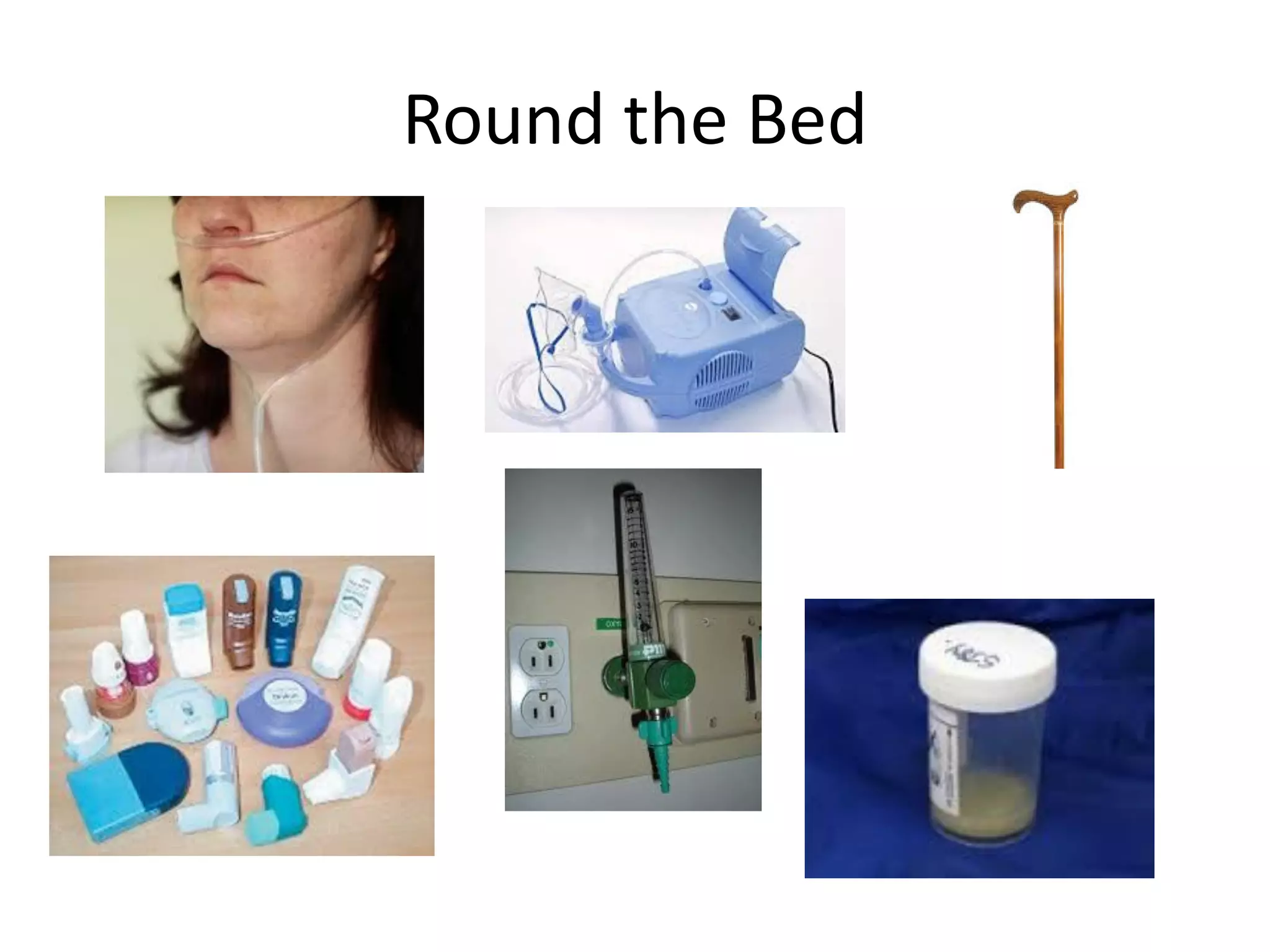
This document provides an overview of conducting a respiratory OSCE station, including examining common signs, conditions, investigations, and management strategies. Key areas of focus are the respiratory examination, common signs and causes of conditions like clubbing, crackles, and reduced breath sounds, investigating and treating issues like pneumonia, COPD, and lung cancer. The goal is to thoroughly examine patients and utilize clinical clues and testing to accurately diagnose and manage respiratory diseases.
Interstitial lung disease is a general category that includes many different lung conditions. All interstitial lung diseases affect the interstitium, a part of the lungs' anatomic structure.
Some of the types of interstitial lung disease include:
Interstitial pneumonia: Bacteria, viruses, or fungi may infect the interstitium of the lung. A bacterium called Mycoplasma pneumonia is the most common cause.
Idiopathic pulmonary fibrosis : A chronic, progressive form of fibrosis (scarring) of the interstitium. Its cause is unknown.
Nonspecific interstitial pneumonitis: Interstitial lung disease that's often present with autoimmune conditions (such as rheumatoid arthritis or scleroderma).
Brief Presentation on clinical examination of Cardio Vascular System with Report of Normal case
references:
macleod's clinical examination 13th edition
hutchinson clinical methods
Apparently a lengthy presentation actually very good for junior physicians as it covers all aspects of assessment, diagnosis and treatment of pleural effusion
Brief Presentation on clinical examination of Respiratory System with Report of Normal case
references:
macleod's clinical examination 13th edition
hutchinson clinical methods
R Alagappan - Manual of Practical Medicine, 4th Edition
Pulmonary route used to treat different respiratory diseases from last decade.
The inhalation therapies involved the use of leaves from plants, vapours from aromatic plants, balsams, and myhrr.
Pulmonary drug delivery is primarily used to treat conditions of the airways, delivering locally acting drugs directly to their site of action.
Delivery of drugs directly to their site of action reduces the dose needed to produce a pharmacological effect.
SHORT TALK ABOUT DIFFERENTIAL DIAGNOSIS BILATERAL HYPERLUCENT LUNGS , COMMON AND LESS COMMON CAUSES WITH CLUES TO DIAGNOSIS AND SOME EXAMPLES
HOPPING YOU LIKE IT
DR HISHAM ALKHATIB
CONSULTANT RADIOLOGIST
This ppt presents the schematic way to read chest X-rays in pediatric and adult patients. Very useful for Clinicians in daily practice and for students who are appearing in practical exams.
7. Breathing Pattern
• Count respiratory rate
• Tachypnoea
• Pursed lipped breathing
• Use of accessory muscles
8. Cough
• Do first as part of inspection
• Dry Cough
– Pulmonary fibrosis
– Pleural effusion
• Purulent cough/productive
– Bronchiectasis/CF
– Pneumonia
18. Pleural Effusion
• Signs
– Reduced expansion
– Trachea away from effusion
– Stony dull percussion note
– Absent tactile vocal fremitus
– Reduced air entry and breath sounds
• Signs to identify cause
– Cancer: clubbing and lymphadenopathy
– CCF: Raised JVP
– Chronic liver disease: spider naevi, leuconychia
– Chronic renal failure: AV fistula
– Connective tissue disease: rheumatoid hands
19. Pleural Effusion
• CXR
• Pleural Aspiration – Ultrasound guidance
– Protein
– LDH
– pH – if < 7.2 consistent with empyema
• Transudate (protein <30g/L)
– CCF
– Chronic renal failure
– Chronic liver failure
• Exudate (protein >30g/L)
– Malignancy – primary or secondary
– Infection
– Infarction
– Inflammation: RA and SLE
20. Pleural Effusion Treatment
• Transudate
– Treat the cause
• Exudate
– Pleural fluid cytology
– May need CT thorax
– Intercostal drainage may be appropriate
– Consider pleurodesis
23. Bronchiectasis
• Signs
– Cachexia and tachypnoea
– Clubbing
– Mixed crackles that alter with coughing
– Occasional squeaks and wheeze
– Sputum +++
• Investigations
– Sputum culture
– CXR
– High resolution CT thorax
• Treatment
– Physiotherapy
– Prompt antibiotic thearpy
– Bronchodilators
24. Pulmonary Fibrosis
• Signs
– Clubbing, central cyanosis and tachypnoea
– Fine end inspiratory crackles
– No sputum
• Investigations
– CXR
– Lung function tests: Restrictive pattern, Low TLC, Low KCO
– High resolution CT
– Lung biopsy
• Treatment
– Immunosuppression, eg. Steroids and azathioprine
– Single lung transplant
– Beware:- Unilateral fine crackles and contralateral thoracotomy
scar with normal breath sounds
32. If you get stuck!
• Say what you hear
• Don’t make up a diagnosis
• Look for bedside clues
• Common respiratory investigations:
– CXR
– CT thorax/high resolution CT thorax
– Lung function tests – obstructive or restrictive
– Peak flow – asthma only
– Sputum culture