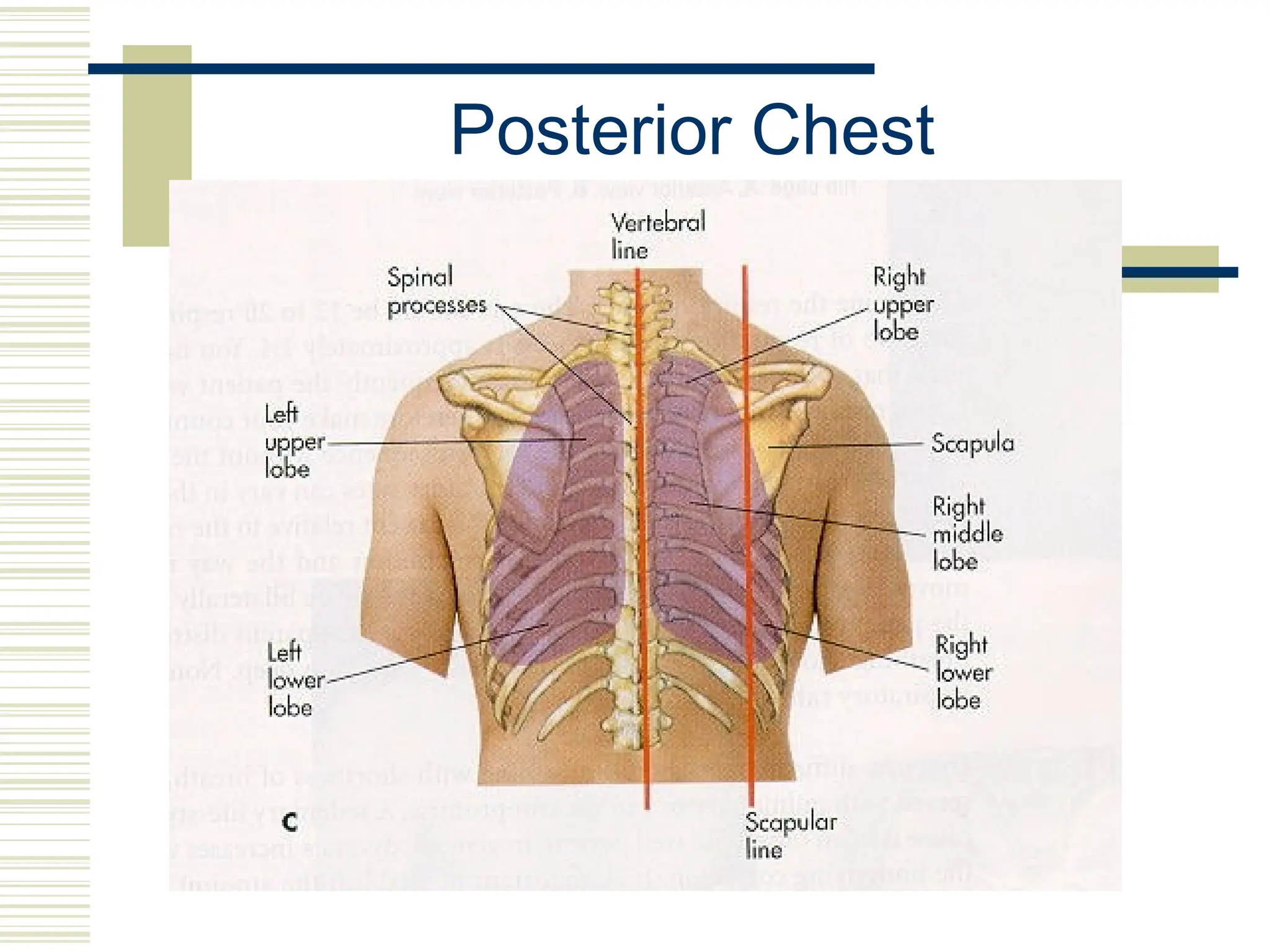
ASSESSMENT OF THE
CHEST AND LUNGS
DR.QURATULAIN MUGHAL
ISRA UNIVERSITY
1
Reference Lines
 Anterior Chest
 Midsternal line
 Anterior axillary lines
 Midclavicular lines
 Posterior Chest
 Vertebral line
 Midscapular lines
 Axilla
 Anterior axillary lines
 Midaxillary lines
 Posterior axillary lines
Anterior Chest
Posterior Chest
Axilla
Anatomy Points to Remember
 Lungs are symmetric
 Lungs are divided into lobes
 Right lung = 3 lobes
 Left lung = 2 lobes
 Primary muscles of respiration
 Diaphragm – divides chest from abdomen
 External intercostal muscles
 Accessory muscles
Anatomy Points to Remember
 Upper Airway
 Nose, pharynx, larynx, intrathoracic trachea
 Functions in respiration

Conduct air to lower airway

Filter to protect lower airway

Warm and humidify inspired air
Anatomy Points to Remember
 Lower Airway
 Trachea, bronchi, bronchioles

Functions in respiration
 Conduct air to alveoli
 Clear mucociliary structures
 Alveoli

Functional unit
 Gas exchange
 Production of surfactant
Anatomy Points to Remember
 Lower Airway
 Trachea splits into left and right mainstem
bronchi which are further subdivided into
bronchioles

Right bronchus is shorted, wider and more
upright than the left

Functions in respiration
 Conduct air to alveoli
 Clear mucociliary structures
History
 Chief Complaint and HPI
 Cough
 Shortness of breath/Dyspnea
Cough
 Onset – sudden, gradual
 Duration
 Nature – dry, moist, hacking, barking
 Sputum – amount, color, odor
 Severity – disrupts activities
 Associated symptoms – sneezing, dyspnea, fever, chills,
congestion, gagging
 What brings it on? – anxiety, talking, activity
 What makes it better?
 What has been tried? – medications, treatments
 Anything similar in the past?
Shortness of Breath (SOB) /
Dyspnea
 Onset – sudden, gradual
 Duration
 Severity – disrupts activities
 Associated symptoms – night sweats, pain, chest
pressure, discomfort, ankle edema, diaphoresis, cyanosis
 What brings it on? – position, time of day, exercise,
allergens, emotions
 What makes it better?
 What has been tried? – medications, inhalers, oxygen
 Anything similar in the past?
History
 Past Health History
 Lung disease or breathing problems

Frequent severe colds, asthma, emphysema,
bronchitis, pneumonia, tuberculosis
 Last PPD and/or chest x-ray
 Allergies
 Medication use
 Family History
History
 Personal and Social History
 Tobacco
 Alcohol
 Drugs
 Home environment
 Occupational environment
 Travel
 Health Promotional Activities
PHYSICAL
EXAMINATION
15
Equipment and Techniques
 Equipment
 Stethoscope
 Techniques
 Inspection
 Palpation
 Percussion
 Auscultation
Inspection
 General
 Appearance
 Posturing
 Breathing effort
 Trachea position

Midline
Inspection
 Chest Wall Configuration
 Form
 Symmetry
 Muscle development
 Anterior-Posterior (AP) diameter

Approximately ½ the transverse diameter

Transverse: Anterior-Posterior = 2:1
 Costal angle

90 degrees or less
Inspection
 Oxygenation: cyanosis
 Nails
 Skin
 Lips
 Respiratory Effort
 Respiratory rate and depth
 Breathing pattern
 Chest expansion
Palpation
 Trachea – for position
 Chest wall – for symmetry
Palpation
 Thoracic Expansion (Excursion)
 Place both thumbs at about 7th
rib
posteriorly along the spinal process
 Extend the fingers of both hands
outward over the posterior chest wall
 Have the person take a deep breath
and observe for bilateral outward
movement of thumbs

Normal: bilateral, symmetric
expansion

Abnormal: unilateral or unequal
Click on the pictures to view video
Palpation
 Vocal (Tactile) Fremitus
 Use palmar or ulnar surfaces of hands
 Systematically position hands over both sides of
posterior chest
 Have person repeat “1 – 2 – 3” or “99” as you
move from the apices to the bases

Normal: bilaterally symmetrical vibrations

Decreased or absent: obstruction of transmission
0bronchitis, emphysema)

Increased: consolidation (compression) of lung
tissue (pneumonia)
Auscultation
 Auscultate in a systematic manner
 Compare one side to the other
 Listen one full respiration at each spot
 Displace breast tissue to listen directly over
chest wall
 DO NOT
DO NOT listen through gowns, clothes, etc.
 Place your stethoscope over bare skin
Auscultation
 Evaluate posterior, lateral, and anterior chest
 Instruct person to sit upright and breathe in
and out slowly through the mouth
 This makes it easier to hear the air movement
 Use the diaphragm of the stethoscope
 Move from the apices to the bases
Auscultation
 Evaluate for normal sounds
Sound Pitch Intensity Quality I:E Location
Bronchial High Loud Blowing/ hollow I < E Trachea
Bronchovesicula
r
Moderate Moderate Combination I = E Between scapulae,
1st
& 2nd
ICS lateral to
sternum
Vesicular Low Soft Gentle rustling/
breezy
I > E Peripheral lung
Auscultation
 Evaluate for adventitious sounds
Sound Intensity/ Pitch I/E Quality Clear with Cough
Crackles/
Rales
Soft (fine)/ High
Loud (coarse)/ Low
I Discontinuous,
nonmusical, brief
Possibly
Wheeze High E Continuous musical
sounds
Possibly
Ronchi Low E Continuous snoring
sounds
Possibly
Pleural
Friction Rub
I & E Continuous or
discontinuous creaking or
brushing sounds
Never
Stridor I Continuous, crowing Never
Auscultation
Copy this URL into your Web browser to hear normal and abnormal lung sounds :
http://medocs.ucdavis.edu/IMD/420C/sounds/lngsound.htm
Developmental Variations
 Neonates
 Measure the chest circumference

Usually 2-3 cm smaller than head circumference

Chest is round (i.e. AP diameter = transverse)
 Obligate nose breathers
 Periodic breathing is common

Sequence of vigorous breathing followed by apnea
for 10-15 seconds

Only concern if it is prolonged or baby becomes
cyanotic
Developmental Variations
 Neonates
 Breathing is diaphragmatic and abdominal
 Signs of compromise

Stridor (“crowing”)

Grunting

Central cyanosis

Flaring nares
Developmental Variations
 Infants and Young Children
 Roundness of the chest persist for first 2 years
 Chest walls are thinner than the adult’s

Breath sounds may sound louder, and more
bronchial than the adult
 Bronchovesicular sounds may be heard
throughout the chest
Developmental Variations
 Pregnancy
 Costal angle increases to about 105 degrees in
the third trimester
 Dyspnea and orthopnea are common
 Breathes more deeply
Developmental Variations
 Older Adult
 Chest expansion is often decreased
 Bony prominences are marked
 AP diameter is increased with respect to
transverse (but not 1:1)
respiratoryassessment and it's examination

respiratoryassessment and it's examination

  • 1.
    ASSESSMENT OF THE CHESTAND LUNGS DR.QURATULAIN MUGHAL ISRA UNIVERSITY 1
  • 2.
    Reference Lines  AnteriorChest  Midsternal line  Anterior axillary lines  Midclavicular lines  Posterior Chest  Vertebral line  Midscapular lines  Axilla  Anterior axillary lines  Midaxillary lines  Posterior axillary lines
  • 3.
  • 4.
  • 5.
  • 6.
    Anatomy Points toRemember  Lungs are symmetric  Lungs are divided into lobes  Right lung = 3 lobes  Left lung = 2 lobes  Primary muscles of respiration  Diaphragm – divides chest from abdomen  External intercostal muscles  Accessory muscles
  • 7.
    Anatomy Points toRemember  Upper Airway  Nose, pharynx, larynx, intrathoracic trachea  Functions in respiration  Conduct air to lower airway  Filter to protect lower airway  Warm and humidify inspired air
  • 8.
    Anatomy Points toRemember  Lower Airway  Trachea, bronchi, bronchioles  Functions in respiration  Conduct air to alveoli  Clear mucociliary structures  Alveoli  Functional unit  Gas exchange  Production of surfactant
  • 9.
    Anatomy Points toRemember  Lower Airway  Trachea splits into left and right mainstem bronchi which are further subdivided into bronchioles  Right bronchus is shorted, wider and more upright than the left  Functions in respiration  Conduct air to alveoli  Clear mucociliary structures
  • 10.
    History  Chief Complaintand HPI  Cough  Shortness of breath/Dyspnea
  • 11.
    Cough  Onset –sudden, gradual  Duration  Nature – dry, moist, hacking, barking  Sputum – amount, color, odor  Severity – disrupts activities  Associated symptoms – sneezing, dyspnea, fever, chills, congestion, gagging  What brings it on? – anxiety, talking, activity  What makes it better?  What has been tried? – medications, treatments  Anything similar in the past?
  • 12.
    Shortness of Breath(SOB) / Dyspnea  Onset – sudden, gradual  Duration  Severity – disrupts activities  Associated symptoms – night sweats, pain, chest pressure, discomfort, ankle edema, diaphoresis, cyanosis  What brings it on? – position, time of day, exercise, allergens, emotions  What makes it better?  What has been tried? – medications, inhalers, oxygen  Anything similar in the past?
  • 13.
    History  Past HealthHistory  Lung disease or breathing problems  Frequent severe colds, asthma, emphysema, bronchitis, pneumonia, tuberculosis  Last PPD and/or chest x-ray  Allergies  Medication use  Family History
  • 14.
    History  Personal andSocial History  Tobacco  Alcohol  Drugs  Home environment  Occupational environment  Travel  Health Promotional Activities
  • 15.
  • 16.
    Equipment and Techniques Equipment  Stethoscope  Techniques  Inspection  Palpation  Percussion  Auscultation
  • 17.
    Inspection  General  Appearance Posturing  Breathing effort  Trachea position  Midline
  • 18.
    Inspection  Chest WallConfiguration  Form  Symmetry  Muscle development  Anterior-Posterior (AP) diameter  Approximately ½ the transverse diameter  Transverse: Anterior-Posterior = 2:1  Costal angle  90 degrees or less
  • 19.
    Inspection  Oxygenation: cyanosis Nails  Skin  Lips  Respiratory Effort  Respiratory rate and depth  Breathing pattern  Chest expansion
  • 20.
    Palpation  Trachea –for position  Chest wall – for symmetry
  • 21.
    Palpation  Thoracic Expansion(Excursion)  Place both thumbs at about 7th rib posteriorly along the spinal process  Extend the fingers of both hands outward over the posterior chest wall  Have the person take a deep breath and observe for bilateral outward movement of thumbs  Normal: bilateral, symmetric expansion  Abnormal: unilateral or unequal Click on the pictures to view video
  • 22.
    Palpation  Vocal (Tactile)Fremitus  Use palmar or ulnar surfaces of hands  Systematically position hands over both sides of posterior chest  Have person repeat “1 – 2 – 3” or “99” as you move from the apices to the bases  Normal: bilaterally symmetrical vibrations  Decreased or absent: obstruction of transmission 0bronchitis, emphysema)  Increased: consolidation (compression) of lung tissue (pneumonia)
  • 23.
    Auscultation  Auscultate ina systematic manner  Compare one side to the other  Listen one full respiration at each spot  Displace breast tissue to listen directly over chest wall  DO NOT DO NOT listen through gowns, clothes, etc.  Place your stethoscope over bare skin
  • 24.
    Auscultation  Evaluate posterior,lateral, and anterior chest  Instruct person to sit upright and breathe in and out slowly through the mouth  This makes it easier to hear the air movement  Use the diaphragm of the stethoscope  Move from the apices to the bases
  • 25.
    Auscultation  Evaluate fornormal sounds Sound Pitch Intensity Quality I:E Location Bronchial High Loud Blowing/ hollow I < E Trachea Bronchovesicula r Moderate Moderate Combination I = E Between scapulae, 1st & 2nd ICS lateral to sternum Vesicular Low Soft Gentle rustling/ breezy I > E Peripheral lung
  • 26.
    Auscultation  Evaluate foradventitious sounds Sound Intensity/ Pitch I/E Quality Clear with Cough Crackles/ Rales Soft (fine)/ High Loud (coarse)/ Low I Discontinuous, nonmusical, brief Possibly Wheeze High E Continuous musical sounds Possibly Ronchi Low E Continuous snoring sounds Possibly Pleural Friction Rub I & E Continuous or discontinuous creaking or brushing sounds Never Stridor I Continuous, crowing Never
  • 27.
    Auscultation Copy this URLinto your Web browser to hear normal and abnormal lung sounds : http://medocs.ucdavis.edu/IMD/420C/sounds/lngsound.htm
  • 28.
    Developmental Variations  Neonates Measure the chest circumference  Usually 2-3 cm smaller than head circumference  Chest is round (i.e. AP diameter = transverse)  Obligate nose breathers  Periodic breathing is common  Sequence of vigorous breathing followed by apnea for 10-15 seconds  Only concern if it is prolonged or baby becomes cyanotic
  • 29.
    Developmental Variations  Neonates Breathing is diaphragmatic and abdominal  Signs of compromise  Stridor (“crowing”)  Grunting  Central cyanosis  Flaring nares
  • 30.
    Developmental Variations  Infantsand Young Children  Roundness of the chest persist for first 2 years  Chest walls are thinner than the adult’s  Breath sounds may sound louder, and more bronchial than the adult  Bronchovesicular sounds may be heard throughout the chest
  • 31.
    Developmental Variations  Pregnancy Costal angle increases to about 105 degrees in the third trimester  Dyspnea and orthopnea are common  Breathes more deeply
  • 32.
    Developmental Variations  OlderAdult  Chest expansion is often decreased  Bony prominences are marked  AP diameter is increased with respect to transverse (but not 1:1)