Downloaded 1,037 times


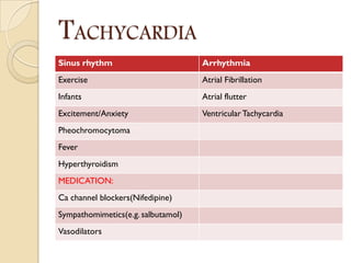
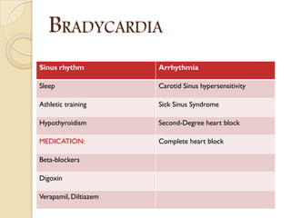
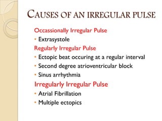


Abnormal findings can occur in the pulse rate, rhythm, volume, character, vessel walls, and radiofemoral delay. Tachycardia is a pulse rate over 100 bpm and can be caused by sinus rhythm, arrhythmias, medications, and medical conditions. Bradycardia is a pulse rate under 60 bpm and can be caused by sinus rhythm, arrhythmias, medications, and medical conditions. An irregular pulse can be occasionally, regularly, or irregularly irregular and caused by conditions like extrasystole, ectopic beats, arrhythmias, and atrial fibrillation. Other abnormalities include high or low pulse volume caused by physiological or pathological conditions, varying volume seen with


