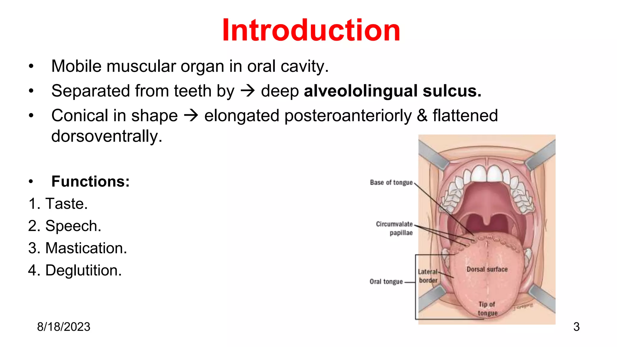
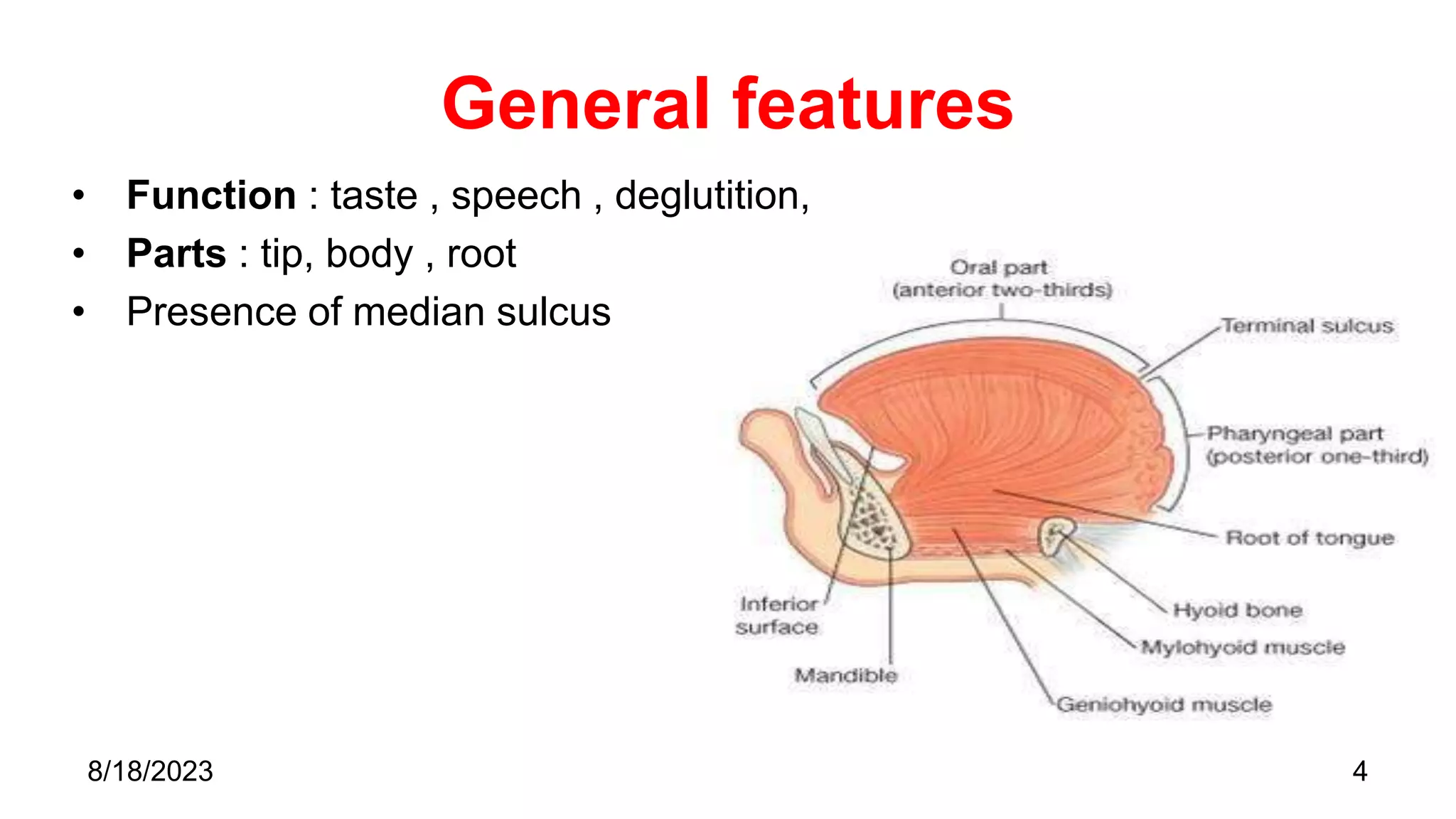
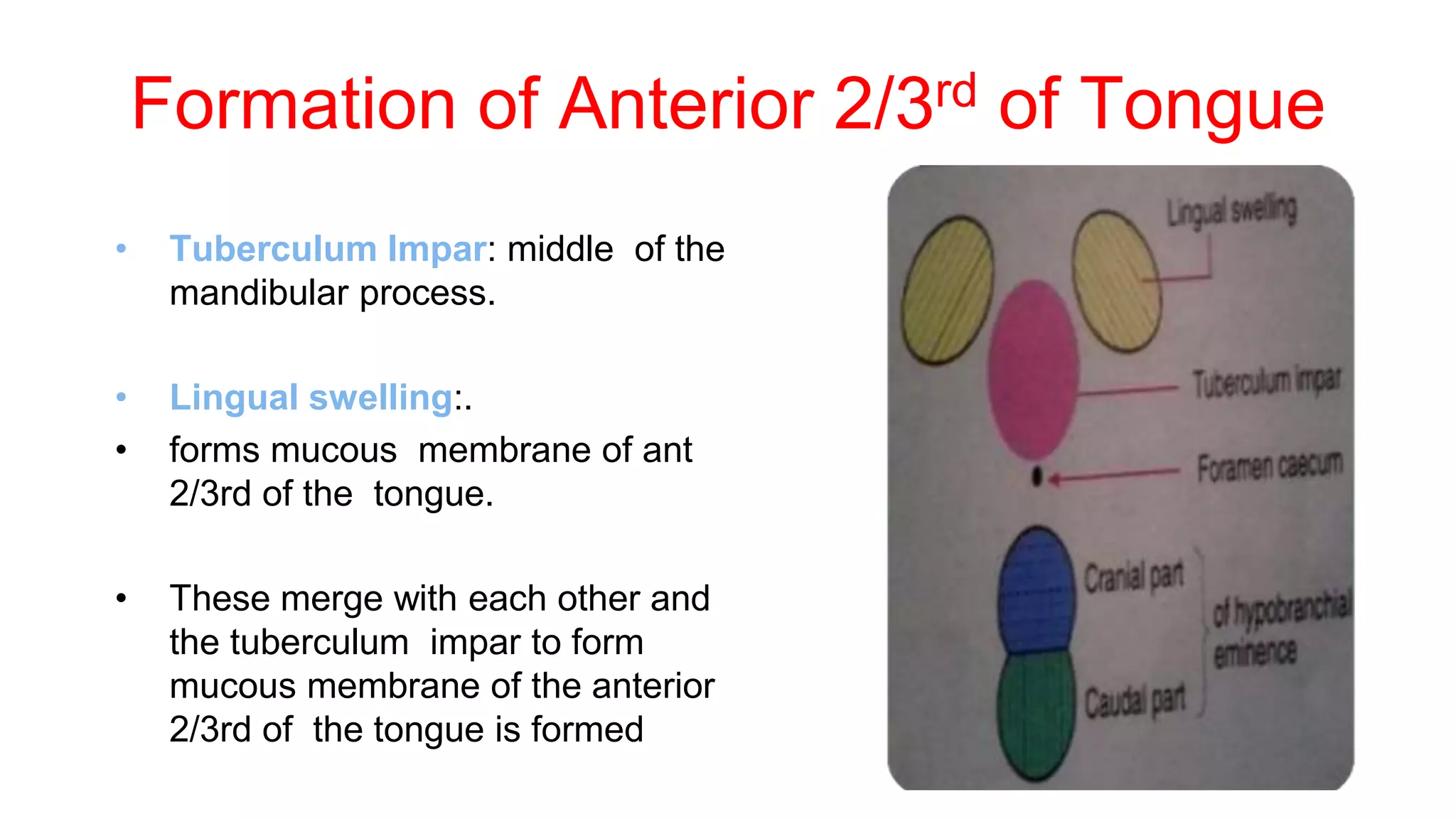
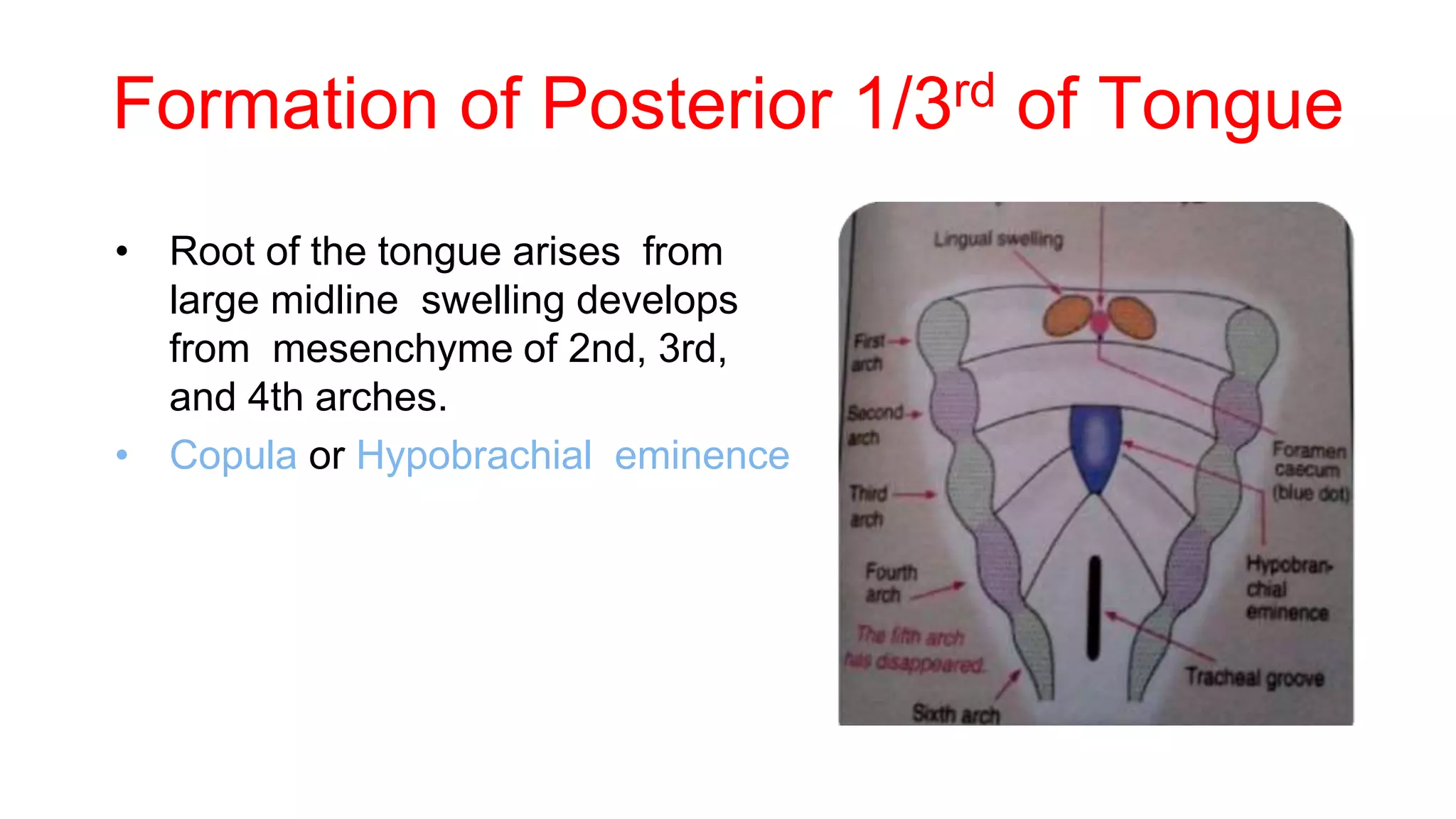
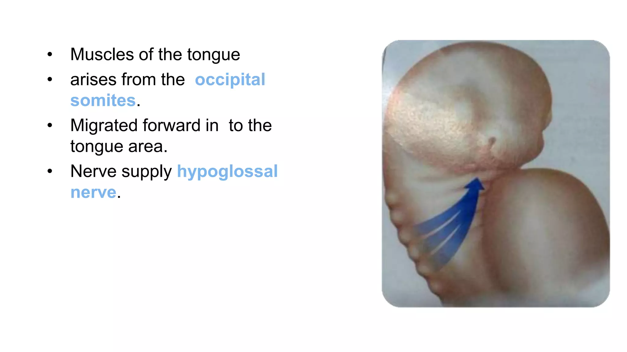
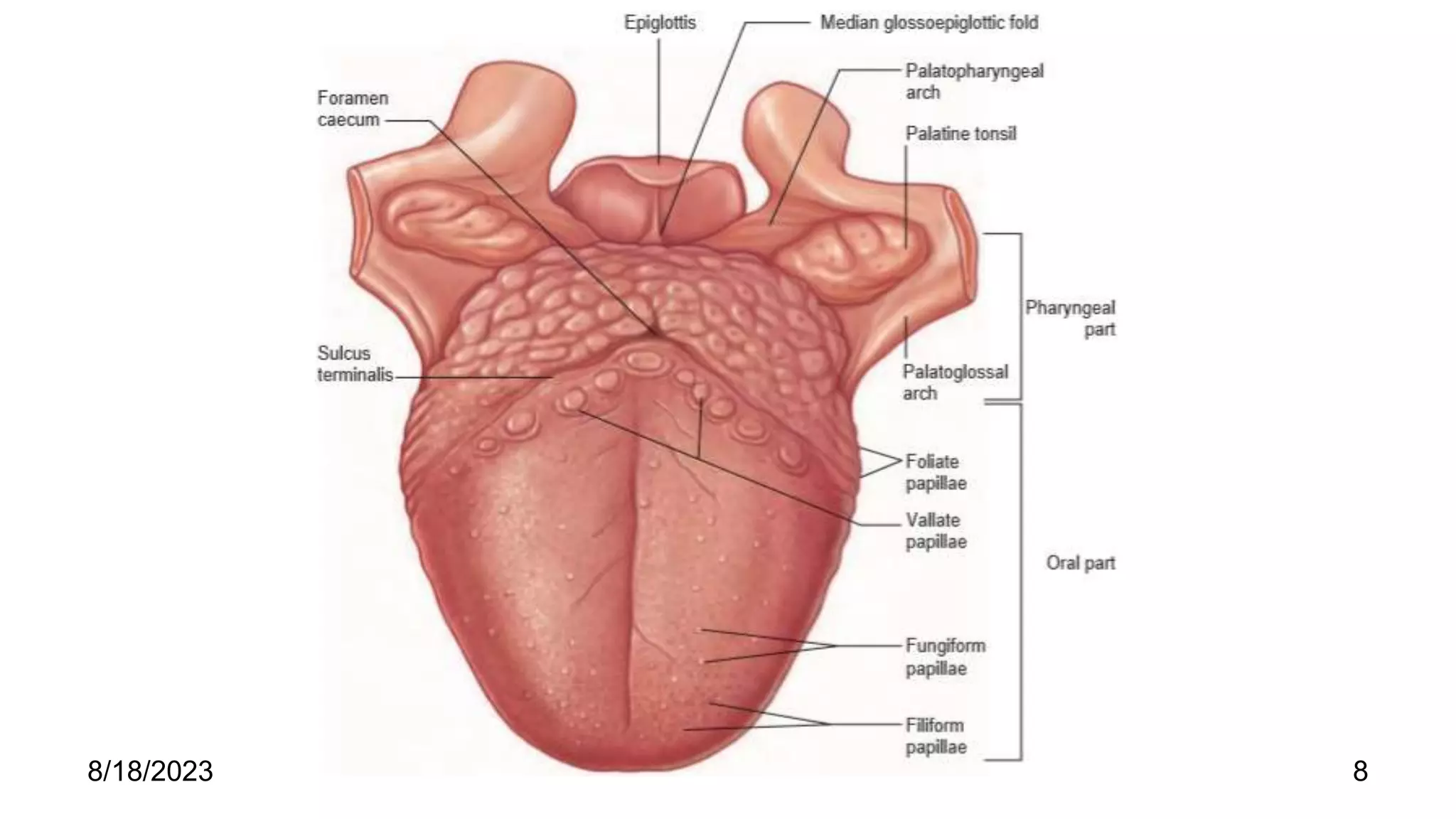
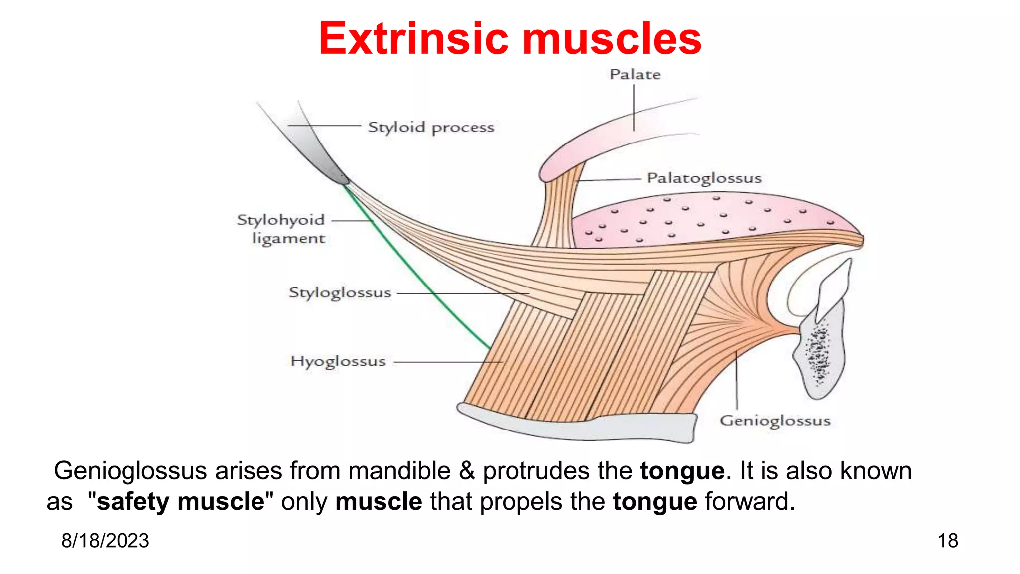
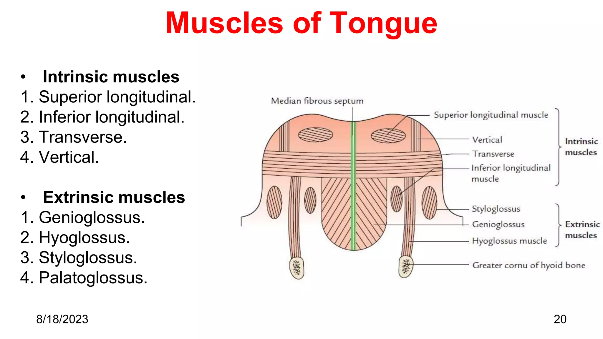
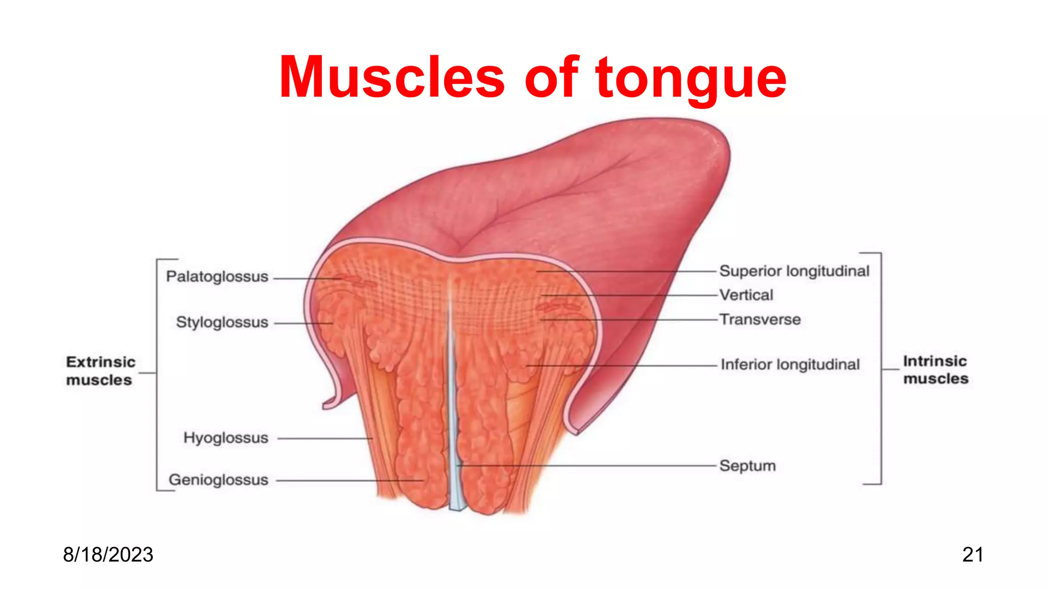
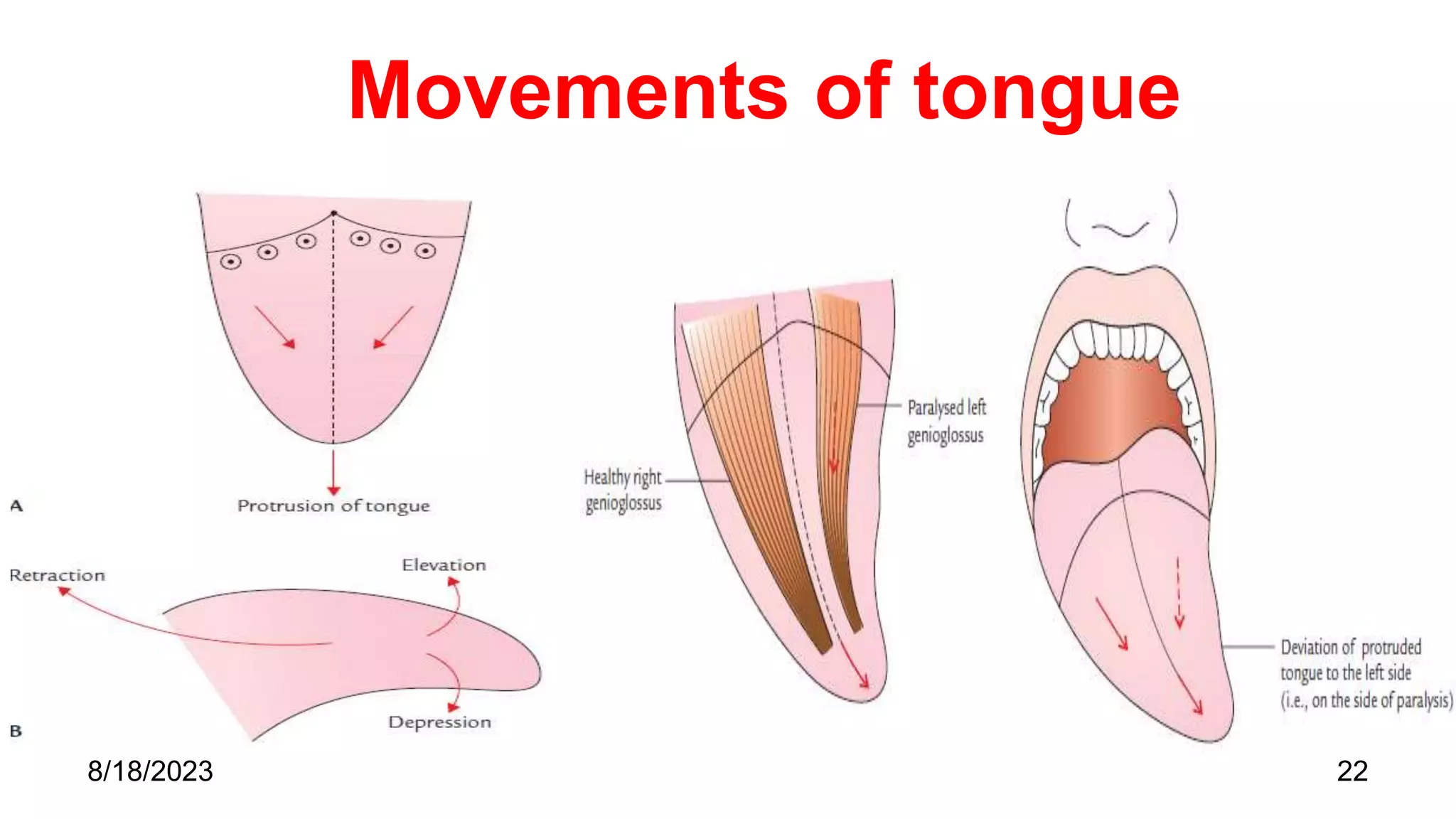
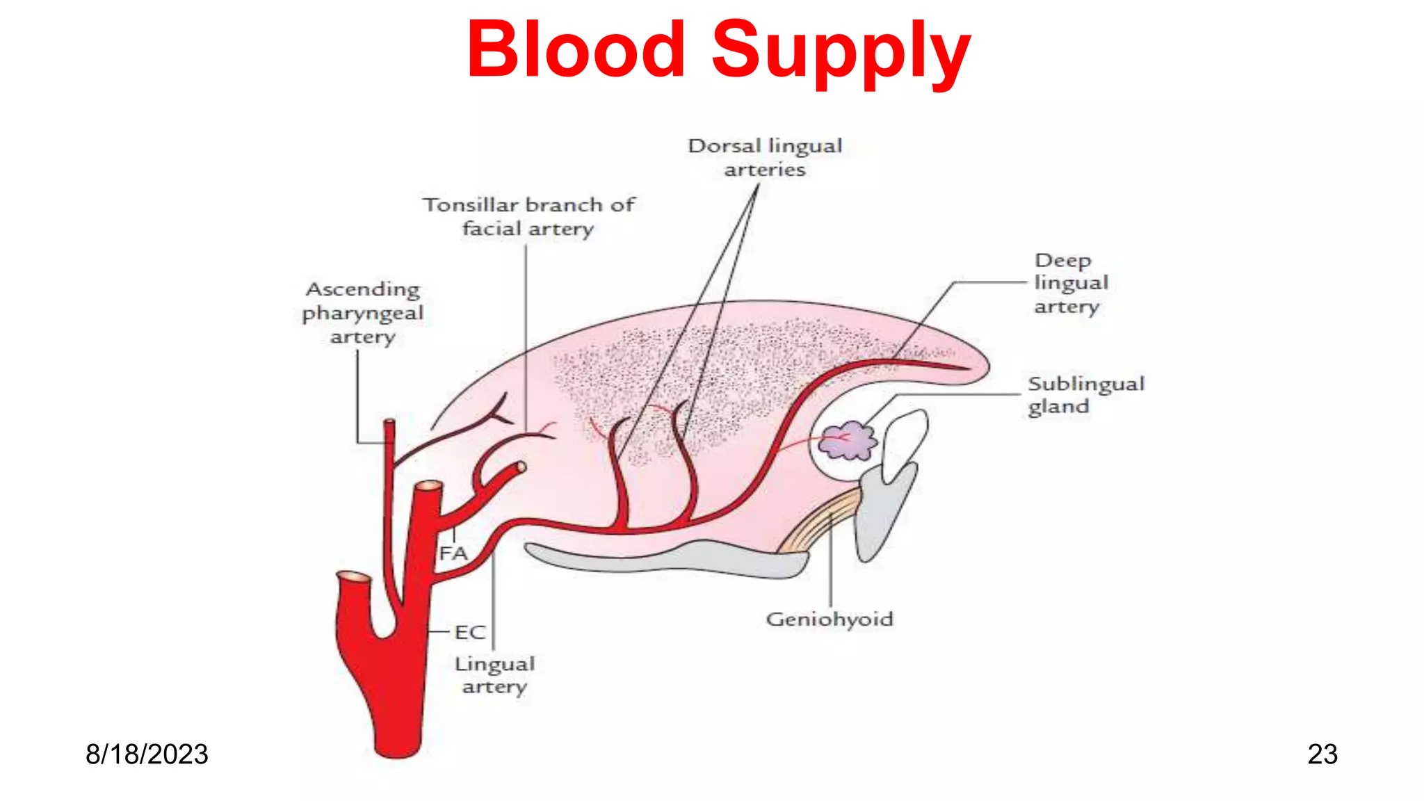
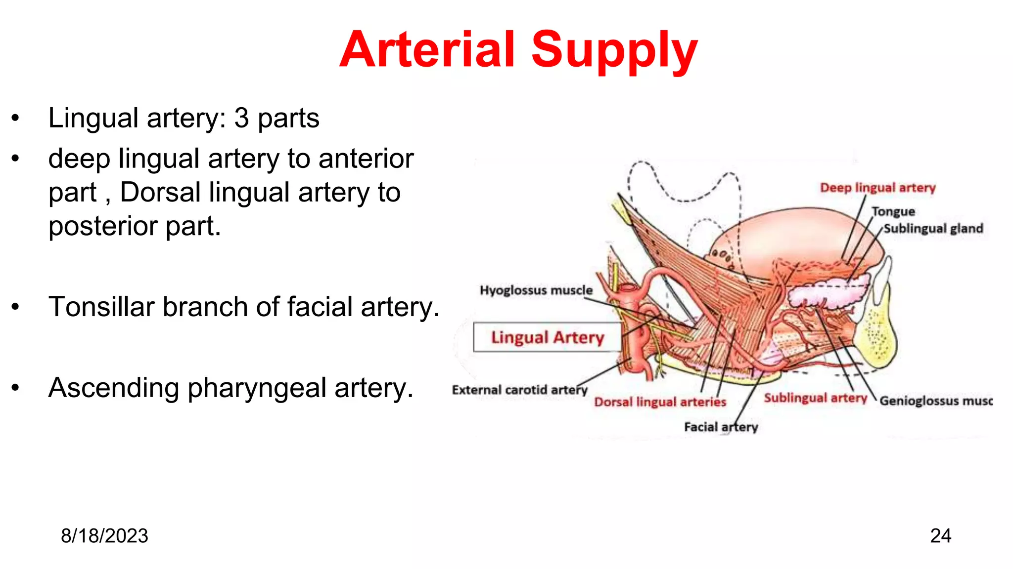
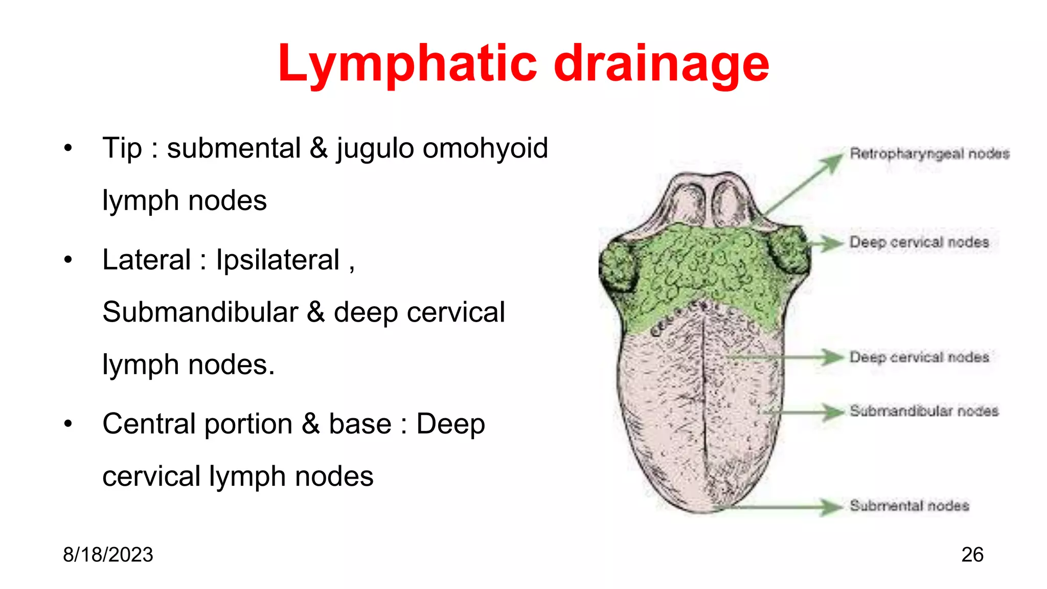
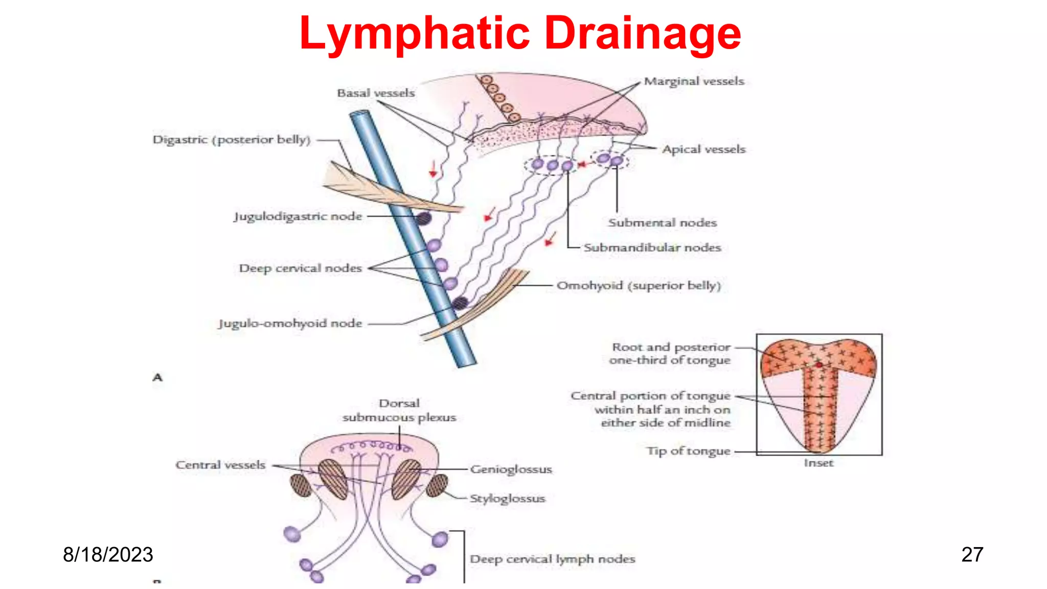
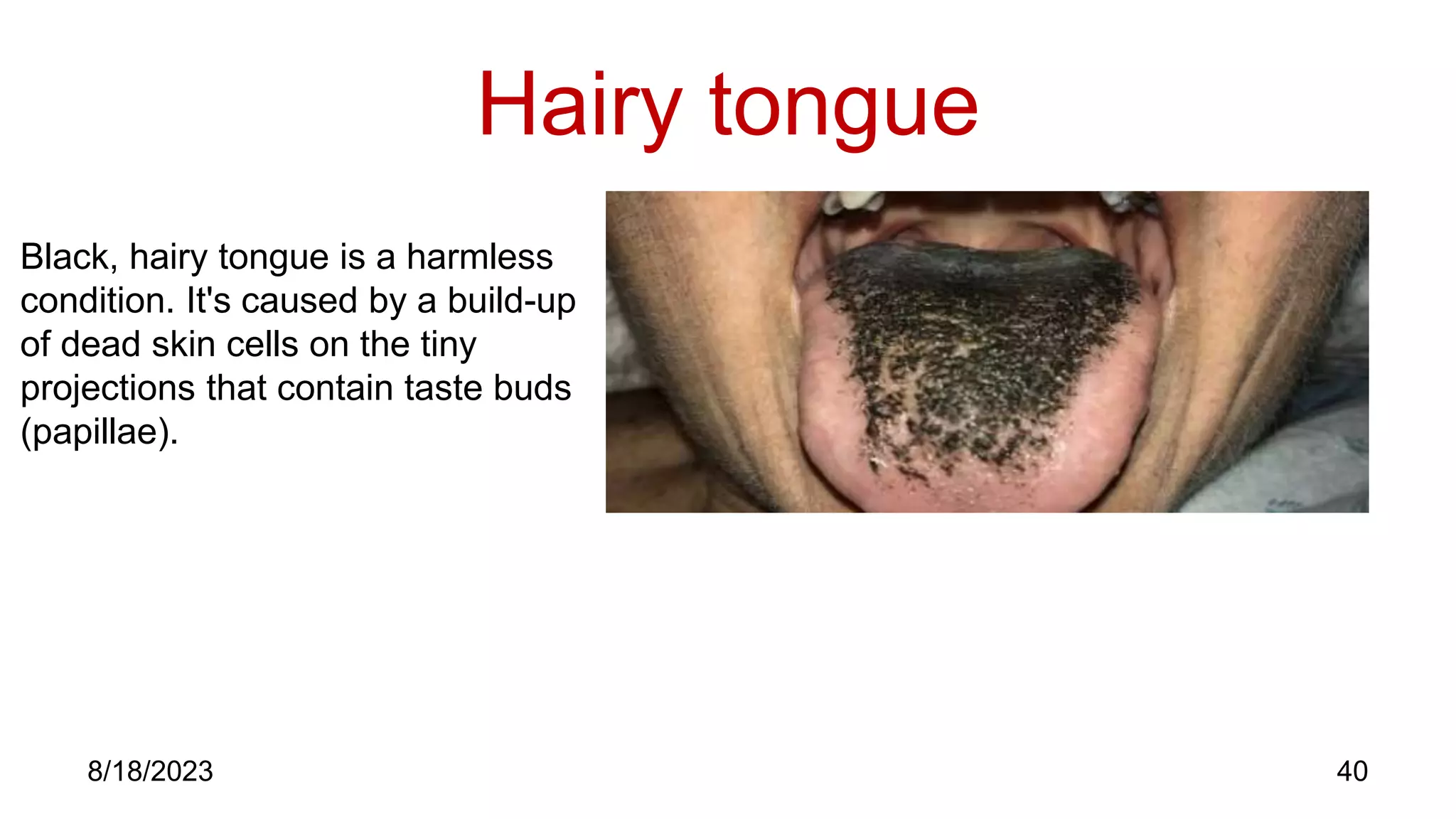
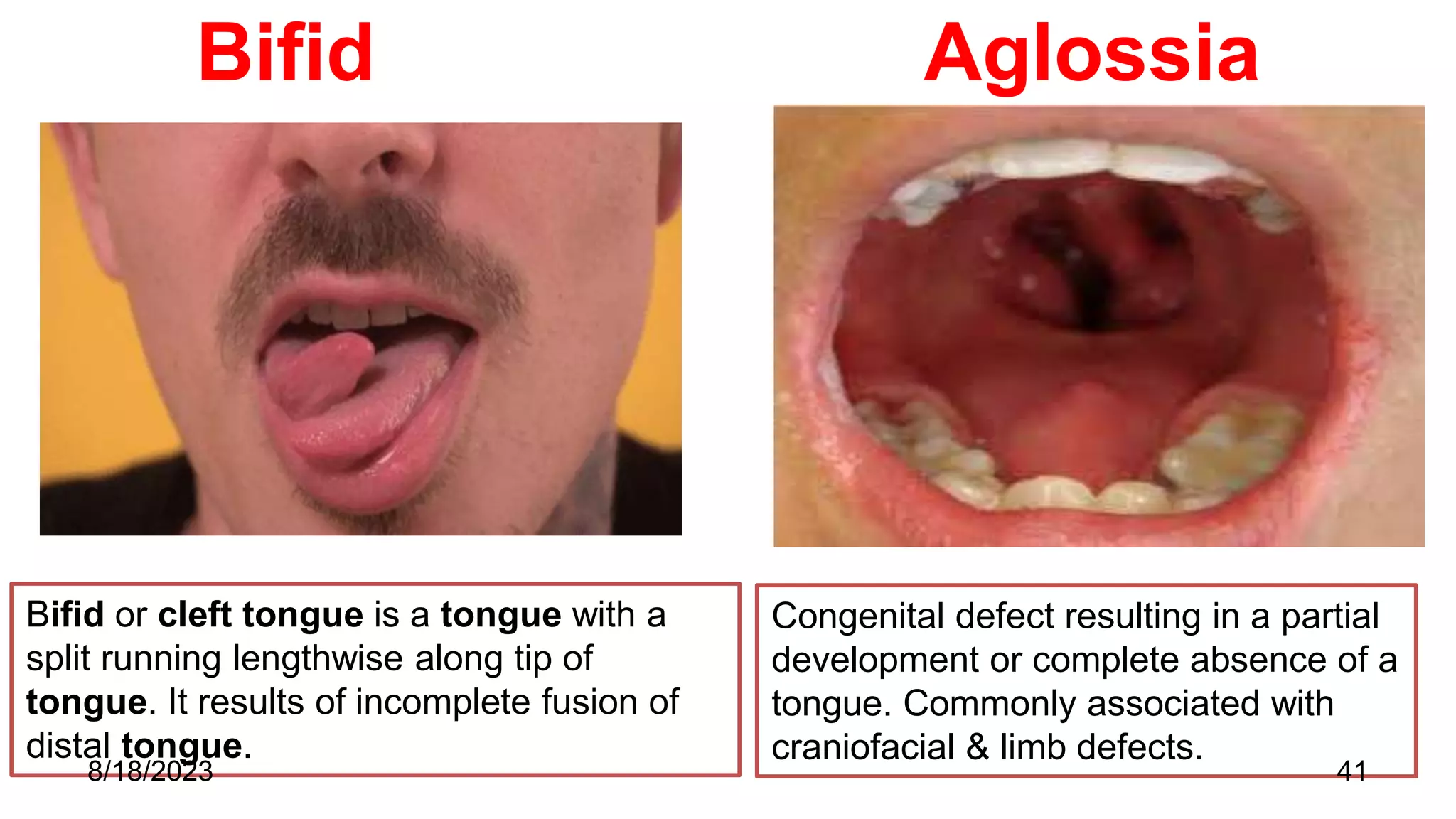
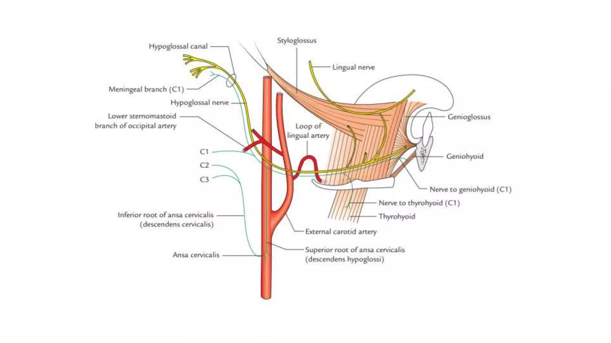
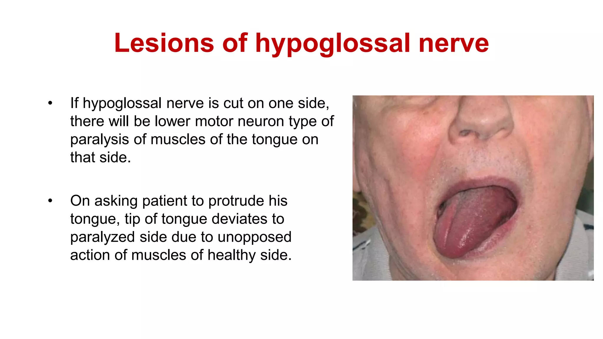
Tongue and hypoglossal nerve
Presenter- Yashu Bhardwaj
8/18/2023
Contents
• Introduction
• External Features
• Development of tongue
• Gross features
• Nerve Supply
• Muscles of Tongue
• Movement of tongue
• Blood supply
• Lymphatic drainage
• Histology of tongue
• Clinical aspects
• Hypoglossal nerve
• Clinical corelations
• MCQ
• References
8/18/2023 2
Introduction
• Mobile muscular organ in oral cavity.
• Separated from teeth by  deep alveololingual sulcus.
• Conical in shape  elongated posteroanteriorly & flattened
dorsoventrally.
• Functions:
1. Taste.
2. Speech.
3. Mastication.
4. Deglutition.
3
8/18/2023
General features
• Function : taste , speech , deglutition,
• Parts : tip, body , root
• Presence of median sulcus
4
8/18/2023
Formation of Anterior 2/3rd of Tongue
• Tuberculum Impar: middle of the
mandibular process.
• Lingual swelling:.
• forms mucous membrane of ant
2/3rd of the tongue.
• These merge with each other and
the tuberculum impar to form
mucous membrane of the anterior
2/3rd of the tongue is formed
Formation of Posterior 1/3rd of Tongue
• Root of the tongue arises from
large midline swelling develops
from mesenchyme of 2nd, 3rd,
and 4th arches.
• Copula or Hypobrachial eminence
• Muscles of the tongue
• arises from the occipital
somites.
• Migrated forward in to the
tongue area.
• Nerve supply hypoglossal
nerve.
8
8/18/2023
Dorsal surface
 Convex
 Presence of median furrow.
 Sulcus termanalis divides it into 2 parts :
• Anterior 2/3rd (oral /presulcal part)
• Posterior 1/3rd (pharyngeal / postsulcal part )
9
8/18/2023
External Features
Anterior two-third: oral
part.
Posterior one-third:
pharyngeal part.
10
8/18/2023
Presulcal Part (Oral)
 Anterior 2/3rd develops from 1st
pharyngeal arch.
 Epithelium : Keratinised stratified
squamous epithelium
 Features:-
• A median furrow representing bilateral
origin of tongue.
 Four types of papillae (lingual papillae)-
• Filiform papillae
• Fungiform papillae
• Circumvallate papillae
• Foliate papillae
11
8/18/2023
Postsulcal Part (Pharyngeal)
• Posterior 1/3rd develops from 3rd and
4th pharyngeal arches.
• Epithelium : Non keratinised st.
squamous epithelium.
• Forms base of tongue.
• Forms anterior wall of oropharynx.
• Lingual tonsil:
Lymphoid follicles are present in
mucosa.
12
8/18/2023
Ventral surface
• Reflected on floor of mouth. Frenulum linguae : May be seen through mucus
membrane on either sides of frenulum linguae( lingual nerve and lingual artery
are medial to vein but not visible)
• Plica Fimbriata : lateral to lingual vein directed forward ,medial and towards
tip of tongue.
13
8/18/2023
14
8/18/2023
15
Nerve Supply
8/18/2023
Nerve Supply
16
8/18/2023
Motor Supply-
• All muscles of tongue are supplied by Hypoglossal Nerve except
Palatoglossus.
• Palatoglossus is supplied by cranial root of Accessory Nerve.
Sensory Supply-
Anterior 2/3rd –
General Sensations- Lingual Nerve
Special Sensations of Taste- Chorda Tympani Nerve [except circumvallate
papillae which are supplied by Glossopharyngeal Nerve.
Posterior 2/3rd –
General Sensations & Special Sensations of Taste- Glossopharyngeal Nerve.
Posterior-most part- Internal Laryngeal Nerve.
8/18/2023 17
Extrinsic muscles
18
Genioglossus arises from mandible & protrudes the tongue. It is also known
as "safety muscle" only muscle that propels the tongue forward.
8/18/2023
Extrinsic muscles of tongue
19
8/18/2023
Muscles of Tongue
• Intrinsic muscles
1. Superior longitudinal.
2. Inferior longitudinal.
3. Transverse.
4. Vertical.
• Extrinsic muscles
1. Genioglossus.
2. Hyoglossus.
3. Styloglossus.
4. Palatoglossus.
20
8/18/2023
Muscles of tongue
21
8/18/2023
Movements of tongue
22
8/18/2023
Blood Supply
23
8/18/2023
Arterial Supply
• Lingual artery: 3 parts
• deep lingual artery to anterior
part , Dorsal lingual artery to
posterior part.
• Tonsillar branch of facial artery.
• Ascending pharyngeal artery.
24
8/18/2023
Venous drainage
• Deep lingual vein:
principle vein
• Venae comitantes with
lingual artery which
adjoins dorsal lingual
vein.
• Venae comitantes
accompanying
hypoglossal nerve.
25
8/18/2023
Lymphatic drainage
• Tip : submental & jugulo omohyoid
lymph nodes
• Lateral : Ipsilateral ,
Submandibular & deep cervical
lymph nodes.
• Central portion & base : Deep
cervical lymph nodes
26
8/18/2023
Lymphatic Drainage
27
8/18/2023
Drawing H&E 40x
1. Stratified squamous
epithelium
2. Lamina propria
3. Skeletal muscle
4. Serous gland
5. Mucous gland
6. Adipose tissue
7. Smooth ventral
surface of tongue
P. Papillae
A. Filliform; B. Fungiform
C. Circumvallate; D. Foliate
Taste Buds
Clinical Aspects
• Glossitis
• Ankyloglossia ( tongue tie)
• Lichen planus
• Geographical tongue
• Hairy tongue
• Fissured tongue
• Aglossia
• Microglossia
• Oral cancer
36
8/18/2023
Glossitis
Affected Normal
37
8/18/2023
tongue is swollen and
inflamed
Lichen Planus
38
8/18/2023
• Appear as white, lacy patches
• Red, swollen tissues
• These lesions may cause
burning, pain or other discomfort.
Fissured tongue
39
8/18/2023
A benign condition
affecting the top surface of
the tongue
Hairy tongue
40
8/18/2023
Black, hairy tongue is a harmless
condition. It's caused by a build-up
of dead skin cells on the tiny
projections that contain taste buds
(papillae).
Bifid Aglossia
Bifid or cleft tongue is a tongue with a
split running lengthwise along tip of
tongue. It results of incomplete fusion of
distal tongue.
Congenital defect resulting in a partial
development or complete absence of a
tongue. Commonly associated with
craniofacial & limb defects.
8/18/2023 41
Clinical
42
Ankyloglossia/ tongue-tie
Paralysis
8/18/2023
Ludwig’s angina
• Cellulitis of floor of mouth usually
due to infection from a tooth,
causing inflammatory edema of
floor of mouth. Tongue is pushed
upwards due to edema with pain.
43
8/18/2023
Hypoglossal Nerve
• Motor nerve, supply muscles of
tongue except Palatoglossus.
• Arises from hypoglossal nuclei.
• Functional components: GSE
(hypoglossal nuclei)
 Branches containing C1 fibres:
• Meningeal branch : duramatar of posterior cranial fossa.
• Nerve to thyrohyoid : crosses greater cornu of hyoid bone
• Nerve to geniohyoid : arises from above hyoid bone.
• Descendens hypoglossi & upper root of ansa cervicalis: joins
inferior root of ansa cervicalis at the level of cricoid cartilage.
Lesions of hypoglossal nerve
• If hypoglossal nerve is cut on one side,
there will be lower motor neuron type of
paralysis of muscles of the tongue on
that side.
• On asking patient to protrude his
tongue, tip of tongue deviates to
paralyzed side due to unopposed
action of muscles of healthy side.
MCQ
1.) There are intrinsic and extrinsic group of muscle which helps in
movement of tongue. Which muscle is considered as safety muscle
of tongue?
A.) Genioglossus.
B.) Hyoglossus.
C.) Styloglossus.
D.) Palatoglossus
Ans: A
Ref: Vishram Singh Textbook of Anatomy Head & Neck, Pg- 192
8/18/2023 48
2.) Cellulitis of floor of mouth usually due to infection from a carious molar
tooth, causing inflammatory edema of floor of mouth. Tongue is pushed
upwards due to edema with pain. Such condition is known as:
A.) Winsons angina
B.) Ludwig’s angina
C.) Ranula
D.) Quinsy
Ans: B
Ref: Vishram Singh Textbook of Anatomy Head & Neck, Pg- 189
8/18/2023 49
3.) Which clinical condition is showing in given picture?
A.) Microglossia
B.) Macroglossia
C.) Ankyloglossia
D.) Bifid of tongue
Ans: C
Ref: Vishram Singh Textbook of Anatomy Head & Neck, Pg- 191
8/18/2023 50
4.) A patient reached to the clinic having right side deviated tongue. Which
diagnosis related to this condition is correct?
A.) Paralysis of left genioglossus.
B.) Paralysis of right genioglossus.
C.) Paralysis of left Hyoglossus
D.) Paralysis of right Hyoglossus
Ans: B
Ref: Vishram Singh Textbook of Anatomy Head & Neck, Pg- 189
8/18/2023 51
5 In which nucleus, special visceral afferent fibre from post 1/3rd of
tongue relay?
A.) Spinal nucleus of trigeminal
B.) Nucleus ambiguus
C.) Nucleus tractus solitarius
D.) Dorsal nucleus of vagus
Ans: C
8/18/2023 52

tongue and hypoglossal nerve.ppt

  • 1.
    Tongue and hypoglossalnerve Presenter- Yashu Bhardwaj 8/18/2023
  • 2.
    Contents • Introduction • ExternalFeatures • Development of tongue • Gross features • Nerve Supply • Muscles of Tongue • Movement of tongue • Blood supply • Lymphatic drainage • Histology of tongue • Clinical aspects • Hypoglossal nerve • Clinical corelations • MCQ • References 8/18/2023 2
  • 3.
    Introduction • Mobile muscularorgan in oral cavity. • Separated from teeth by  deep alveololingual sulcus. • Conical in shape  elongated posteroanteriorly & flattened dorsoventrally. • Functions: 1. Taste. 2. Speech. 3. Mastication. 4. Deglutition. 3 8/18/2023
  • 4.
    General features • Function: taste , speech , deglutition, • Parts : tip, body , root • Presence of median sulcus 4 8/18/2023
  • 5.
    Formation of Anterior2/3rd of Tongue • Tuberculum Impar: middle of the mandibular process. • Lingual swelling:. • forms mucous membrane of ant 2/3rd of the tongue. • These merge with each other and the tuberculum impar to form mucous membrane of the anterior 2/3rd of the tongue is formed
  • 6.
    Formation of Posterior1/3rd of Tongue • Root of the tongue arises from large midline swelling develops from mesenchyme of 2nd, 3rd, and 4th arches. • Copula or Hypobrachial eminence
  • 7.
    • Muscles ofthe tongue • arises from the occipital somites. • Migrated forward in to the tongue area. • Nerve supply hypoglossal nerve.
  • 8.
  • 9.
    Dorsal surface  Convex Presence of median furrow.  Sulcus termanalis divides it into 2 parts : • Anterior 2/3rd (oral /presulcal part) • Posterior 1/3rd (pharyngeal / postsulcal part ) 9 8/18/2023
  • 10.
    External Features Anterior two-third:oral part. Posterior one-third: pharyngeal part. 10 8/18/2023
  • 11.
    Presulcal Part (Oral) Anterior 2/3rd develops from 1st pharyngeal arch.  Epithelium : Keratinised stratified squamous epithelium  Features:- • A median furrow representing bilateral origin of tongue.  Four types of papillae (lingual papillae)- • Filiform papillae • Fungiform papillae • Circumvallate papillae • Foliate papillae 11 8/18/2023
  • 12.
    Postsulcal Part (Pharyngeal) •Posterior 1/3rd develops from 3rd and 4th pharyngeal arches. • Epithelium : Non keratinised st. squamous epithelium. • Forms base of tongue. • Forms anterior wall of oropharynx. • Lingual tonsil: Lymphoid follicles are present in mucosa. 12 8/18/2023
  • 13.
    Ventral surface • Reflectedon floor of mouth. Frenulum linguae : May be seen through mucus membrane on either sides of frenulum linguae( lingual nerve and lingual artery are medial to vein but not visible) • Plica Fimbriata : lateral to lingual vein directed forward ,medial and towards tip of tongue. 13 8/18/2023
  • 14.
  • 15.
  • 16.
  • 17.
    Motor Supply- • Allmuscles of tongue are supplied by Hypoglossal Nerve except Palatoglossus. • Palatoglossus is supplied by cranial root of Accessory Nerve. Sensory Supply- Anterior 2/3rd – General Sensations- Lingual Nerve Special Sensations of Taste- Chorda Tympani Nerve [except circumvallate papillae which are supplied by Glossopharyngeal Nerve. Posterior 2/3rd – General Sensations & Special Sensations of Taste- Glossopharyngeal Nerve. Posterior-most part- Internal Laryngeal Nerve. 8/18/2023 17
  • 18.
    Extrinsic muscles 18 Genioglossus arisesfrom mandible & protrudes the tongue. It is also known as "safety muscle" only muscle that propels the tongue forward. 8/18/2023
  • 19.
    Extrinsic muscles oftongue 19 8/18/2023
  • 20.
    Muscles of Tongue •Intrinsic muscles 1. Superior longitudinal. 2. Inferior longitudinal. 3. Transverse. 4. Vertical. • Extrinsic muscles 1. Genioglossus. 2. Hyoglossus. 3. Styloglossus. 4. Palatoglossus. 20 8/18/2023
  • 21.
  • 22.
  • 23.
  • 24.
    Arterial Supply • Lingualartery: 3 parts • deep lingual artery to anterior part , Dorsal lingual artery to posterior part. • Tonsillar branch of facial artery. • Ascending pharyngeal artery. 24 8/18/2023
  • 25.
    Venous drainage • Deeplingual vein: principle vein • Venae comitantes with lingual artery which adjoins dorsal lingual vein. • Venae comitantes accompanying hypoglossal nerve. 25 8/18/2023
  • 26.
    Lymphatic drainage • Tip: submental & jugulo omohyoid lymph nodes • Lateral : Ipsilateral , Submandibular & deep cervical lymph nodes. • Central portion & base : Deep cervical lymph nodes 26 8/18/2023
  • 27.
  • 28.
    Drawing H&E 40x 1.Stratified squamous epithelium 2. Lamina propria 3. Skeletal muscle 4. Serous gland 5. Mucous gland 6. Adipose tissue 7. Smooth ventral surface of tongue P. Papillae
  • 30.
    A. Filliform; B.Fungiform C. Circumvallate; D. Foliate
  • 33.
  • 36.
    Clinical Aspects • Glossitis •Ankyloglossia ( tongue tie) • Lichen planus • Geographical tongue • Hairy tongue • Fissured tongue • Aglossia • Microglossia • Oral cancer 36 8/18/2023
  • 37.
  • 38.
    Lichen Planus 38 8/18/2023 • Appearas white, lacy patches • Red, swollen tissues • These lesions may cause burning, pain or other discomfort.
  • 39.
    Fissured tongue 39 8/18/2023 A benigncondition affecting the top surface of the tongue
  • 40.
    Hairy tongue 40 8/18/2023 Black, hairytongue is a harmless condition. It's caused by a build-up of dead skin cells on the tiny projections that contain taste buds (papillae).
  • 41.
    Bifid Aglossia Bifid orcleft tongue is a tongue with a split running lengthwise along tip of tongue. It results of incomplete fusion of distal tongue. Congenital defect resulting in a partial development or complete absence of a tongue. Commonly associated with craniofacial & limb defects. 8/18/2023 41
  • 42.
  • 43.
    Ludwig’s angina • Cellulitisof floor of mouth usually due to infection from a tooth, causing inflammatory edema of floor of mouth. Tongue is pushed upwards due to edema with pain. 43 8/18/2023
  • 44.
    Hypoglossal Nerve • Motornerve, supply muscles of tongue except Palatoglossus. • Arises from hypoglossal nuclei. • Functional components: GSE (hypoglossal nuclei)
  • 46.
     Branches containingC1 fibres: • Meningeal branch : duramatar of posterior cranial fossa. • Nerve to thyrohyoid : crosses greater cornu of hyoid bone • Nerve to geniohyoid : arises from above hyoid bone. • Descendens hypoglossi & upper root of ansa cervicalis: joins inferior root of ansa cervicalis at the level of cricoid cartilage.
  • 47.
    Lesions of hypoglossalnerve • If hypoglossal nerve is cut on one side, there will be lower motor neuron type of paralysis of muscles of the tongue on that side. • On asking patient to protrude his tongue, tip of tongue deviates to paralyzed side due to unopposed action of muscles of healthy side.
  • 48.
    MCQ 1.) There areintrinsic and extrinsic group of muscle which helps in movement of tongue. Which muscle is considered as safety muscle of tongue? A.) Genioglossus. B.) Hyoglossus. C.) Styloglossus. D.) Palatoglossus Ans: A Ref: Vishram Singh Textbook of Anatomy Head & Neck, Pg- 192 8/18/2023 48
  • 49.
    2.) Cellulitis offloor of mouth usually due to infection from a carious molar tooth, causing inflammatory edema of floor of mouth. Tongue is pushed upwards due to edema with pain. Such condition is known as: A.) Winsons angina B.) Ludwig’s angina C.) Ranula D.) Quinsy Ans: B Ref: Vishram Singh Textbook of Anatomy Head & Neck, Pg- 189 8/18/2023 49
  • 50.
    3.) Which clinicalcondition is showing in given picture? A.) Microglossia B.) Macroglossia C.) Ankyloglossia D.) Bifid of tongue Ans: C Ref: Vishram Singh Textbook of Anatomy Head & Neck, Pg- 191 8/18/2023 50
  • 51.
    4.) A patientreached to the clinic having right side deviated tongue. Which diagnosis related to this condition is correct? A.) Paralysis of left genioglossus. B.) Paralysis of right genioglossus. C.) Paralysis of left Hyoglossus D.) Paralysis of right Hyoglossus Ans: B Ref: Vishram Singh Textbook of Anatomy Head & Neck, Pg- 189 8/18/2023 51
  • 52.
    5 In whichnucleus, special visceral afferent fibre from post 1/3rd of tongue relay? A.) Spinal nucleus of trigeminal B.) Nucleus ambiguus C.) Nucleus tractus solitarius D.) Dorsal nucleus of vagus Ans: C 8/18/2023 52