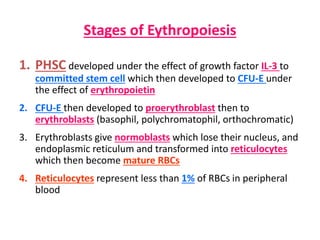
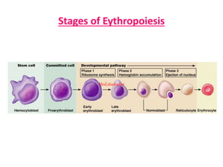
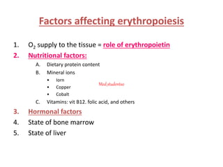
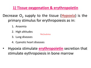
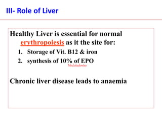
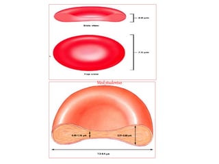
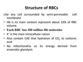
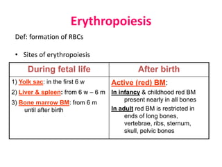
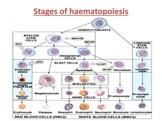
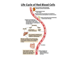
Red Blood Corpuscles
(RBCs)
Med_students0
At the end of the session the students should
be able to:
Describe the structure of RBCs
Describe in detail erythropoiesis mechanism
Describe the life-span of RBC (circulation and
their breakdown)
 Explain the factors affecting erythropoiesis
Guyton and Hall “Textbook of Medical Physiology”12th ed. (413-420)
Med_students0
Red blood cells RBCs
• Named red blood corpuscles as it not a true
cell as it do not contain nuclei and other cell
organells as mitochonderia
Normal RBCs count:
– Adult male 5 -5.5 million/ mm3
– Adult female 4.5 – 5 million/ mm3
– Being higher in new born and person living at high
altitude
RBCs size and shape:
• Normal RBCs are circular and biconcave discs
• Diameter 7.8 micron
• Thickness 2.5 micron
• Volume 90 cubic micron
Significance of RBCs shape
1. Larger surface area
2. Allow higher flexibility that allow RBCs to squeeze in
small capillaries
RBCs life span about 120 days
Med_students0
Med_students0
Structure of RBCs
Like any cell surrounded by semi-permeable cell
membrane
• Hb is its main content represent about 33% of RBC
volume.
• Each RBC has 200 million Hb molecules
• K+ is the main intracellular cation
• Also contain CAE that hydration of CO2 to carbonic
acid
• No mitochondria so its energy derived from
anaerobic glycolysis
Erythropoiesis
Def: formation of RBCs
• Sites of erythropoiesis
After birthDuring fetal life
Active (red) BM:
In infancy & childhood red BM
present nearly in all bones
In adult red BM is restricted in
ends of long bones,
vertebrae, ribs, sternum,
skull, pelvic bones
1) Yolk sac: in the first 6 w
2) Liver & spleen: from 6 w – 6 m
3) Bone marrow BM: from 6 m
until after birth
Stages of haematopoiesis
Stages of Eythropoiesis
1. PHSC developed under the effect of growth factor IL-3 to
committed stem cell which then developed to CFU-E under
the effect of erythropoietin
2. CFU-E then developed to proerythroblast then to
erythroblasts (basophil, polychromatophil, orthochromatic)
3. Erythroblasts give normoblasts which lose their nucleus, and
endoplasmic reticulum and transformed into reticulocytes
which then become mature RBCs
4. Reticulocytes represent less than 1% of RBCs in peripheral
blood
Stages of Eythropoiesis
Med_students0
Factors affecting erythropoiesis
1. O2 supply to the tissue = role of erythropoietin
2. Nutritional factors:
A. Dietary protein content
B. Mineral ions
• Iorn
• Copper
• Cobalt
C. Vitamins: vit B12. folic acid, and others
3. Hormonal factors
4. State of bone marrow
5. State of liver
Med_students0
1) Tissue oxygenation & erythropoietin
Decrease O2 supply to the tissue (Hypoxia) is the
primary stimulus for erythropoiesis as in:
1. Anaemia
2. High altitudes
3. Lung diseases
4. Cyanotic heart diseases
• Hypoxia stimulate erythropoietin secretion that
stimulate eythropoiesis in bone marrow
Med_students0
Erythropoietin
A glycoprotein hormone (mw 34000 d)
Source:
– 90% from the kidney (renal tubular epithelium or endothelial cells of
peritubular capillaries) and 10% form the liver (but mainly from the liver in
fetal life).
Function:
– Stimulates the production of proerythroblasts from stem cells
– Speeds up all stages of development of erythroblasts into mature RBCs
Regulation (control of secretion):
1. Hypoxia the main stimulus
2. Adrenaline, noradernaline and some PGs
3. Androgens
4. Adenosine (adenosine antagonist decrease EPO secretion)
5. Cobalt salts
Clinical uses:
1. Chronic renal failure
2. Aplastic anaemia
3. Anaemia with chronic diseases
Erythropoietin Mechanism
Reduces O2 levels
in blood
Erythropoietin
stimulates red bone
marrow
Enhanced
erythropoiesis
increases RBC
count
Normal blood oxygen levels Stimulus: Hypoxia due to
decreased RBC count,
decreased availability of O2 to
blood, or increased tissue
demands for O2
Start
Kidney (and liver to a
smaller extent) releases
erythropoietin
Increases
O2-carrying
ability of blood
90% of EPO is renal
2) Dietary factors
A) proteins:
– Proteins of high biological value are essential for erythropoeisis (for the
formation of globin part of Hb.
– Prolonged protein under nutrition lead to anaemia
B) Minerals:
1. Iron (Fe) is essential for formation of haeme part of Hb.
2. Copper (Cu)
– Cu essential for erythropoeisis, transported in the plasma by ceruloplasmin
(which catalyze the oxidation of ferrous iron to ferric)
– Co-factors in Hb synthesis
3. Cobalt (Co)
– Stimulate erythropoeisis though stimulation of erythropoeitin secretion from
the kdney
– enters in synthesis of Vit. B12
C) Vitamins:
• Vit B12, folic acid, others vit C
• Vitamin B12 & Folic acid; essential for DNA synthesis & maturation of bone
marrow cells
Med_students0
Iron (Fe+2)
• Iron is essential for formation of haeme part of Hb (also in
other heme containing particles as myoglbin, cytochrome
oxidase, catalase,perioxidase)
• decrease iron supply leads to iron deficiency anaemia
Iron metabolism:
• The total body iron content is 4 – 5 gm.
– 65% in Hb, 15-30% stored as ferritin in RES in the liver, 4% in
myoglobin, 1% in enzymes, 0.1% in transferrin in plasma.
– Normal serum iron level. (90-150 ug/dL) is bound to Transferrin
Daily requirement:
– 0.6 mg / day for male
– 1.3 mg /day for female
Absorption of Iron
Iron actively transported mainly in the upper small intestine (Duodenum &
Jejunum)
1) Dietary Ferric (Fe3+) reduced to ferrous (Fe2+)
2) Fe2+ or Heme transported at brush border by different carrier proteins (IT,
iron transporter & HT, heme transporter)
3) Intracellularly Fe2+ released from Heme by heme oxygenase
Most of intracellular Fe2+ actively transported (AT) across the basolateral
membrane to enter the blood.
Some Fe2+ oxidize to Fe3+ & bound apoferritin forming ferritin
Decrease Fe2+ AbsorptionIncrease Fe2+ Absorption
1. Oxalates & Phosphates
2. Phytates & Tannin
1. Vit C
2. Gastric HCl
Iron absorption occurs according to body needs when all apoferritin become
saturated iron absorption from enterocytes is inhibited
Transport:
In blood Fe2+ oxidize to Fe3+ & bound apotransferrin forming
transferrin reach various iron tissues store.
Storage:
Excess iron in blood is stored in cells of RE system (liver mainly
and spleen) it combind with apoferritin forming tissue ferritin
Med_students0
Feedback regulation of iron absorption
The rate of iron absorption from GIT depend on the iron stores in
the body:
– increased 5 times or more when iron stores in the body
become depleted
– Greatly decrease when the body iron stores are saturated
in the form of ferritin due to:
• The transferrin become fully saturated with iron (decrease the iron
binding capacity of the blood) that leads to accumulation of
ferritin in enterocytes that depress the active absorption of iron
from the intestinal lumen
• The liver decrease the synthesis of apotransferrin required for iron
absorption.
Iron Transport & Metabolism
III- Dietary vitamins
• Vitamin B12 & Folic acid; essential for DNA
synthesis & maturation of bone marrow cells
(maturation factors).
Deficiency of B12 & Folic acid leads to failure
of maturation of erythroblasts leading to
formation of fragile larger cells with shorter
life span (Macrocytic or Megaloblastic
anaemia)
Med_students0
Vit B12
Source: animal source liver, meat, egg, fish, vegetables are poor for vit B12
• Daily requirement: 5 µg.
Absorption:
• Vit B12 combined with intrinsic factor (a glycoprotein secreted by
parietal cells of gastric gland
• Intrinsic factor-vit B12 complex absorbed in the terminal ileum by
pinocytosis
Transport:
• Vit B12 is carried in the blood by PP transcobalamin to the site of storage
or use
Storage:
• Liver store large quantities of vit B12 (5 mg) that sufficient to supply vit
B12 requirement for about 3 years
Vit B12 deficiency:
1. Megaloblastic anaemia
2. Neurological manifestations
III- Role of Liver
Healthy Liver is essential for normal
erythropoiesis as it the site for:
1. Storage of Vit. B12 & iron
2. synthesis of 10% of EPO
Chronic liver disease leads to anaemia
Med_students0
IV- Hormones
• Thyroid H
• Glucocorticoides
• Androgens
All stimulate Erythropoiesis as they
promote tissue metabolism
Med_students0
V- State of bone marrow
• Healthy bone marrow is essential for normal
erythropoiesis
Destruction of BM by irradiation, drugs
toxins leads to aplastic anaemia
Life Cycle of Red Blood Cells
Life span and fate of RBCs:
Erythrocytes live in the circulation for an average of 120 days.
* As the cells grow older, they become more fragile and rupture
during their passage through narrow spots in the circulation
specially in the spleen.
* The released Hb from ruptured RBC’s is phagocytized by the
macrophage cells.
* Inside the macrophage cells:
Hb breaks into  globin + heme.
Globin → amino acids
Heme → iron + biliverdin → bilirubin
biliruin (yellow, pigment excreted by the liver in bile).
Med_students0

Red Blood Corpuscles

  • 1.
  • 2.
    At the endof the session the students should be able to: Describe the structure of RBCs Describe in detail erythropoiesis mechanism Describe the life-span of RBC (circulation and their breakdown)  Explain the factors affecting erythropoiesis Guyton and Hall “Textbook of Medical Physiology”12th ed. (413-420) Med_students0
  • 3.
    Red blood cellsRBCs • Named red blood corpuscles as it not a true cell as it do not contain nuclei and other cell organells as mitochonderia Normal RBCs count: – Adult male 5 -5.5 million/ mm3 – Adult female 4.5 – 5 million/ mm3 – Being higher in new born and person living at high altitude
  • 4.
    RBCs size andshape: • Normal RBCs are circular and biconcave discs • Diameter 7.8 micron • Thickness 2.5 micron • Volume 90 cubic micron Significance of RBCs shape 1. Larger surface area 2. Allow higher flexibility that allow RBCs to squeeze in small capillaries RBCs life span about 120 days Med_students0
  • 5.
  • 6.
    Structure of RBCs Likeany cell surrounded by semi-permeable cell membrane • Hb is its main content represent about 33% of RBC volume. • Each RBC has 200 million Hb molecules • K+ is the main intracellular cation • Also contain CAE that hydration of CO2 to carbonic acid • No mitochondria so its energy derived from anaerobic glycolysis
  • 7.
    Erythropoiesis Def: formation ofRBCs • Sites of erythropoiesis After birthDuring fetal life Active (red) BM: In infancy & childhood red BM present nearly in all bones In adult red BM is restricted in ends of long bones, vertebrae, ribs, sternum, skull, pelvic bones 1) Yolk sac: in the first 6 w 2) Liver & spleen: from 6 w – 6 m 3) Bone marrow BM: from 6 m until after birth
  • 8.
  • 9.
    Stages of Eythropoiesis 1.PHSC developed under the effect of growth factor IL-3 to committed stem cell which then developed to CFU-E under the effect of erythropoietin 2. CFU-E then developed to proerythroblast then to erythroblasts (basophil, polychromatophil, orthochromatic) 3. Erythroblasts give normoblasts which lose their nucleus, and endoplasmic reticulum and transformed into reticulocytes which then become mature RBCs 4. Reticulocytes represent less than 1% of RBCs in peripheral blood
  • 10.
  • 11.
    Factors affecting erythropoiesis 1.O2 supply to the tissue = role of erythropoietin 2. Nutritional factors: A. Dietary protein content B. Mineral ions • Iorn • Copper • Cobalt C. Vitamins: vit B12. folic acid, and others 3. Hormonal factors 4. State of bone marrow 5. State of liver Med_students0
  • 12.
    1) Tissue oxygenation& erythropoietin Decrease O2 supply to the tissue (Hypoxia) is the primary stimulus for erythropoiesis as in: 1. Anaemia 2. High altitudes 3. Lung diseases 4. Cyanotic heart diseases • Hypoxia stimulate erythropoietin secretion that stimulate eythropoiesis in bone marrow Med_students0
  • 13.
    Erythropoietin A glycoprotein hormone(mw 34000 d) Source: – 90% from the kidney (renal tubular epithelium or endothelial cells of peritubular capillaries) and 10% form the liver (but mainly from the liver in fetal life). Function: – Stimulates the production of proerythroblasts from stem cells – Speeds up all stages of development of erythroblasts into mature RBCs Regulation (control of secretion): 1. Hypoxia the main stimulus 2. Adrenaline, noradernaline and some PGs 3. Androgens 4. Adenosine (adenosine antagonist decrease EPO secretion) 5. Cobalt salts Clinical uses: 1. Chronic renal failure 2. Aplastic anaemia 3. Anaemia with chronic diseases
  • 16.
    Erythropoietin Mechanism Reduces O2levels in blood Erythropoietin stimulates red bone marrow Enhanced erythropoiesis increases RBC count Normal blood oxygen levels Stimulus: Hypoxia due to decreased RBC count, decreased availability of O2 to blood, or increased tissue demands for O2 Start Kidney (and liver to a smaller extent) releases erythropoietin Increases O2-carrying ability of blood 90% of EPO is renal
  • 17.
    2) Dietary factors A)proteins: – Proteins of high biological value are essential for erythropoeisis (for the formation of globin part of Hb. – Prolonged protein under nutrition lead to anaemia B) Minerals: 1. Iron (Fe) is essential for formation of haeme part of Hb. 2. Copper (Cu) – Cu essential for erythropoeisis, transported in the plasma by ceruloplasmin (which catalyze the oxidation of ferrous iron to ferric) – Co-factors in Hb synthesis 3. Cobalt (Co) – Stimulate erythropoeisis though stimulation of erythropoeitin secretion from the kdney – enters in synthesis of Vit. B12 C) Vitamins: • Vit B12, folic acid, others vit C • Vitamin B12 & Folic acid; essential for DNA synthesis & maturation of bone marrow cells Med_students0
  • 18.
    Iron (Fe+2) • Ironis essential for formation of haeme part of Hb (also in other heme containing particles as myoglbin, cytochrome oxidase, catalase,perioxidase) • decrease iron supply leads to iron deficiency anaemia Iron metabolism: • The total body iron content is 4 – 5 gm. – 65% in Hb, 15-30% stored as ferritin in RES in the liver, 4% in myoglobin, 1% in enzymes, 0.1% in transferrin in plasma. – Normal serum iron level. (90-150 ug/dL) is bound to Transferrin Daily requirement: – 0.6 mg / day for male – 1.3 mg /day for female
  • 19.
    Absorption of Iron Ironactively transported mainly in the upper small intestine (Duodenum & Jejunum) 1) Dietary Ferric (Fe3+) reduced to ferrous (Fe2+) 2) Fe2+ or Heme transported at brush border by different carrier proteins (IT, iron transporter & HT, heme transporter) 3) Intracellularly Fe2+ released from Heme by heme oxygenase Most of intracellular Fe2+ actively transported (AT) across the basolateral membrane to enter the blood. Some Fe2+ oxidize to Fe3+ & bound apoferritin forming ferritin Decrease Fe2+ AbsorptionIncrease Fe2+ Absorption 1. Oxalates & Phosphates 2. Phytates & Tannin 1. Vit C 2. Gastric HCl Iron absorption occurs according to body needs when all apoferritin become saturated iron absorption from enterocytes is inhibited
  • 20.
    Transport: In blood Fe2+oxidize to Fe3+ & bound apotransferrin forming transferrin reach various iron tissues store. Storage: Excess iron in blood is stored in cells of RE system (liver mainly and spleen) it combind with apoferritin forming tissue ferritin Med_students0
  • 21.
    Feedback regulation ofiron absorption The rate of iron absorption from GIT depend on the iron stores in the body: – increased 5 times or more when iron stores in the body become depleted – Greatly decrease when the body iron stores are saturated in the form of ferritin due to: • The transferrin become fully saturated with iron (decrease the iron binding capacity of the blood) that leads to accumulation of ferritin in enterocytes that depress the active absorption of iron from the intestinal lumen • The liver decrease the synthesis of apotransferrin required for iron absorption.
  • 22.
    Iron Transport &Metabolism
  • 23.
    III- Dietary vitamins •Vitamin B12 & Folic acid; essential for DNA synthesis & maturation of bone marrow cells (maturation factors). Deficiency of B12 & Folic acid leads to failure of maturation of erythroblasts leading to formation of fragile larger cells with shorter life span (Macrocytic or Megaloblastic anaemia) Med_students0
  • 24.
    Vit B12 Source: animalsource liver, meat, egg, fish, vegetables are poor for vit B12 • Daily requirement: 5 µg. Absorption: • Vit B12 combined with intrinsic factor (a glycoprotein secreted by parietal cells of gastric gland • Intrinsic factor-vit B12 complex absorbed in the terminal ileum by pinocytosis Transport: • Vit B12 is carried in the blood by PP transcobalamin to the site of storage or use Storage: • Liver store large quantities of vit B12 (5 mg) that sufficient to supply vit B12 requirement for about 3 years Vit B12 deficiency: 1. Megaloblastic anaemia 2. Neurological manifestations
  • 25.
    III- Role ofLiver Healthy Liver is essential for normal erythropoiesis as it the site for: 1. Storage of Vit. B12 & iron 2. synthesis of 10% of EPO Chronic liver disease leads to anaemia Med_students0
  • 26.
    IV- Hormones • ThyroidH • Glucocorticoides • Androgens All stimulate Erythropoiesis as they promote tissue metabolism Med_students0
  • 27.
    V- State ofbone marrow • Healthy bone marrow is essential for normal erythropoiesis Destruction of BM by irradiation, drugs toxins leads to aplastic anaemia
  • 28.
    Life Cycle ofRed Blood Cells
  • 31.
    Life span andfate of RBCs: Erythrocytes live in the circulation for an average of 120 days. * As the cells grow older, they become more fragile and rupture during their passage through narrow spots in the circulation specially in the spleen. * The released Hb from ruptured RBC’s is phagocytized by the macrophage cells. * Inside the macrophage cells: Hb breaks into  globin + heme. Globin → amino acids Heme → iron + biliverdin → bilirubin biliruin (yellow, pigment excreted by the liver in bile). Med_students0