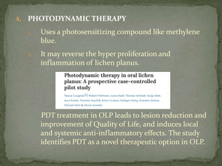
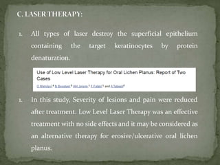
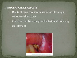
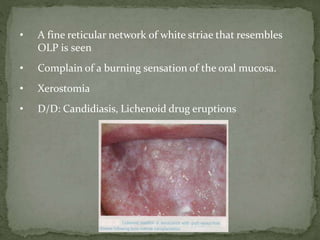
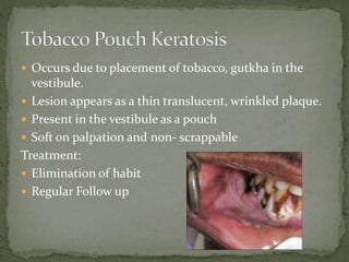
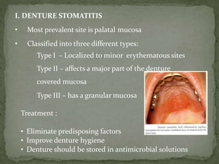
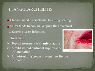
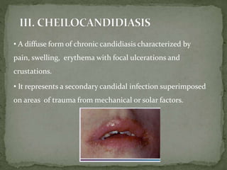
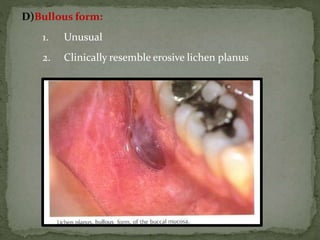
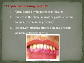
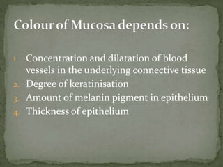
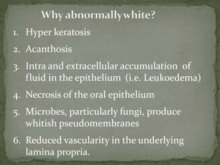
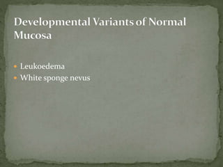
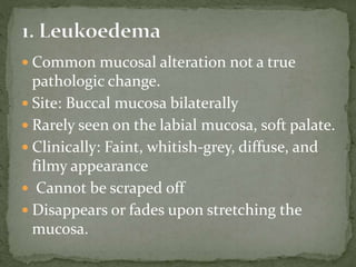
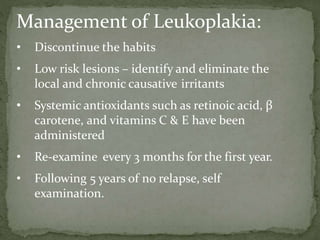
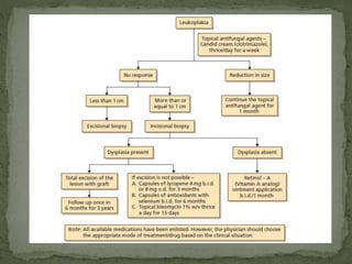
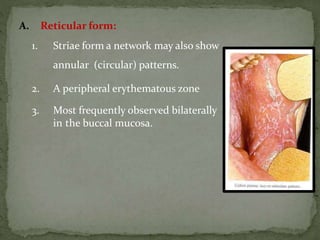
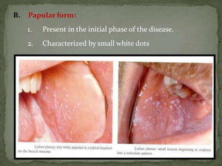
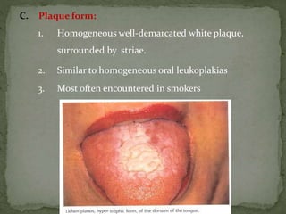
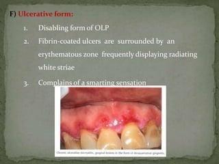
Dr. Arun Panwar
03/06/2020
 Introduction
 Why Mucosa appears white ?
 Classification
 Premalignant lesion
 Immunopathological diseases
 Allergic reactions
 Infectious diseases
 Reaction to mechanical trauma
 Conclusion
1. Concentration and dilatation of blood
vessels in the underlying connective tissue
2. Degree of keratinisation
3. Amount of melanin pigment in epithelium
4. Thickness of epithelium
1. Hyper keratosis
2. Acanthosis
3. Intra and extracellular accumulation of
fluid in the epithelium (i.e. Leukoedema)
4. Necrosis of the oral epithelium
5. Microbes, particularly fungi, produce
whitish pseudomembranes
6. Reduced vascularity in the underlying
lamina propria.
 Leukoedema
 White sponge nevus
 Common mucosal alteration not a true
pathologic change.
 Site: Buccal mucosa bilaterally
 Rarely seen on the labial mucosa, soft palate.
 Clinically: Faint, whitish-grey, diffuse, and
filmy appearance
 Cannot be scraped off
 Disappears or fades upon stretching the
mucosa.
 Investigation: Not required
 Treatment: No treatment is required since it
is a variation of the normal condition.
 No malignant change has been reported.
 D/D: Leukoplakia, White sponge nevus
 Rare genetic disorder
 Mutation of keratin 13 gene.
 Usually involves oral mucosa
and rarely mucous
membranes of the nose,
esophagus.
 Lesions may be present at
birth or manifest in early
childhood
 Clinical features:
 Presents bilaterally as symmetric white,
soft, “spongy,” or velvety thick plaques on
the buccal mucosa.
 Usually asymptomatic and has no
tendency toward malignant change.
 Investigation: Biopsy
 Treatment: Not required
 D/D: Chronic cheek biting, Leukoplakia,
Pachyonchia Congenita
PSEUDOMEMBRANOUS CANDIDIASIS
HYPERPLASTIC CANDIDIASIS
HAIRY LEUKOPLAKIA
 Also known as Candidosis, Thrush
 Opportunistic infection affecting the oral mucosa.
 Caused by candida albicans.
 Predisposing factors convert candida from the normal
commensal flora (saprophytic stage) to a pathogenic
organism (Parasitic stage).
 Affects patients medicated with
antibiotics, immunosuppressant drugs
or a disease
 Clinically: Oral lesions are white,
slightly elevated plaques occurring on
the buccal mucosa, tongue, palate,
gingiva and floor of the mouth.
 “Curdly white” Plaques can be wiped
away with gauze leaving an
erythamatous area.
 D/D: Chemical Burns, Traumatic Ulcers
 Also called as Candidal leukoplakia
 Characterized by a non scrappable white, firm
and raised plaque similar to leukoplakia.
 Site: Lips, tongue and cheeks
 Rarely, have been associated with malignant
transformation.
1. Smear: Useful in Pseudomembranous oral candidiasis.
2. Cell Culture: Sterile foam pads submerged in sabouraud broth,
placed on the infected surface for 60 seconds and then Cultivated at 37oC.
Result is expressed as colony forming units per cubicmillimeter.
(CFU/mm3).
Valuable in erythematous candidiasis.
3.Histopathological Examination:- Chronic plaque-type
and nodular candidiasis, identify the presence of epithelial dysplasia.
Treatment
 Also called as Green span lesion
 First reported by Greenspan and coworkers in 1984
in young homosexual males
 Etiology: EBV, Immunocompromised patients like
AIDS.
 Second most common HIV associated lesion
 Not pathognomonic for HIV since cancer drugs,
chemotherapy are also associated with OHL.
• Most commonly involves the lateral border of the
tongue but may extend to the ventral or dorsal
surfaces.
• Occasionally located on the buccal mucosa.
• Typical clinical appearance is white vertical streaks,
furrowed, corrugated white areas.
• Non scrappable,
• Always found bilateral.
• Asymptomatic
 Biopsy – Histological examination
 ELISA
Treatment:
 No treatment required.
 If associated with HIV, antiviral medications such
as zidovudine, acyclovir are given.
Differential Diagnosis:
 Hyperplastic candidiasis
 Leukoplakia
 Lichen planus
 The World Health Organization (WHO) in 1978
defined leukoplakia as - “a white patch or plaque that
cannot be characterized, clinically, or pathologically as
any other disease.”
 According to this definition, diagnosis of leukoplakia
was to be arrived by exclusion and histopathology is
not to be considered.
 In 1984, the definition was changed to “leukoplakia is a
whitish patch or plaque that cannot be characterized
clinically or pathologically as any other disease and it
is not associated with any physical or chemical
causative agent except the use of tobacco.”
 In 1994, leukoplakia was defined as - “a predominantly
white lesion of the oral mucosa that cannot be
characterized as any other definable disease, some oral
leukoplakias will transform into cancer.”
 In 2005, WHO defined leukoplakia as - “a white plaque
of questionable risk having excluded (other) known
diseases or disorders that carry no increased risk for
cancer.”
 Leukoplakia commences as a protective reaction
against a chronic irritant.
Clinical Findings:
• Asymptomatic
• Etiology: Chronic irritation
• Tobacco with other precipitating factors: Alcohol, Occlusal
trauma, Sharp prosthesis
• Site on involvement: Buccal mucosa, gingiva, tongue, Lip.
• Solitary or multiple plaques scattered through the mouth
• Floor of the mouth and the lateral borders of the tongue are
high-risk sites for malignant transformation.
• “ Cracked mud or Wrinkled” appearance
• Divided into Homogenous type and Non-Homogenous type
Homogenous Leukoplakia
a. White, well-demarcated plaque with an identical
reaction pattern throughout the entire lesion.
b. No red component
c. A smooth thin surface to a leathery appearance
with surface fissures as cracked mud.
2. Verrucous leukoplakia:-
• Verrucous, papillary (nodular) or exophytic
components
• Etiology: Idiopathic, may be associated with HPV,
EBV
• More aggressive proliferation pattern
• Lower gingiva is a predilection site.
• Malignant potential is high in PVL.
Differential Diagnosis:
1) Hyperplastic candidiasis
2) White sponge nevus.
3) Hairy leukoplakia
Management of Leukoplakia:
• Discontinue the habits
• Low risk lesions – identify and eliminate the
local and chronic causative irritants
• Systemic antioxidants such as retinoic acid, β
carotene, and vitamins C & E have been
administered
• Re-examine every 3 months for the first year.
• Following 5 years of no relapse, self
examination.
• Chronic insidious disease caused by areca nut use, and is
associated with increased risk for malignancy.
History:
1. First described by schwartz in 1952 coined the term
“atrophia idiopathica (trophica) mucosae oris”
2. In 1953, Joshi from Mumbai redesignated the
condition as oral submucous fibrosis.
• Etiology: Areca nut with precipitating factors like
chilli, Nutritional deficiency
• Genetic susceptibility
Clinical Features:
Early OSMF (Prodromal Symptoms)
1. Burning sensation in the mouth
2. Ulcerations/generalized inflammation
3. Defective gustatory sensation
4. Dryness of the mouth
5. Appearance of small vesicles in the cheek and palate.
6. Petechiae on the tongue, labial and buccal mucosa
7. Pain where submucous fibrotic bands are developing
Advanced Symptoms:
1. White marbling
2. Fibrous bands appear beneath an atrophic epithelium in
the buccal mucosa run in a vertical direction
3. Circular band around the entire rima oris (mouth orifice)
4. Impairment of tongue movement
5. Difficulty in opening the mouth
6. Inability to whistle or blow out a candle
7. Difficulty in swallowing
8. Increased fibrosis interferes with speech
9. 25% patients exhibit oral leukoplakias.
For the Clinical diagnosis of OSMF
a.Palpable fibrous bands
b.Mucosal texture feels tough and leathery
c.Blanching of mucosa
1. Elimination of the habit
2. Management strategies are
A. Nutritional support:
High protein and calories, vitamin B
complex and minerals
B. Immunomodulatory drugs:
1. Local and systemic application of
glucocorticoids and placental extract
2.Glucocorticoids act as
immunosuppressive agents.
3.Also prevent or suppress inflammatory
reactions.
 Turmeric, Tulsi, Aloe vera
Physiotherapy:
1. Kneading is a massage therapy used to improve the elasticity of
fibrous tissues and used in mobilizing scar tissues. Soft tissue
manipulation is extensively used in physiotherapy for improving
tissue extensibility.
2. Muscle stretching exercises for the mouth may be helpful to
prevent further restriction of mouth movements and to prevent
relapse.
Mouth gag, acrylic surgical stent, ballooning of mouth, hot water
gargling, inter positioning spatula between the teeth and adding a
new spatula every 5 - 10 days can be used for performing these
stretching exercises .
1.Application of heat in the form of lukewarm water, hot rinses or
selective deep heating therapies like short wave or micro wave
diathermy is another intervention used in physiotherapy to reduce
trismus caused by OSMF
C. Local Drug Delivery
1. Local injections of dexamethasone,
hyaluronidase and placental extract
Hyaluronidase:
•Breaks down hyaluronic acid
•Lowers the viscosity of the intercellular
cement substances
•Decreases collagen formation
3. Placental extracts:
a. Local injections as well as parental form
b. Placenta acts as a biogenous stimulator
c. Other actions of placental extract are
1. Anti-inflammatory
2. Increase in blood circulation
3. Arrest of tissue growth stagnation,
metabolic degenerative conditions and
lowered immunity response

 lnj. Placenta extract was given locally and effects were
monitored in reducing the severity of the disease.
 There was significant improvement in mouth opening,
colour of oral mucosa, burning sensation, and
reduction of fibrous bands.
Surgical Management:
1. Submucosal resection of fibrotic bands and
replacement with a partial-thickness skin or
mucosal graft
2. Bilateral temporalis myotomy
3. New treatment regimen composed of surgical
excision of the fibrotic bands with submucosal
placement of fresh human placental grafts,
followed by local injections of dexamethasone
for advanced cases.
 Introduction:
 Chronic inflammatory disease that affects the skin and
the mucous membrane.
 First described by Erasmus wilson in 1869.
 Epidemiology:
1. Prevalence is 0.5 – 2.2%
2. Commonly seen in women than men.
3. Mean age at the time of diagnosis is
approximately 55 years.
 Classic appearance of skin lesions - pruritic
erythematous to violaceous papules which are
flat topped, small (only a few millimeters) in diameter.
 Covered by a fine, glistening scale.
 Surface is covered by characteristic, very fine grayish-
white lines, wickham’s striae.
 Occur in a bilaterally symmetrical pattern, on the
flexor surfaces of the wrist and forearms, the inner
aspect of the knees and thighs, and the trunk.
 Primary symptom is a severe pruritis
 Trauma may aggravate the disease, referred to as a
Koebner phenomenon.
 Most frequent extra oral mucosal site is the genital
mucosa. Classic skin lesions described as
 Purplish
 Polygonal
 Planar
 Pruritic
 Papules
 Plaques
• Classically characterized by lesion consisting of
radiating white or gray, velvety, thread – like papules in
a linear, annular, or retiform arrangement
• A tiny white elevated dot present at the intersection of
the white lines, known as the striae of wickham.
• OLP may contain both red and white components with
different textures.
a. Reticulum
b. Papules
c. Plaque-like
d. Bullous
e. Erythematous
f. Ulcerative
A. Reticular form:
1. Striae form a network may also show
annular (circular) patterns.
2. A peripheral erythematous zone
3. Most frequently observed bilaterally
in the buccal mucosa.
B. Papular form:
1. Present in the initial phase of the disease.
2. Characterized by small white dots
C. Plaque form:
1. Homogeneous well-demarcated white plaque,
surrounded by striae.
2. Similar to homogeneous oral leukoplakias
3. Most often encountered in smokers
1. Lichenoid contact reactions
2. Oral Lichenoid drug reactions
3. Oral Graft Versus Host Disease
4. Discoid Lupus Erythematosus
Striae in DLE are typically more prominent with a more
marked hyperkeratinization which terminates against a
sharp demarcation.
5. Leukoplakia
-Leukoplakia is not featured with papular or reticular
elements.
Lavanya N, Jayanthi P, Rao UK, RanganathanK.
Oral lichen planus: An update on pathogenesisand treatment. J Oral Maxillofac Pathol
2011;15:127-32.
Management
 I PHARMACOLOGICAL MODALITIES
 A) CORTICOSTEROIDS
 Ability to modulate inflammation and immune
response
 Topical triamcinolone acetonide, betamethasone and
clobetasol are used.
 Antifungal therapy for erosive or erythamatous LP
 Systemic prednisolone (40-80mg for 5-7 days) (Tab.
Prednis. Tab Amipred)
2. TACROLIMUS:
• Immunosuppressive agent for the treatment of
atrophic dermatitis
• Successfully used in recalcitrant OLP cases
• Has a potential cancer risk from the prolonged use
• Available as Tacrolimus ointment 0.1%
3. RETINOIDS
• Topical retionoids - tretinoin, isotretinoin
• Has immunomodulatory properties and effective in OLP
• Reversal of white striae
• Systemic retinoids used in severe lichen planus.
• Retin-A cream 0.05%
A. PUVA THERAPY:
1. Uses photochemotherapy with 8-methoxypsoralen and long
wave ultraviolet light (PUVA)
2. Methoxypsoralen is given orally, followed by administration of
2 hours of UV radiation intraorally in the affected sites.
3. Successfully used in the treatment of severe cases of OLP.
1. The conclusion of this study is that PUVA seems to be effective
in the treatment of OLP and should be considered in severe
cases of OLP.
A. PHOTODYNAMIC THERAPY
1. Uses a photosensitizing compound like methylene
blue.
2. It may reverse the hyper proliferation and
inflammation of lichen planus.
PDT treatment in OLP leads to lesion reduction and
improvement of Quality of Life, and induces local
and systemic anti-inflammatory effects. The study
identifies PDT as a novel therapeutic option in OLP.
1. All types of laser destroy the superficial epithelium
containing the target keratinocytes by protein
denaturation.
1. In this study, Severity of lesions and pain were reduced
after treatment. Low Level Laser Therapy was an effective
treatment with no side effects and it may be considered as
an alternative therapy for erosive/ulcerative oral lichen
planus.
Reactive Lesions
Frictional Keratosis
Morsicatio buccarum
Tobacco pouch Keratosis
Lichenoid Reaction
1. FRICTIONAL KERATOSIS
• Due to chronic mechanical irritation like rough
denture or sharp cusp
• Characterized by a rough white lesion without any
red element.
Etiology and Pathogenesis:
1. Increased abrasion stimulates the epithelium to respond with an
increased production of keratin.
2. Regarded as a physiologic response to trauma
Clinical Findings:
1. Seen in edentulous areas of the alveolar ridge
2. Asymptomatic
Management:
1. No surgical intervention
2. Reduce predisposing factors.
• Due to chronic irritation because of repeated sucking,
nibbling or chewing.
• Seen in the buccal mucosa, labial mucosa and lateral
border of tongue
• F>M
• Management is by assurance
Lichenoid reactions include the following
1) Lichenoid contact reactions
2) Lichenoid drug eruptions
3) Lichenoid reactions of graft-versus-host disease (GVHD)
Aetologic agents: Hyper sensitivity reaction
to constituents derived from dental
materials likes mercury (Hg), Composite
resins, tobacco
Clinical Findings:
• Same reaction patterns as seen in OLP
• Appear as mixed white red lesion with
peripheral striae
• Major clinical difference between OLP
and LCR is the extensions of lesions
• Asymptomatic
Diagnosis:
• Replacement of the dental material may assist to
discriminate between LCR and OLP.
• Patch test
Management:
• Replacement of dental materials result in cure in 90%
of the cases.
• Lesions expected to heal within 1 to 2 months.
• Clinically, little to distinguish drug-induced
lichenoid reactions from lichen planus.
• Lichenoid lesions that include the lip,
symmetric in distribution and also involve
skin are more likely to be drug-related.
• Drug-induced Lichenoid reactions may
resolve promptly when the offending drug is
eliminated.
• NSAIDS, Diuretics and antihypertensives
Diagnosis:
1) Oral lesions comprise a white reticulum (or) papules
concurrently with erythematous and ulcerative lesions
2) A correct diagnosis is easier when a patient develops DILR
after starting a new drug.
Management:
1) Discontinuance of the drug and symptomatic treatment
with topical steroids.
•Characterized by interaction of immuno competent cells
from one individual (the donor) to a host (the recipient).
•Etiology and Pathogenesis: Major cause is allogenic
hematopoietic cell transplantation
Clinical Findings:
1) Lesions of GVHD are indistinguishable from the lesions
of OLP.
2) The lesions of GVHD associated with a more widespread
involvement of the oral mucosa.
3) Involves the cheeks, tongue, lips and gingivae
• A fine reticular network of white striae that resembles
OLP is seen
• Complain of a burning sensation of the oral mucosa.
• Xerostomia
• D/D: Candidiasis, Lichenoid drug eruptions
 Occurs due to placement of tobacco, gutkha in the
vestibule.
 Lesion appears as a thin translucent, wrinkled plaque.
 Present in the vestibule as a pouch
 Soft on palpation and non- scrappable
Treatment:
 Elimination of habit
 Regular Follow up
 Differential Diagnosis of Oral and Maxillofacial
lesions: Wood and Goaz
 Burkett’s Oral Medicine 12th edition
 Shafer’s textbook of Oral Pathology 5th edition
I. DENTURE STOMATITIS
• Most prevalent site is palatal mucosa
• Classified into three different types:
Type I – Localized to minor erythematous sites
Type II – affects a major part of the denture
covered mucosa
Type III – has a granular mucosa
Treatment :
• Eliminate predisposing factors
• Improve denture hygiene
• Denture should be stored in antimicrobial solutions
Characterized by erythema, fissuring, scaling
•Saliva tends to pool in, keeping the area moist
& favoring yeast infection.
•Treatment:
 Topical treatment with miconazole
 A mild steroid ointment suppress the
inflammation
 A moisturizing cream prevent new fissure
formation
II. ANGULARCHEILITIS
• A diffuse form of chronic candidiasis characterized by
pain, swelling, erythema with focal ulcerations and
crustations.
• It represents a secondary candidal infection superimposed
on areas of trauma from mechanical or solar factors.
D)Bullous form:
1. Unusual
2. Clinically resemble erosive lichen planus
E) Erythematous (Atrophic) OLP:
1. Characterized by homogeneous red area
2. Present in the buccal mucosa or palate, striae are
frequently seen in the periphery
3. Exclusively affecting attached gingiva presents
as desquamative gingivitis.
F) Ulcerative form:
1. Disabling form of OLP
2. Fibrin-coated ulcers are surrounded by an
erythematous zone frequently displaying radiating
white striae
3. Complains of a smarting sensation
Red and White lesions Part 1

Red and White lesions Part 1

  • 1.
  • 2.
     Introduction  WhyMucosa appears white ?  Classification  Premalignant lesion  Immunopathological diseases  Allergic reactions  Infectious diseases  Reaction to mechanical trauma  Conclusion
  • 3.
    1. Concentration anddilatation of blood vessels in the underlying connective tissue 2. Degree of keratinisation 3. Amount of melanin pigment in epithelium 4. Thickness of epithelium
  • 4.
    1. Hyper keratosis 2.Acanthosis 3. Intra and extracellular accumulation of fluid in the epithelium (i.e. Leukoedema) 4. Necrosis of the oral epithelium 5. Microbes, particularly fungi, produce whitish pseudomembranes 6. Reduced vascularity in the underlying lamina propria.
  • 8.
  • 9.
     Common mucosalalteration not a true pathologic change.  Site: Buccal mucosa bilaterally  Rarely seen on the labial mucosa, soft palate.  Clinically: Faint, whitish-grey, diffuse, and filmy appearance  Cannot be scraped off  Disappears or fades upon stretching the mucosa.
  • 10.
     Investigation: Notrequired  Treatment: No treatment is required since it is a variation of the normal condition.  No malignant change has been reported.  D/D: Leukoplakia, White sponge nevus
  • 11.
     Rare geneticdisorder  Mutation of keratin 13 gene.  Usually involves oral mucosa and rarely mucous membranes of the nose, esophagus.  Lesions may be present at birth or manifest in early childhood
  • 12.
     Clinical features: Presents bilaterally as symmetric white, soft, “spongy,” or velvety thick plaques on the buccal mucosa.  Usually asymptomatic and has no tendency toward malignant change.  Investigation: Biopsy  Treatment: Not required  D/D: Chronic cheek biting, Leukoplakia, Pachyonchia Congenita
  • 13.
  • 15.
     Also knownas Candidosis, Thrush  Opportunistic infection affecting the oral mucosa.  Caused by candida albicans.  Predisposing factors convert candida from the normal commensal flora (saprophytic stage) to a pathogenic organism (Parasitic stage).
  • 16.
     Affects patientsmedicated with antibiotics, immunosuppressant drugs or a disease  Clinically: Oral lesions are white, slightly elevated plaques occurring on the buccal mucosa, tongue, palate, gingiva and floor of the mouth.  “Curdly white” Plaques can be wiped away with gauze leaving an erythamatous area.  D/D: Chemical Burns, Traumatic Ulcers
  • 17.
     Also calledas Candidal leukoplakia  Characterized by a non scrappable white, firm and raised plaque similar to leukoplakia.  Site: Lips, tongue and cheeks  Rarely, have been associated with malignant transformation.
  • 18.
    1. Smear: Usefulin Pseudomembranous oral candidiasis. 2. Cell Culture: Sterile foam pads submerged in sabouraud broth, placed on the infected surface for 60 seconds and then Cultivated at 37oC. Result is expressed as colony forming units per cubicmillimeter. (CFU/mm3). Valuable in erythematous candidiasis. 3.Histopathological Examination:- Chronic plaque-type and nodular candidiasis, identify the presence of epithelial dysplasia.
  • 19.
  • 20.
     Also calledas Green span lesion  First reported by Greenspan and coworkers in 1984 in young homosexual males  Etiology: EBV, Immunocompromised patients like AIDS.  Second most common HIV associated lesion  Not pathognomonic for HIV since cancer drugs, chemotherapy are also associated with OHL.
  • 21.
    • Most commonlyinvolves the lateral border of the tongue but may extend to the ventral or dorsal surfaces. • Occasionally located on the buccal mucosa. • Typical clinical appearance is white vertical streaks, furrowed, corrugated white areas. • Non scrappable, • Always found bilateral. • Asymptomatic
  • 22.
     Biopsy –Histological examination  ELISA Treatment:  No treatment required.  If associated with HIV, antiviral medications such as zidovudine, acyclovir are given. Differential Diagnosis:  Hyperplastic candidiasis  Leukoplakia  Lichen planus
  • 24.
     The WorldHealth Organization (WHO) in 1978 defined leukoplakia as - “a white patch or plaque that cannot be characterized, clinically, or pathologically as any other disease.”  According to this definition, diagnosis of leukoplakia was to be arrived by exclusion and histopathology is not to be considered.  In 1984, the definition was changed to “leukoplakia is a whitish patch or plaque that cannot be characterized clinically or pathologically as any other disease and it is not associated with any physical or chemical causative agent except the use of tobacco.”
  • 25.
     In 1994,leukoplakia was defined as - “a predominantly white lesion of the oral mucosa that cannot be characterized as any other definable disease, some oral leukoplakias will transform into cancer.”  In 2005, WHO defined leukoplakia as - “a white plaque of questionable risk having excluded (other) known diseases or disorders that carry no increased risk for cancer.”  Leukoplakia commences as a protective reaction against a chronic irritant.
  • 26.
    Clinical Findings: • Asymptomatic •Etiology: Chronic irritation • Tobacco with other precipitating factors: Alcohol, Occlusal trauma, Sharp prosthesis • Site on involvement: Buccal mucosa, gingiva, tongue, Lip. • Solitary or multiple plaques scattered through the mouth • Floor of the mouth and the lateral borders of the tongue are high-risk sites for malignant transformation. • “ Cracked mud or Wrinkled” appearance • Divided into Homogenous type and Non-Homogenous type
  • 27.
    Homogenous Leukoplakia a. White,well-demarcated plaque with an identical reaction pattern throughout the entire lesion. b. No red component c. A smooth thin surface to a leathery appearance with surface fissures as cracked mud.
  • 29.
    2. Verrucous leukoplakia:- •Verrucous, papillary (nodular) or exophytic components • Etiology: Idiopathic, may be associated with HPV, EBV • More aggressive proliferation pattern • Lower gingiva is a predilection site. • Malignant potential is high in PVL.
  • 30.
    Differential Diagnosis: 1) Hyperplasticcandidiasis 2) White sponge nevus. 3) Hairy leukoplakia
  • 31.
    Management of Leukoplakia: •Discontinue the habits • Low risk lesions – identify and eliminate the local and chronic causative irritants • Systemic antioxidants such as retinoic acid, β carotene, and vitamins C & E have been administered • Re-examine every 3 months for the first year. • Following 5 years of no relapse, self examination.
  • 33.
    • Chronic insidiousdisease caused by areca nut use, and is associated with increased risk for malignancy. History: 1. First described by schwartz in 1952 coined the term “atrophia idiopathica (trophica) mucosae oris” 2. In 1953, Joshi from Mumbai redesignated the condition as oral submucous fibrosis. • Etiology: Areca nut with precipitating factors like chilli, Nutritional deficiency • Genetic susceptibility
  • 35.
    Clinical Features: Early OSMF(Prodromal Symptoms) 1. Burning sensation in the mouth 2. Ulcerations/generalized inflammation 3. Defective gustatory sensation 4. Dryness of the mouth 5. Appearance of small vesicles in the cheek and palate. 6. Petechiae on the tongue, labial and buccal mucosa 7. Pain where submucous fibrotic bands are developing
  • 36.
    Advanced Symptoms: 1. Whitemarbling 2. Fibrous bands appear beneath an atrophic epithelium in the buccal mucosa run in a vertical direction 3. Circular band around the entire rima oris (mouth orifice) 4. Impairment of tongue movement 5. Difficulty in opening the mouth 6. Inability to whistle or blow out a candle 7. Difficulty in swallowing 8. Increased fibrosis interferes with speech 9. 25% patients exhibit oral leukoplakias.
  • 37.
    For the Clinicaldiagnosis of OSMF a.Palpable fibrous bands b.Mucosal texture feels tough and leathery c.Blanching of mucosa
  • 38.
    1. Elimination ofthe habit 2. Management strategies are A. Nutritional support: High protein and calories, vitamin B complex and minerals B. Immunomodulatory drugs: 1. Local and systemic application of glucocorticoids and placental extract 2.Glucocorticoids act as immunosuppressive agents. 3.Also prevent or suppress inflammatory reactions.
  • 40.
  • 41.
    Physiotherapy: 1. Kneading isa massage therapy used to improve the elasticity of fibrous tissues and used in mobilizing scar tissues. Soft tissue manipulation is extensively used in physiotherapy for improving tissue extensibility. 2. Muscle stretching exercises for the mouth may be helpful to prevent further restriction of mouth movements and to prevent relapse. Mouth gag, acrylic surgical stent, ballooning of mouth, hot water gargling, inter positioning spatula between the teeth and adding a new spatula every 5 - 10 days can be used for performing these stretching exercises . 1.Application of heat in the form of lukewarm water, hot rinses or selective deep heating therapies like short wave or micro wave diathermy is another intervention used in physiotherapy to reduce trismus caused by OSMF
  • 42.
    C. Local DrugDelivery 1. Local injections of dexamethasone, hyaluronidase and placental extract Hyaluronidase: •Breaks down hyaluronic acid •Lowers the viscosity of the intercellular cement substances •Decreases collagen formation
  • 43.
    3. Placental extracts: a.Local injections as well as parental form b. Placenta acts as a biogenous stimulator c. Other actions of placental extract are 1. Anti-inflammatory 2. Increase in blood circulation 3. Arrest of tissue growth stagnation, metabolic degenerative conditions and lowered immunity response
  • 44.
      lnj. Placentaextract was given locally and effects were monitored in reducing the severity of the disease.  There was significant improvement in mouth opening, colour of oral mucosa, burning sensation, and reduction of fibrous bands.
  • 45.
    Surgical Management: 1. Submucosalresection of fibrotic bands and replacement with a partial-thickness skin or mucosal graft 2. Bilateral temporalis myotomy 3. New treatment regimen composed of surgical excision of the fibrotic bands with submucosal placement of fresh human placental grafts, followed by local injections of dexamethasone for advanced cases.
  • 46.
     Introduction:  Chronicinflammatory disease that affects the skin and the mucous membrane.  First described by Erasmus wilson in 1869.  Epidemiology: 1. Prevalence is 0.5 – 2.2% 2. Commonly seen in women than men. 3. Mean age at the time of diagnosis is approximately 55 years.
  • 47.
     Classic appearanceof skin lesions - pruritic erythematous to violaceous papules which are flat topped, small (only a few millimeters) in diameter.  Covered by a fine, glistening scale.  Surface is covered by characteristic, very fine grayish- white lines, wickham’s striae.  Occur in a bilaterally symmetrical pattern, on the flexor surfaces of the wrist and forearms, the inner aspect of the knees and thighs, and the trunk.
  • 48.
     Primary symptomis a severe pruritis  Trauma may aggravate the disease, referred to as a Koebner phenomenon.  Most frequent extra oral mucosal site is the genital mucosa. Classic skin lesions described as  Purplish  Polygonal  Planar  Pruritic  Papules  Plaques
  • 49.
    • Classically characterizedby lesion consisting of radiating white or gray, velvety, thread – like papules in a linear, annular, or retiform arrangement • A tiny white elevated dot present at the intersection of the white lines, known as the striae of wickham. • OLP may contain both red and white components with different textures. a. Reticulum b. Papules c. Plaque-like d. Bullous e. Erythematous f. Ulcerative
  • 50.
    A. Reticular form: 1.Striae form a network may also show annular (circular) patterns. 2. A peripheral erythematous zone 3. Most frequently observed bilaterally in the buccal mucosa.
  • 51.
    B. Papular form: 1.Present in the initial phase of the disease. 2. Characterized by small white dots
  • 52.
    C. Plaque form: 1.Homogeneous well-demarcated white plaque, surrounded by striae. 2. Similar to homogeneous oral leukoplakias 3. Most often encountered in smokers
  • 53.
    1. Lichenoid contactreactions 2. Oral Lichenoid drug reactions 3. Oral Graft Versus Host Disease 4. Discoid Lupus Erythematosus Striae in DLE are typically more prominent with a more marked hyperkeratinization which terminates against a sharp demarcation. 5. Leukoplakia -Leukoplakia is not featured with papular or reticular elements.
  • 54.
    Lavanya N, JayanthiP, Rao UK, RanganathanK. Oral lichen planus: An update on pathogenesisand treatment. J Oral Maxillofac Pathol 2011;15:127-32. Management
  • 55.
     I PHARMACOLOGICALMODALITIES  A) CORTICOSTEROIDS  Ability to modulate inflammation and immune response  Topical triamcinolone acetonide, betamethasone and clobetasol are used.  Antifungal therapy for erosive or erythamatous LP  Systemic prednisolone (40-80mg for 5-7 days) (Tab. Prednis. Tab Amipred)
  • 56.
    2. TACROLIMUS: • Immunosuppressiveagent for the treatment of atrophic dermatitis • Successfully used in recalcitrant OLP cases • Has a potential cancer risk from the prolonged use • Available as Tacrolimus ointment 0.1% 3. RETINOIDS • Topical retionoids - tretinoin, isotretinoin • Has immunomodulatory properties and effective in OLP • Reversal of white striae • Systemic retinoids used in severe lichen planus. • Retin-A cream 0.05%
  • 57.
    A. PUVA THERAPY: 1.Uses photochemotherapy with 8-methoxypsoralen and long wave ultraviolet light (PUVA) 2. Methoxypsoralen is given orally, followed by administration of 2 hours of UV radiation intraorally in the affected sites. 3. Successfully used in the treatment of severe cases of OLP. 1. The conclusion of this study is that PUVA seems to be effective in the treatment of OLP and should be considered in severe cases of OLP.
  • 58.
    A. PHOTODYNAMIC THERAPY 1.Uses a photosensitizing compound like methylene blue. 2. It may reverse the hyper proliferation and inflammation of lichen planus. PDT treatment in OLP leads to lesion reduction and improvement of Quality of Life, and induces local and systemic anti-inflammatory effects. The study identifies PDT as a novel therapeutic option in OLP.
  • 59.
    1. All typesof laser destroy the superficial epithelium containing the target keratinocytes by protein denaturation. 1. In this study, Severity of lesions and pain were reduced after treatment. Low Level Laser Therapy was an effective treatment with no side effects and it may be considered as an alternative therapy for erosive/ulcerative oral lichen planus.
  • 60.
    Reactive Lesions Frictional Keratosis Morsicatiobuccarum Tobacco pouch Keratosis Lichenoid Reaction
  • 61.
    1. FRICTIONAL KERATOSIS •Due to chronic mechanical irritation like rough denture or sharp cusp • Characterized by a rough white lesion without any red element.
  • 62.
    Etiology and Pathogenesis: 1.Increased abrasion stimulates the epithelium to respond with an increased production of keratin. 2. Regarded as a physiologic response to trauma Clinical Findings: 1. Seen in edentulous areas of the alveolar ridge 2. Asymptomatic Management: 1. No surgical intervention 2. Reduce predisposing factors.
  • 63.
    • Due tochronic irritation because of repeated sucking, nibbling or chewing. • Seen in the buccal mucosa, labial mucosa and lateral border of tongue • F>M • Management is by assurance
  • 64.
    Lichenoid reactions includethe following 1) Lichenoid contact reactions 2) Lichenoid drug eruptions 3) Lichenoid reactions of graft-versus-host disease (GVHD)
  • 65.
    Aetologic agents: Hypersensitivity reaction to constituents derived from dental materials likes mercury (Hg), Composite resins, tobacco Clinical Findings: • Same reaction patterns as seen in OLP • Appear as mixed white red lesion with peripheral striae • Major clinical difference between OLP and LCR is the extensions of lesions • Asymptomatic
  • 66.
    Diagnosis: • Replacement ofthe dental material may assist to discriminate between LCR and OLP. • Patch test Management: • Replacement of dental materials result in cure in 90% of the cases. • Lesions expected to heal within 1 to 2 months.
  • 67.
    • Clinically, littleto distinguish drug-induced lichenoid reactions from lichen planus. • Lichenoid lesions that include the lip, symmetric in distribution and also involve skin are more likely to be drug-related. • Drug-induced Lichenoid reactions may resolve promptly when the offending drug is eliminated. • NSAIDS, Diuretics and antihypertensives
  • 68.
    Diagnosis: 1) Oral lesionscomprise a white reticulum (or) papules concurrently with erythematous and ulcerative lesions 2) A correct diagnosis is easier when a patient develops DILR after starting a new drug. Management: 1) Discontinuance of the drug and symptomatic treatment with topical steroids.
  • 69.
    •Characterized by interactionof immuno competent cells from one individual (the donor) to a host (the recipient). •Etiology and Pathogenesis: Major cause is allogenic hematopoietic cell transplantation Clinical Findings: 1) Lesions of GVHD are indistinguishable from the lesions of OLP. 2) The lesions of GVHD associated with a more widespread involvement of the oral mucosa. 3) Involves the cheeks, tongue, lips and gingivae
  • 70.
    • A finereticular network of white striae that resembles OLP is seen • Complain of a burning sensation of the oral mucosa. • Xerostomia • D/D: Candidiasis, Lichenoid drug eruptions
  • 71.
     Occurs dueto placement of tobacco, gutkha in the vestibule.  Lesion appears as a thin translucent, wrinkled plaque.  Present in the vestibule as a pouch  Soft on palpation and non- scrappable Treatment:  Elimination of habit  Regular Follow up
  • 72.
     Differential Diagnosisof Oral and Maxillofacial lesions: Wood and Goaz  Burkett’s Oral Medicine 12th edition  Shafer’s textbook of Oral Pathology 5th edition
  • 73.
    I. DENTURE STOMATITIS •Most prevalent site is palatal mucosa • Classified into three different types: Type I – Localized to minor erythematous sites Type II – affects a major part of the denture covered mucosa Type III – has a granular mucosa Treatment : • Eliminate predisposing factors • Improve denture hygiene • Denture should be stored in antimicrobial solutions
  • 74.
    Characterized by erythema,fissuring, scaling •Saliva tends to pool in, keeping the area moist & favoring yeast infection. •Treatment:  Topical treatment with miconazole  A mild steroid ointment suppress the inflammation  A moisturizing cream prevent new fissure formation II. ANGULARCHEILITIS
  • 75.
    • A diffuseform of chronic candidiasis characterized by pain, swelling, erythema with focal ulcerations and crustations. • It represents a secondary candidal infection superimposed on areas of trauma from mechanical or solar factors.
  • 76.
    D)Bullous form: 1. Unusual 2.Clinically resemble erosive lichen planus
  • 77.
    E) Erythematous (Atrophic)OLP: 1. Characterized by homogeneous red area 2. Present in the buccal mucosa or palate, striae are frequently seen in the periphery 3. Exclusively affecting attached gingiva presents as desquamative gingivitis.
  • 78.
    F) Ulcerative form: 1.Disabling form of OLP 2. Fibrin-coated ulcers are surrounded by an erythematous zone frequently displaying radiating white striae 3. Complains of a smarting sensation