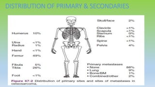
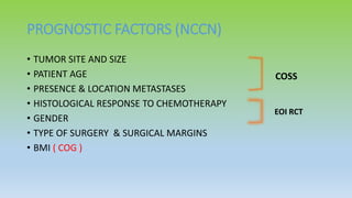
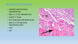
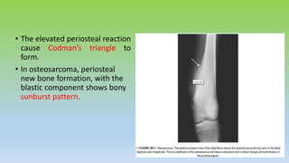
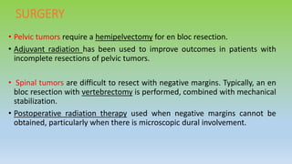
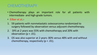
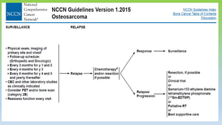
Osteosarcoma is the most common primary bone cancer, often arising in the appendicular skeleton of teenagers and young adults. It is typically a high grade tumor associated with rapid bone proliferation and spread. Treatment involves complete surgical resection with limb-sparing surgery when possible, along with chemotherapy both before and after surgery. Radiation therapy may be used for unresectable or incompletely resected tumors. Prognostic factors include tumor size, location of metastases, and response to preoperative chemotherapy. While survival has improved with modern multimodal treatment, new strategies are still needed given the risk of recurrence and lung metastases.
Radiation therapy plays an important role in the management of many bone tumors as an adjunct to surgery or as primary treatment for inoperable tumors. Newer radiation techniques like IMRT and proton beam therapy allow for more conformal dose distributions that improve local tumor control while reducing damage to surrounding healthy tissues. Radiation is used as primary treatment, post-operatively, or palliatively depending on the tumor type, location, surgical margins, and other factors.
Bone is commonly affected by metastasis. Radiation therapy is effective for relieving bone pain from metastases. Shorter fractionation schedules like single 8 Gy fractions provide pain relief but have higher retreatment rates compared to longer schedules like 30 Gy in 10 fractions. Newer techniques like SBRT and hemibody irradiation also effectively palliate bone pain with acceptable toxicity. Bisphosphonates combined with radiation can further improve pain relief and increase bone density. Surgery to stabilize fractures is recommended for high risk or impending fractures to allow early mobility.
1) Preoperative chemoradiotherapy improves local control rates and tumor downstaging for rectal cancer compared to postoperative chemoradiotherapy or radiotherapy alone.
2) The addition of chemotherapy to radiotherapy, whether in the preoperative or postoperative setting, improves local control and disease-free survival compared to radiotherapy alone.
3) For patients who achieve a clinical complete response after preoperative chemoradiotherapy, observation without surgery may be feasible, with local recurrence rates of approximately 30% that can often be successfully salvaged.
Radiotherapy plays an important role in the treatment of soft tissue sarcomas by improving local control rates when used adjuvantly with surgery. Post-operative radiotherapy reduces local recurrence rates compared to surgery alone, even for low-grade tumors. Pre-operative radiotherapy may provide a better chance of limb salvage for large or unresectable tumors but risks delaying wound healing. Positive surgical margins are associated with higher local recurrence rates, but margins within 1mm do not significantly impact outcomes. Adjuvant radiotherapy should be considered for all high-grade soft tissue sarcomas based on its ability to improve local control.
Radiotherapy is used as primary treatment for early-stage Hodgkin lymphoma or as part of combined modality treatment with chemotherapy. Historically, large mantle fields covering lymph node regions from the skull to the pelvis were used. More modern approaches use smaller involved field radiotherapy targeting only initially involved lymph node regions after chemotherapy based on imaging. Proper delineation of clinical target volumes requires pre-chemotherapy imaging ideally with PET/CT to define original disease extent.
This document provides information about total body irradiation (TBI). It discusses that TBI uses megavoltage photon beams to destroy the recipient's bone marrow and tumor cells prior to bone marrow transplantation. It is used to treat various diseases like leukemia, lymphoma, and multiple myeloma. TBI can be delivered at high or low doses, to half the body, or total nodes. Techniques include parallel opposed beams from linear accelerators or cobalt-60 machines. Dosimetry and in vivo dosimetry are important due to the large fields and difficulty achieving uniform dose. Complications can include sterility, secondary cancers, and growth issues.
Palliation brain, spinal and bone metsDrAyush Garg
The document summarizes guidelines for palliation of brain, spinal cord, and bone metastases. It addresses epidemiology and clinical presentation of brain metastases and recommendations for various treatment approaches including whole brain radiation therapy, surgical resection, radiosurgery, chemotherapy, and management of recurrent metastases. It also covers prophylactic use of anticonvulsants and steroids. Key recommendations include that surgical resection plus WBRT is superior to WBRT alone for single brain metastases, and radiosurgery is effective for lesions under 3cm and not causing significant midline shift.
ROLE OF RADIATION IN BONE TUMORS FOR ORTHOPEDICSKanhu Charan
This document discusses soft tissue sarcomas. It begins by listing various histologic diagnoses of soft tissue sarcomas categorized by tissue of origin. It then discusses imaging with MRI and CT to evaluate tumors. Key points regarding surgery include performing limb-sparing over amputation when possible and obtaining negative margins of at least 2cm if no post-op radiation is planned. Adjuvant radiation improves local control but not necessarily survival. Pre-op radiation may increase wound complications compared to post-op.
Learn about the process of radiation therapy to treat soft tissue sarcoma, and how new radiation technology has improved treatment of the disease.
This presentation was given by Elizabeth H. Baldini, MD, MPH, radiation oncology director for the Center for Sarcoma and Bone Oncology at Dana-Farber Cancer Institute. It was originally presented as part of the "15 Years of GIST/Soft Tissue Sarcoma Symposium," held on Sept. 12, 2015 at Dana-Farber in Boston, Mass.
Radioisotopes in the management of bone metastasesSneha George
Radionuclides can be used to treat bone metastases by accumulating in areas of bone involvement, allowing localized treatment to minimize side effects. Common radionuclides used include strontium-89, samarium-153, and rhenium-186. They are indicated for diffuse skeletal metastases, painful lesions not adequately treated with other methods, or hormone-insensitive prostate cancer. Potential side effects include increased pain, bone marrow suppression, and fractures. New strategies to enhance treatment include combining radionuclide therapy with chemotherapy, repeating radionuclide injections, or using high-dose radionuclide therapy with bone marrow support.
RADIOTHERAPY IN CARCINOMA BREAST (EARLY AND LOCALLY ADVANCED)DrAnkitaPatel
This document discusses radiation therapy for breast cancer. It begins by outlining the important role of radiation therapy at various stages of breast cancer, including as part of breast conservation and after mastectomy. It then discusses indications for adjuvant radiation therapy based on factors like tumor size and lymph node involvement. The document reviews evidence from clinical trials demonstrating the benefits of radiation therapy after breast-conserving surgery in reducing recurrence rates and improving survival. It also discusses techniques, dosing, and toxicity considerations for radiation therapy delivery.
This document summarizes a panel discussion on oligometastatic disease. It defines oligometastatic disease as having a solitary or few detectable metastatic lesions confined to a single organ or more than one organ. There is ongoing debate around how many lesions constitute oligometastatic disease. The document discusses various theories on metastasis patterns and improving treatments like stereotactic radiosurgery that have led to reclassification of some metastatic tumors as oligometastatic. Ongoing trials are exploring more aggressive local treatment of oligometastatic lesions combined with systemic therapies to improve long-term survival.
The document discusses the role of radiation therapy in treating soft tissue sarcoma. It notes that radiation reduces local recurrence rates after surgery and allows for limb salvage procedures. Both preoperative and postoperative radiation are discussed, with advantages and disadvantages listed for each approach. Intensity modulated radiation therapy and brachytherapy are presented as techniques that may improve local control while reducing toxicity. The timing, dose, and volume of radiation treatment are important considerations. Overall, the document evaluates the evidence for integrating radiation therapy with surgery and chemotherapy to effectively treat soft tissue sarcomas.
The document discusses craniospinal irradiation (CSI), which delivers radiation to the entire cranial-spinal axis to treat intracranial tumors. It was pioneered in the 1950s and is commonly used to treat tumors that may spread through the cerebrospinal fluid such as medulloblastoma. The document outlines the techniques, challenges, indications, and evolving approaches for CSI such as reduced dose protocols and hyperfractionated regimens. It discusses topics like patient positioning, target volumes, critical structures, field arrangements, and the use of newer technologies like virtual simulation.
Altered Fractionation Radiotherapy in Head-Neck CancerJyotirup Goswami
Altered fractionation radiotherapy has been shown to improve outcomes for head and neck cancer patients compared to conventional fractionation. Meta-analyses demonstrate significant benefits including improved 5-year locoregional control and overall survival. However, most modern trials do not address fractionation. Hypofractionation shows promise with comparable tumor control and toxicity but reduced treatment time. Ongoing research combines altered fractionation with chemotherapy and radiosensitizers to further improve outcomes while minimizing toxicity.
This document discusses dose-volume histograms (DVHs) which are used to analyze and compare radiation dose distributions in radiotherapy treatment planning. It describes how DVHs are generated by counting the number of voxels receiving different dose levels. DVHs can be displayed cumulatively or differentially and show the volume of structures receiving particular doses. The document outlines some limitations of DVHs including their insensitivity to small hot or cold spots and lack of spatial information. It emphasizes that DVHs should be used along with visual analysis of dose distributions and dose-volume statistics when evaluating treatment plans.
Hippocampal sparing whole brain radiation therapy- Making a case!VIMOJ JANARDANAN NAIR
- A 58-year-old female nurse presented with headache, nausea, and mental status changes. Imaging showed 3 brain metastases with no known primary site.
- She had a KPS of 90, age <60, controlled primary (pending further workup), and no extracranial mets, placing her in RPA class I and GPA group with a predicted median survival of 7.1-11.3 months.
- Treatment options included WBRT alone or WBRT with boost to reduce risk of neurocognitive toxicity based on the RTOG 0933 trial, which showed reduced memory decline with hippocampal avoidance. Patient selection focused on tumor types likely to benefit and survival duration to gain
The document discusses various radiation fractionation schedules used in cancer treatment. It begins with an overview of conventional fractionation, which divides the total radiation dose into smaller daily doses to allow healthy cells to repair sublethal damage between fractions. It then explores the radiobiological rationale of the 5 R's of fractionation - repair, redistribution, reoxygenation, repopulation, and radiosensitivity. The document discusses various altered fractionation schedules including hyperfractionation, accelerated fractionation, split-course, and hypofractionation, explaining how each schedule aims to improve the therapeutic ratio for cancer patients.
1. SABR has emerged as an alternative to surgery for medically operable early-stage NSCLC based on case-control studies showing equivalence in outcomes.
2. Randomized trials are still needed to provide level 1 evidence of equivalence since residual differences remain between surgery and SABR cohorts in existing studies.
3. Ongoing trials such as STABLE-MATES, VALOR, and SABRTOOTH aim to address this evidence gap through randomized comparisons of SABR to surgery.
IORT uses a high single dose of radiation delivered during surgery to treat cancer remnants after tumor removal. It has two objectives: increase local tumor control and increase the ratio of tumor control to damage of nearby healthy tissues. IORT can be delivered via two methods - IOERT uses electron beams from a LINAC and IOHDR uses radioactive sources. Treatment planning requires a multidisciplinary team to determine applicator positioning and appropriate dose to maximize tumor coverage while minimizing radiation to other organs. IORT provides local tumor control comparable to conventional fractionated radiotherapy but with faster treatment time and less damage to surrounding tissues.
Bone metastases occur when cancer spreads from its original site, such as the breast, prostate, or lungs, to the bone. Common sites for bone metastases include the spine, pelvis, and ribs. Bone metastases can be either osteoblastic, causing abnormal bone growth, or lytic, creating holes in the bone. Imaging tests like MRI, bone scans, and PET scans can detect bone metastases. Radiation therapy is effective at reducing pain from bone metastases and can provide lasting symptom relief for many patients.
Ewing sarcoma is the second most common bone tumor in children. Radiotherapy plays an important role in the treatment of both localized and metastatic Ewing sarcoma. For localized disease, radiotherapy is recommended for patients who cannot undergo surgery or have unresectable tumors. It is also used post-operatively if there is residual disease. For metastatic disease, radiotherapy can help control the primary tumor and reduce pulmonary metastases when combined with chemotherapy. Advances in radiotherapy planning and techniques have improved outcomes while reducing long-term side effects.
This document discusses a trial investigating the role of local radiation therapy for metastatic prostate cancer. The main findings were:
1. No overall survival benefit was seen with radiation therapy, but survival improved in patients with low metastatic burden.
2. Failure-free survival improved with radiation therapy overall and in the low metastatic burden group.
3. Adverse effects from radiation therapy were modest.
The trial provides evidence that radiation therapy to the prostate improves outcomes for men with metastatic prostate cancer who have a low metastatic burden and does not negatively impact side effects.
Management of cacrinoma cervix: Techniques of radiotherapy (2D conventional, 3D Conformal radiotherapy (3DCRT) and IMRT with a review of various contouring guidelines.
This document discusses the use of radiotherapy in the treatment of acute lymphoblastic leukemia (ALL). It provides an overview of ALL, including classification, risk groups, and treatment approaches involving induction, intensification, maintenance, and central nervous system prophylaxis. It then focuses on the role of radiotherapy, describing protocols for cranial irradiation to prevent central nervous system relapse, including dose schedules. It also discusses radiotherapy for meningeal leukemia at diagnosis, testicular irradiation, and total body irradiation used for bone marrow transplantation conditioning.
The combined use of radiation therapy and chemotherapy in cancer treatment is a logical and reasonable approach that has already proven beneficial for several malignancies.
Management of ewings sarcoma & osteosarcomaPRARABDH95
EBRT can play an important role in the management of Ewing sarcoma and osteosarcoma.
For Ewing sarcoma, radiotherapy is commonly used pre-operatively to sterilize the tumor bed, post-operatively for positive or close margins, or definitively when surgery is not possible. Treatment planning aims to cover the pre-treatment tumor volume plus a 2-2.5cm margin using IMRT or 3D-CRT.
For osteosarcoma, radiotherapy can be used definitively for unresectable tumors or adjuvantly after surgery if margins were positive. A dose of 70.2Gy is typically prescribed for definitive cases and 64.8Gy for
Externalbeam rt in ews3.12.20 - frida yseminar-finallllPRARABDH95
1) Ewing sarcoma and osteosarcoma are rare bone cancers that typically affect children and young adults. Ewing sarcoma is the second most common primary bone cancer while osteosarcoma most commonly presents as a primary bone malignancy.
2) Both cancers are diagnosed through imaging, biopsy and staging workup. Management involves chemotherapy along with local therapy through surgery and/or radiation therapy.
3) Radiation therapy planning aims to adequately cover the tumor volume while sparing nearby organs at risk. Techniques such as 3D conformal radiation therapy and intensity modulated radiation therapy (IMRT) allow for improved dose distribution over conventional radiation.
1) Chemotherapy has improved survival rates for osteosarcoma dramatically over the past 30 years from less than 20% to between 40-60% through the use of effective combination chemotherapy and neoadjuvant treatment.
2) Key trials showed that neoadjuvant chemotherapy followed by surgery and adjuvant chemotherapy improved relapse-free and overall survival compared to surgery alone.
3) The combination of doxorubicin and cisplatin administered every 3 weeks is now considered the standard first-line chemotherapy regimen based on results from large cooperative trials.
Neoadjuvant therapy in colorectal carcinomaAnkita Singh
- Several studies have shown that neoadjuvant therapy decreases local recurrence rates in colorectal cancer when compared to surgery alone. One study showed a statistically significant decrease in local recurrence with the addition of chemotherapy to preoperative radiotherapy.
- Evidence indicates that long-course chemoradiotherapy, induction chemotherapy followed by long-course chemoradiotherapy, and short-course radiotherapy are the three accepted neoadjuvant approaches, with long-course chemoradiotherapy being the most commonly used currently. Short-course radiotherapy has also shown non-inferior oncologic outcomes compared to long-course chemoradiotherapy in some studies.
This document summarizes key information about cancer of the esophagus:
1) The esophagus spans from the cricopharyngeus to the GE junction. Esophageal cancer risk factors include smoking, alcohol, caustic injury, HPV infection, obesity, and Barrett's esophagus.
2) The most common types of esophageal cancer are squamous cell carcinoma (SCC) in the upper two-thirds and adenocarcinoma in the lower third. Distant metastases are most common in the lung, liver, and bone.
3) Treatment options discussed include surgery, chemotherapy, radiation therapy, and their combinations in neoadjuvant and adjuvant settings. Several large randomized controlled trials
This document discusses the management of intermediate and high risk prostate cancer. It begins by providing background on prostate cancer epidemiology and risk stratification. It then covers various treatment options including observation, active surveillance, radical prostatectomy, radiotherapy, and androgen deprivation therapy. Several studies comparing the efficacy of radiotherapy alone versus radiotherapy with short or long-term ADT are summarized. For intermediate risk prostate cancer, the document recommends 4-6 months of ADT with radiotherapy based on trial results. For high risk prostate cancer, 2-3 years of ADT with radiotherapy is recommended.
Uveal melanoma commonly spreads to the liver. This document discusses uveal melanoma (MUM) that has metastasized to the liver. It provides background on MUM, noting that half of patients develop metastases, usually first appearing in the liver. It describes genetic risk factors for metastasis and different risk classifications. The document advocates for locoregional therapies for liver metastases since there are no effective systemic therapies. It presents evidence that liver-directed therapies may prolong survival more than systemic treatments or surveillance alone.
retroperitoneal tumors esp. retroperitoneal sarcoma is most challenging condition to treat in retroperitoneal region inspite of using all treatment modalities.here is brief description of its management acc. to nccn , and other text book ref.
1) The study analyzed outcomes of 728 soft tissue sarcoma patients, 281 of whom underwent unplanned excisions prior to referral to specialty centers.
2) Patients who had unplanned excisions were found to have smaller and more superficial tumors on average.
3) After adjusting for factors like tumor grade and size, unplanned excisions were not found to provide any survival benefit compared to planned surgery, and resulted in more complex subsequent procedures.
4) The study concludes that while unplanned excisions do not seem to directly harm prognosis, they should still be avoided due to increased treatment complexity.
Soft tissue sarcomas are rare malignant tumors that can arise in any soft tissue of the body. They are characterized by their genetic alterations and histological grade. Diagnosis is made through biopsy and imaging is used to stage the tumor. Treatment typically involves complete surgical resection with negative margins, along with possible adjuvant radiation and chemotherapy depending on tumor grade and size. Prognosis depends on factors like tumor size, grade, depth, and completeness of resection. Recurrence rates remain high, especially for retroperitoneal and visceral soft tissue sarcomas.
GISTs are rare tumors that arise from interstitial cells of Cajal in the gastrointestinal tract. The most common sites are the stomach, small intestine, and colon/rectum. Immunohistochemistry shows positivity for CD117 in 80-90% of cases. Surgery is the standard treatment for localized resectable GISTs, while imatinib is the standard first-line treatment for unresectable or metastatic GISTs due to its inhibitory effects on c-KIT and PDGFR-α mutations. Close monitoring with imaging is important after surgery or medical treatment due to the risk of recurrence.
Evaluation and Management of Retroperitoneal Sarcoma.pptxmasoom parwez
Retroperitoneal sarcomas (RPS) are rare tumors that are best managed by an experienced multidisciplinary team using standardized staging and preoperative assessment methods. Complete gross resection is the cornerstone of management and should aim to achieve macroscopically complete resection by resecting the tumor with adjacent structures even if not overtly infiltrated. Liposarcoma is the most common subtype of RPS and complete resection of all retroperitoneal fatty tissue at risk of harboring tumor is ideal.
Osteosarcoma is a rare bone cancer that is most common in adolescents and older adults. It typically presents as a painful bone mass near the knee, upper arm, or thigh bone. Diagnosis involves imaging and biopsy of the tumor. Treatment consists of chemotherapy before and after surgery to remove the tumor, with the goal of complete resection. Post-treatment surveillance is important due to the risk of recurrence or metastasis, especially to the lungs. Prognosis depends on tumor stage, size, and response to initial chemotherapy.
This document discusses bladder preservation as an alternative to radical cystectomy for muscle-invasive bladder cancer. It describes trimodality therapy (TMT) involving maximal transurethral resection of bladder tumor (TURBT), radiation therapy with concurrent chemotherapy. Observational studies have found no significant difference in survival outcomes between TMT and radical cystectomy. Patient selection factors for TMT include clinical T2-T3a disease, absence of hydronephrosis or extensive carcinoma in situ. Concurrent chemoradiation typically involves cisplatin-based chemotherapy with 40-45Gy radiation to the bladder and pelvis, followed by a boost to the bladder. Follow up involves frequent cystoscopy and urine cytology.
This patient is a 13-year-old girl presenting with right thigh pain and elevated markers. Imaging shows erosion of the tumor through the cortex with no metastases. Biopsy reveals malignant spindle cells producing osteoid. This represents Stage IIb osteosarcoma - a high grade tumor with extra-compartmental growth and no metastases. The next steps are neoadjuvant chemotherapy followed by wide excision and reconstruction with adjuvant chemotherapy to treat micrometastatic disease.
- The tumor showed no MYCN amplification or loss of chromosome 1p, which are associated with a poorer prognosis. It had a near triploid number of chromosomes.
- Persisting residual tumors in patients with intermediate-risk neuroblastoma were not associated with progression despite imaging findings, as long as catecholamine levels were not elevated.
- Infants under 18 months with Stage 3 neuroblastoma had a significantly better 5-year event-free and overall survival than patients over 18 months.
The document discusses the role of radiation therapy in treating oligometastatic prostate cancer, noting that radiation can potentially achieve durable responses or even cure in some cases when metastases are limited. It reviews definitions of oligometastatic prostate cancer, the rationale for local and metastasis-directed radiation therapy, clinical evidence from studies on the use of external beam radiation therapy and stereotactic body radiation therapy to treat the primary tumor and metastases, and outcomes from these studies including local control rates, progression-free survival, and overall survival. The document concludes that radiation therapy plays an important role in the treatment of oligometastatic prostate cancer.
This document provides information on the management of diffuse gliomas, including:
1. It defines diffuse gliomas and discusses their WHO classification, typically involving infiltration of normal brain tissue without clear borders.
2. Symptoms can include raised intracranial pressure, seizures, focal neurological deficits, and others depending on the tumor location.
3. Managing diffuse gliomas requires a multidisciplinary team including radiologists, neurosurgeons, oncologists and others.
4. Trial evidence is discussed regarding the use of radiotherapy and chemotherapy at different doses and timings for diffuse low-grade gliomas.
This document summarizes several landmark trials in breast cancer treatment. It describes trials evaluating chemoprevention using tamoxifen and raloxifene, surgery including lumpectomy versus mastectomy, sentinel node biopsy versus axillary dissection, and the role of radiation therapy. Key findings include that lumpectomy with radiation was found to have similar survival as mastectomy, sentinel node biopsy reduced arm morbidity compared to axillary dissection, and radiation therapy after lumpectomy lowered the risk of recurrence in early-stage breast cancer patients.
Similar to Osteosarcoma and Radiation therapy (20)
Basavarajeeyam is a Sreshta Sangraha grantha (Compiled book ), written by Neelkanta kotturu Basavaraja Virachita. It contains 25 Prakaranas, First 24 Chapters related to Rogas& 25th to Rasadravyas.
Rasamanikya is a excellent preparation in the field of Rasashastra, it is used in various Kushtha Roga, Shwasa, Vicharchika, Bhagandara, Vatarakta, and Phiranga Roga. In this article Preparation& Comparative analytical profile for both Formulationon i.e Rasamanikya prepared by Kushmanda swarasa & Churnodhaka Shodita Haratala. The study aims to provide insights into the comparative efficacy and analytical aspects of these formulations for enhanced therapeutic outcomes.
Integrating Ayurveda into Parkinson’s Management: A Holistic ApproachAyurveda ForAll
Explore the benefits of combining Ayurveda with conventional Parkinson's treatments. Learn how a holistic approach can manage symptoms, enhance well-being, and balance body energies. Discover the steps to safely integrate Ayurvedic practices into your Parkinson’s care plan, including expert guidance on diet, herbal remedies, and lifestyle modifications.
Osteoporosis - Definition , Evaluation and Management .pdfJim Jacob Roy
Osteoporosis is an increasing cause of morbidity among the elderly.
In this document , a brief outline of osteoporosis is given , including the risk factors of osteoporosis fractures , the indications for testing bone mineral density and the management of osteoporosis
Does Over-Masturbation Contribute to Chronic Prostatitis.pptxwalterHu5
In some case, your chronic prostatitis may be related to over-masturbation. Generally, natural medicine Diuretic and Anti-inflammatory Pill can help mee get a cure.
Histololgy of Female Reproductive System.pptxAyeshaZaid1
Dive into an in-depth exploration of the histological structure of female reproductive system with this comprehensive lecture. Presented by Dr. Ayesha Irfan, Assistant Professor of Anatomy, this presentation covers the Gross anatomy and functional histology of the female reproductive organs. Ideal for students, educators, and anyone interested in medical science, this lecture provides clear explanations, detailed diagrams, and valuable insights into female reproductive system. Enhance your knowledge and understanding of this essential aspect of human biology.
share - Lions, tigers, AI and health misinformation, oh my!.pptxTina Purnat
• Pitfalls and pivots needed to use AI effectively in public health
• Evidence-based strategies to address health misinformation effectively
• Building trust with communities online and offline
• Equipping health professionals to address questions, concerns and health misinformation
• Assessing risk and mitigating harm from adverse health narratives in communities, health workforce and health system
NVBDCP.pptx Nation vector borne disease control programSapna Thakur
NVBDCP was launched in 2003-2004 . Vector-Borne Disease: Disease that results from an infection transmitted to humans and other animals by blood-feeding arthropods, such as mosquitoes, ticks, and fleas. Examples of vector-borne diseases include Dengue fever, West Nile Virus, Lyme disease, and malaria.
Promoting Wellbeing - Applied Social Psychology - Psychology SuperNotesPsychoTech Services
A proprietary approach developed by bringing together the best of learning theories from Psychology, design principles from the world of visualization, and pedagogical methods from over a decade of training experience, that enables you to: Learn better, faster!
Adhd Medication Shortage Uk - trinexpharmacy.comreignlana06
The UK is currently facing a Adhd Medication Shortage Uk, which has left many patients and their families grappling with uncertainty and frustration. ADHD, or Attention Deficit Hyperactivity Disorder, is a chronic condition that requires consistent medication to manage effectively. This shortage has highlighted the critical role these medications play in the daily lives of those affected by ADHD. Contact : +1 (747) 209 – 3649 E-mail : sales@trinexpharmacy.com
2. INTRODUCTION
• Osteosarcoma is most common primary bone cancer (35%).
• Osteosarcoma is most common radiation induced sarcoma.
• Osteosarcoma has a bimodal distribution as a function of age, with
cases arising during the teenage years as well as cases associated with
other conditions (Paget Disease, fibrous dysplasia) that arise in an
older (age >65 yrs) population.
• Osteosarcoma is more common in boys (> girls) and in blacks (>
whites).
3. INTRODUCTION
• Osteosarcoma arises most frequently in the appendicular skeleton (80% of
cases) at the metaphyseal portions of the distal femur, tibia, and humerus.
• Osteosarcoma broadly classified in 3 histologic subtypes:-intramedullary,
surface and extra-skeletal.
• High grade intramedullary osteosarcoma (conventional /classic) refers to
the most common (80% of all cases) variant of osteosarcoma, which
typically presents within areas of rapidly proliferating skeletal bone.
• Osteosarcoma is associated with Li-Fraumeni syndrome (germline
inactivation of p53) as well as retinoblastoma.
4. INTRODUCTION
• Other types of less common osteosarcomas include telangiectatic,
small cell, juxtacortical, periosteal, and high-grade surface sarcomas.
• Juxtacortical osteosarcoma refers to a set of more rare osteosarcoma
variants that arise adjacent to the outer surface of cortical bone.
• Osteosarcoma spreads hematogenously, with the lung being most
common metastatic site.
5. CLINICAL PRESENTATION
• Pts with osteosarcoma typically
present with localized bone pain
(often associated with an injury) of
several months duration and a soft
tissue mass/swelling.
• Osteosarcoma is derived from
bone-forming mesenchyme, and is
described as a malignant
sarcomatous stroma associated
with the production of osteoid
bone.
7. PROGNOSTIC FACTORS (NCCN)
• TUMOR SITE AND SIZE
• PATIENT AGE
• PRESENCE & LOCATION METASTASES
• HISTOLOGICAL RESPONSE TO CHEMOTHERAPY
• GENDER
• TYPE OF SURGERY & SURGICAL MARGINS
• BMI ( COG )
COSS
EOI RCT
8. WORKUP/STAGING
• HISTORY AND PHYSICAL
EXAMINATION
• MRI +/- CT OF PRIMARY SITE
• CHEST CT SCAN
• PET SCAN AND /OR BONE SCAN
• MRI or CT OF SKELETAL
METASTATIC SITES
• LDH
• ALP
9. • The elevated periosteal reaction
cause Codman’s triangle to
form.
• In osteosarcoma, periosteal
new bone formation, with the
blastic component shows bony
sunburst pattern.
12. TREATMENT OF OSTEOSARCOMA
SURGERY
• Complete en bloc resection of tumor is the mainstay of local
treatment of osteosarcoma.
• Surgical resection is performed either by amputation or a limb-
sparing approach and has a 5% local failure rate.
• For extremity lesions, limb preservation is preferred and can be
accomplished in the majority of cases.
13. SURGERY
• Pelvic tumors require a hemipelvectomy for en bloc resection.
• Adjuvant radiation has been used to improve outcomes in patients with
incomplete resections of pelvic tumors.
• Spinal tumors are difficult to resect with negative margins. Typically, an en
bloc resection with vertebrectomy is performed, combined with mechanical
stabilization.
• Postoperative radiation therapy used when negative margins cannot be
obtained, particularly when there is microscopic dural involvement.
14. CHEMOTHERAPY
• Chemotherapy plays an important role for all patients with
intermediate- and high-grade tumors.
• Eilber et al.:-
1. 59 patients with nonmetastatic osteosarcoma randomized to
surgery followed by observation versus adjuvant chemotherapy.
2. DFS at 2 years was 55% with chemotherapy and 20% with
observation (p < .01).
3. OS was also superior at 2 years: 80% versus 48% with and without
chemotherapy, respectively (p < .01).
15. CHEMOTHERAPY
• Link et al.:-
1. 36 patients with nonmetastatic, high-grade osteosarcoma
randomized to observation versus adjuvant chemotherapy after
primary surgery.
2. DFS at 2 years was 66% with chemotherapy and 17% with
observation (p < .001).
16. CHEMOTHERAPY
• POG 8651 randomized patients with nonmetastatic, high-grade
osteosarcoma to neoadjuvant chemotherapy followed by surgery or
surgery followed by the same chemotherapy.
• 5-year relapse-free survival was not statistically different between the
two groups (65% vs. 61%, respectively), nor was the rate of limb
salvage (55% vs. 50%, respectively).
• This trial did not show improved outcomes with neoadjuvant
chemotherapy, it did show equivalence and established a benchmark
for comparison with future trials.
17. Memorial Sloan-Kettering Cancer Center T10 regimen is frequently
used for nonprotocol patients :- high-dose methotrexate, doxorubicin,
bleomycin, cyclophosphamide, and actinomycin D.
EURAMOS I (AOST 0331):- This ongoing trial is evaluating the benefit of
additional chemotherapy after preoperative and postoperative
chemotherapy consisting of methotrexate, doxorubicin, and cisplatin.
Patients with a poor response to preoperative chemotherapy are
randomized to the addition of ifosfamide and etoposide, whereas those
with a good response to preoperative chemotherapy are randomized to
the addition of interferon.
18. RADIATION THERAPY
INDICATION
• Unresectable primary tumors
• Incompletely resected tumors with positive margins
• Patients who refuse surgery
• For palliation of symptomatic metastases
19. Radiation Therapy Techniques
• 3-D treatment planning with the aid of presurgical and postsurgical imaging is
used to define gross tumor volumes and areas of subclinical disease.
• Typically, a 2-cm margin is used for axial tumors, which can be extended to 4
to 5 cm for extremity tumors.
• These margins can be restricted at natural tissue and fascial boundaries.
• The radiation technique used, either 3D-CRT or IMRT, should be tailored to
the individual patient.
• Dose to uninvolved organs should be minimized to prevent late organ
dysfunction, as should the integral dose to minimize risk of secondary
malignancy.
20. DOSE
• 60 Gy in 2-Gy fractions used for microscopically involved
margins
• 66 Gy is used for macroscopic residual disease and
• 70 Gy is used for inoperable tumors.
• Chemotherapy should not be interrupted to deliver local
radiation therapy.
• Radiation can be given concurrently but is usually delivered
after chemotherapy due to increased acute toxicity with
concurrent administration.
21. Cooperative Osteosarcoma Study Group (COSS)
• Total of 175 pts with histologically proven osteosarcoma irradiated over the period of
1980−2007. 100 pts were eligible for analysis.
• The median age was 18 (3–66) years.
• Indication for RT was :- a primary tumor in 66, a local recurrence in 11, and
metastases in 23 pts. 94 pts got external photon therapy; 2 pts, proton therapy; 2
pts, neutron therapy; and 2 pts, intraoperative RT.
• In addition, a group of 17 pts received bone-targeted radionuclide therapy by
samarium-153-EDTMP-therapy alone or in combination with external RT.
• The median dose for external RT was 55.8 Gy (30–120). All the pts received
chemotherapy in accordance with different COSS-protocols.
• The median follow-up :- 1.5 (0.2–23) years.
22. Cooperative Osteosarcoma Study Group (COSS)
• Survival and local control rates at 5 years were calculated.
• The overall survival rate after biopsy was 41% at 5 years, while the overall
survival rates after RT for the whole group, for treatment of primary tumors,
local recurrence, and metastases were 36%, 55%, 15%, and 0% respectively.
• Local control for the whole group was 30%. Local control rates for combined
surgery and RT were significantly better than those for RT alone (48% vs. 22%, p
= 0.002).
• Local control for treatment of primary tumors, local recurrence, and metastases
were 40%, 17%, and 0% respectively.
• Local control for pts given an addition of samarium-153-EDTMP was poor,
though not statistically significant . A dose of over 60 Gy had no significant
effect on local control.
23. Schwarz et al.
• Reported on an analysis of 100 patients treated with radiation
therapy in the COSS registry.
• Local control and overall survival for the whole group were 30% and
36%, respectively, at 5 years.
• Local control was significantly better when surgery was combined
with radiation compared to radiation alone: 48% vs. 22%, respectively
(p = .002)
24. Machak et al.
• Reported on a series of 187 patients with nonmetastatic
osteosarcoma treated with induction chemotherapy.
• 31 patients refused surgery and were treated with radiation to a
mean dose of 60 Gy.
• Local control was related to response to induction chemotherapy.
There were no local recurrences in 11 patients who had a good
response to chemotherapy.
• However, local progression-free survival was 31% at 3 years and 0% at
5 years for nonresponders.
25. De Laney et al.
• Reported on 41 pts with osteosarcoma who were either not resected
or were excised with close or positive margins and who underwent RT
with external beam photons (median dose of 66 Gy (10–80 Gy))
and/or protons at the Massachusetts General Hospital (MGH).
• Local control rates, according to the extent of resection, were 78.4%
for gross total resection, 77.8% for subtotal resection, and 40% for
biopsy only.
26. OTHER MODALITIES
• Intraoperative radiation therapy has been used to deliver dose
directly to close or involved surgical margins.
• Radionuclide therapy with rhenium, strontium and samarium has
been used for palliation of extensive bone metastases with good
effects.
• Radium-223 dichloride is bone seeking radiopharmaceutical ,under
investigation for treatment of metastatic or recurrent osteosarcoma.
This agent is approved in US for treating bone mets assoc. with
castration resistant prostate cancer.
27. EXTRACORPOREAL IRRADIATION (ECI)
• It consists of en-bloc removal of the tumor bearing bone segment, removal of the tumor
from the bone ,irradiation, and re-implantation back in the body.
• First reported by Spira et al in 1968, there are limited reports available in the literature.
• ECI has several potential advantages.:-
1. The affected bone segment is removed from the body and irradiated and therefore,
avoidance of radiation injury to the un-irradiated bone, muscles, joint, and other
healthy tissues of the body.
2. The delivery of very high doses of radiation to tumor bearing bone by ECI, which is
otherwise not possible in the intact bone. These higher doses in the range of 50-300 Gy,
are lethal to the remaining tumor cells and therefore, reduce the risk of recurrence.
3. It provides an anatomically size-matched graft for biological reconstruction.
4. It is cost effective as compared to the prosthetic devices.
5. It has psychological advantage as patients feel that their own bone is being used as
prosthesis.
28. Davidson et al (2005)
• Reported a series of 50 patients with different malignant bone tumor
mainly ESFT (21 patients) and OS (16 patients) using en bloc resection
and ECI (50 Gy).
• The mean time of ECI process was 35 min. With a mean follow-up of
38 months (range 12-92), 84% patients were alive without any
disease and only 8% developed LR. The mean MSTS score was 77.
29. Poffyn et al (2011)
• Recently published a retrospective analysis of 107 patients with 108
malignant or locally aggressive bone tumors treated by ECI with 300
Gy, and re-implantation of the bone as an orthotopic autograft.
• At 5 year follow-up, there was no LR and 64% of patients had well
healed graft. The 0% LR rate could be due to relatively very high dose
of ECI (300 Gy) used in their study.
30. • radiological images and operative
photographs of a 10 year old boy diagnosed
with osteosarcoma of lower end of right
femur that underwent extracorporeal
irradiation (ECI).
• (a) (plain radiograph) and
• (b) (reconstructed computed tomography
scan) showing the tumor at the lower end of
femur.
• (c) the resected bone segment from which
the tumor has been removed. The same was
packaged and sent for ECI.
• (d) the operated limb after the resection of
the bone segment