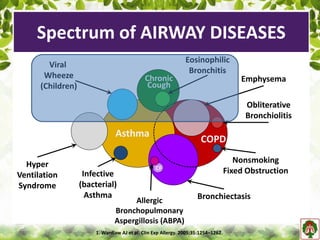
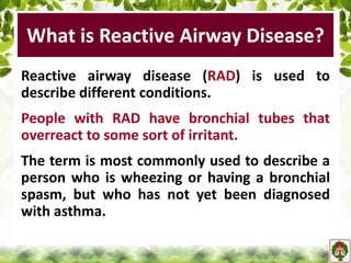
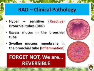
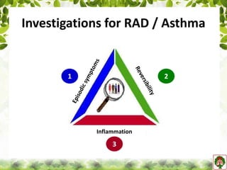
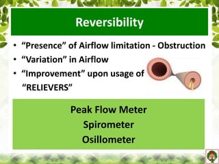
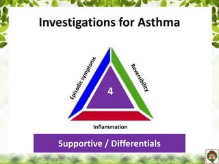
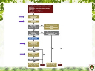
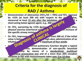
Dr. Parthiv Mehta
Hon. Chief, Pulmonary- Critical Care, UNMICRC
Medical Director, Central United Hospital
Medical Director, Mission Life India
Ahmedabad - GUJARAT
parthivmehta@Hotmail.com
REACTIVE
Airway Diseases
Asthma
COPD
Viral
Wheeze
(Children)
Emphysema
Hyper
Ventilation
Syndrome
Infective
(bacterial)
Asthma
Allergic
Bronchopulmonary
Aspergillosis (ABPA)
Bronchiectasis
Nonsmoking
Fixed Obstruction
Obliterative
Bronchiolitis
Eosinophilic
Bronchitis
CF
Chronic
Cough
1. Wardlaw AJ et al. Clin Exp Allergy. 2005;35:1254–1262.
Spectrum of AIRWAY DISEASES
What is Reactive Airway Disease?
Reactive airway disease (RAD) is used to
describe different conditions.
People with RAD have bronchial tubes that
overreact to some sort of irritant.
The term is most commonly used to describe a
person who is wheezing or having a bronchial
spasm, but who has not yet been diagnosed
with asthma.
RAD used more often for young children, who
are younger than 5 – before stamping it as
ASTHMA!
RAD is not the same as RADS.
RADS is caused by excessive exposure to some
sort of corrosive gas, its fumes or vapours.
RADS occurs just one time and is not chronic.
What is Reactive Airway Disease?
RAD – Clinical Pathology
• Hyper – sensitive (Reactive)
Bronchial tubes (BHR)
• Excess mucus in the bronchial
tube
• Swollen mucous membrane in
the bronchial tube (Inflammation)
FORGET NOT, We are…
REVERSIBLE
RAD – Clinical Presentation
• Wheezing
• Coughing
• Shortness of breath or difficulty breathing
Remember, We are…
EPISODIC - INTERMITTENT
How Do I Catch RAD?
RAD / Asthma
Inflammation
Investigations for RAD / Asthma
Inflammation
1 2
3
Episodic Symptoms
• Skills of clinician to acquire vital information
through patient's medical history and
physical examination.
Episodic Symptoms
• Age of onset
• Symptom free period
• Family history
• Triggers
• Use of Relievers
• Exacerbations
• Most often after an infection.
• It’s caused by some irritant that triggers
the airways to overreact and swell or
narrow.
Pet hair or dander Smoke
Dust and Pollen Exercise
Perfume/Strong odours Stress
Changes in the weather Food
RAD – Pre-disposing FactorsEpisodic Symptoms - TRIGGERS
Episodic Symptoms
• Age of onset
• Symptom free period
• Family history
• Triggers
• Use of Relievers
• Exacerbations
Episodic Symptoms
Reversibility
• “Presence” of Airflow limitation - Obstruction
• “Variation” in Airflow
• “Improvement” upon usage of
“RELIEVERS”
Peak Flow Meter
Spirometer
Osillometer
Peak Flow Meter
• Peak expiratory flow (PEF), also called peak
expiratory flow rate (PEFR) is a person's
maximum speed of expiration
• Directly correlates with “available aperture”
of airway
• Measures the airflow through the bronchi
and thus the degree of obstruction in the
airways
• Effort Dependent
R e v e r s i b i l i t y
Peak Flow Meter
• Simple
• Handheld
• Easy to perform and interpret
• “DO IT YOURSELF”
Weighing Scale
Thermometer
Sphygmomanometer
R e v e r s i b i l i t y
• RAD / Asthma is suspected when there is >/= 20%
diurnal variation on >/= 3 days a week or for 2
weeks in a PEF
• Wide range of ‘normal' values and high degree of
variability >> Not “the recommended test” to
identify asthma
Peak Flow Meter
Good tool to MONITOR RAD/Asthma
Higher the FLOW
Lesser the SEVERITY
Investigations for Asthma
Inflammation
Supportive / Differentials
4
Spirometer
Spirometer
• Spirometry is a vital test to establish “Airway
Obstruction” and its “Reversibility”
objectively and accurately.
R e v e r s i b i l i t y
Spirometer
• Reversibility : Significant reversibility is
indicated by an increase of > 12 % and/or 200
ml in FEV1 after inhaling a short-acting B2
agonist – bronchodilator.
R e v e r s i b i l i t y
Spirometer
• Performing Reversibility:
– Spirometry test (with at least 2 reproducible flow
volume loops)
– Intake of a fast acting bronchodilator (often
Salbutamol) through inhalation
– 15 minutes pause
– Second (Post Bronchodilatation) Spirometry test
(with at least 2 reproducible flow volume loops)
R e v e r s i b i l i t y
• Impulse Osillometery:
– Conventional Spirometry: Effort Dependent
Spirometer
• Impulse Osillometery (IOS): Very
useful for diagnosis of RAD /
Asthma, especially in children
where dependency of Spirometry
and flow volume loop is
questionable.
– IOS uses small amplitude pressure
oscillations to determine the
resistance of the airway.
– It is largely independent of effort
does not require coordination, but
does require cooperation of patient.
Spirometer
Episodic Symptoms
Inflammation
• Inflammation in airways is MUST to confirm
RAD / Asthma.
• Airways – Upper and Lower
• Eosinophilic >> Neutrophilic
• Modalities:
– Direct: Sputum - Eosinophilia
– Indirect: Markers - Exhaled NO, ECP, EPX
Sputum
• Sputum is induced by
proper coughing or using
inhalations of 3% of
hypertonic saline.
• Sample: should look
more opaque and/or
dense and unlike saliva.
• Cell differential counts
are performed.
Inflammation
Sputum
• Eosinophilia in sputum:
Directly linked with the
underlying eosinophilic
airway inflammation.
Management employing
sputum monitoring has
been found useful in
preventing exacerbations
and hospitalizations.
Inflammation
Exhaled Nitric Oxide
• Nitric Oxide (NO) is produced in discrete
concentrations in the healthy human airway
(Respiratory epithelium, Nose, upper and lower
airways) where it is important in physiological
functions such as maintaining airway patency.
• It is responsible for airway inflammation and is also
the product of airway inflammation. So, Evaluation
of NO as “Surrogate marker of Eosinophilic
Inflammation” is gaining acceptance.
Inflammation
Exhaled Nitric Oxide
• Nitric oxide analyzers are used to measure
exhaled nitric oxide (FENO).
Inflammation
Exhaled Nitric Oxide
• NO is over produced in asthmatic individuals.
• Potential of FENO to predict exacerbations of
asthma has been examined in various studies
and levels were found to be elevated before the
fall in lung functions or the development of
clinical symptoms of asthma exacerbations.
• NO is increased in inflammation of lung e.g.
asthma. If bronchospasm without inflammation,
NO is not increased i.e. it is not Asthma.
Inflammation
Exhaled Nitric Oxide
• Ingestion of foods containing nitrates,
smoking status, ambient nitric oxide level,
nasopharyngeal contamination, airway
infections and drugs such as leukotriene
modifiers may affect the actual collection
and quantification of exhaled nitric oxide.
• Patients are asked to take nothing by mouth
for one hour before sample collection.
Inflammation
Markers of Inflammation
• Important especially for diagnosis of Asthma in
Children
• Currently, the noninvasive clinical assessment of
airway inflammation in young children is
limited.
• The detection of raised blood eosinophil levels
or evidence of eosinophil activation proteins in
blood or urine, such as Eosinophil Cationic
Protein (ECP) or Eosinophil Protein X (EPX), can
be used in addition to the examination of
nasopharyngeal secretions.
Inflammation
Markers of Inflammation
• In the near future it is likely that tidal
breathing methods for measuring exhaled
nitric oxide (NO) and other gases will be
validated for use in the younger children.
• The analysis of other constituents of breath,
such as exhaled proteins, is currently being
investigated as potential indicators of airway
pathology.
Inflammation
Supportive / Differentials
• Allergy test
• Blood test
• Radiology: Chest X-Ray, CT Scan
• Endoscopy: Upper-Lower Respiratory, Upper GI
• 2D Echo
Allergy Test
• Allergy test :
– This test is to look for the trigger factors causing
asthma.
– Useful to diagnose occupational asthma and
seasonal asthma by detecting the triggers causing
the attack.
– Supports diagnosis of Asthma
– Identification of allergen triggers can assist in
formulating an avoidance strategy.
– A trial of allergen avoidance may be diagnostic and
therapeutic.
Supportive / Differentials
• Skin tests:
– Main tool in diagnosing allergies all over the
world
– Different allergy profile can be known by the skin
allergy tests.
• Patch test (used mainly for diagnosing contact
dermatitis)
• Scratch test
• Skin prick test
• Intradermal test
• Skin end point titration
• Parasite- kustner test (Passive transfer test).
Allergy Test
Supportive / Differentials
• Mechanism of skin allergy testing:
– Cells and antibodies responsible for allergies are
present under the skin as well as other parts of
the body.
– If an allergen to which patient is allergic is
applied to the skin a reaction occur and a wheal
is formed.
– The size of the wheal is measured to grade the
severity of allergy.
Allergy Test
Supportive / Differentials
• RAST (Radio allegro sorbent technique):
– Detects allergen specific IgE in serum.
– The results of the tests correlate well with the
skin allergy tests.
– One sample of the serum can be used to test
many allergens.
– Benefits: can be used where the skin allergy tests
cannot be performed like young children, severe
atopic dermatitis, dermatographism, history of
extreme sensitivity, patients afraid of multiple
injections.
Allergy Test
Supportive / Differentials
RAST is not influenced by drugs while skin tests are
suppressed by anti allergic drugs and steroids.
There is no risk of anaphylaxis with RAST.
• X-Ray Chest :
– Preferred for routine evaluation
– Helps to establish other diagnosis
• Eosinophilic Lung Disease
• Churg Strauss Syndrome
• Cystic Bronchiectasis
• Interstitial Lung Disease
• Congestive Cardiac failure
– Must for Exacerbation or Uncontrolled
• Consolidation
• Pneumothorax
Radio-Diagnostics
Supportive / Differentials
• CT Scan:
– Useful tool for differentiating other “asthma
alike” or co-existing morbidities for “non-
responders”.
– Role of CT scan has been established
• with suspicion of bronchiectasis
• occupational asthma
• parenchymal infiltrates
• suspicion of Allergic Bronchopulmonary Aspergillosis
and/or invasive aspergillosis
Radio-Diagnostics
Supportive / Differentials
• CT Scan:
Bronchial wall thickening Bronchiectasis / Bronchial dilatation
Radio-Diagnostics
Bronchial wall thickening Bronchiectasis / Bronchial dilatation
Supportive / Differentials
• Laryngoscopy and Bronchoscopy:
Inspection: Allows visual inspection of the upper and
lower airway.
– Help to rule out Vocal cord dysfunction, Laryngomalacia
or Tracheomalacia as an important differential diagnosis
Lavage: Broncho Alveolar Lavage (BAL) is an
important investigation for differentiation of infection
and inflammation.
Biopsy: Performing Endo-Bronchial biopsy (EBLB) or
Trans-Bronchial biopsy (TBLB)
– Will help to establish type of inflammation in airway,
parenchymal infiltration and infection as well as
structural changes in parenchyma. ABPA v/s Invasive
Aspergillosis, DILD
Endoscopy
Supportive / Differentials
Normal Abnormal
Laryngomalacia Tracheomalacia
Endoscopy
Supportive / Differentials
Nearly 20% of patients with refractory asthma referred for
tertiary care have coexisting VCD.
Medical utilization is higher in patients with VCD compared
with age/sex-matched asthma patients.
• Upper GI Scopy:
– Gastro-oesophageal reflux (GERD) is common in
patients of chronic or “difficult” asthma – nearly
twice to normal individuals.
– Though it is hard to identify potential responders
amongst asthmatics having GERD and treatment
often has little effect on asthma symptoms, it is
important to diagnose GERD.
Endoscopy
Supportive / Differentials
• Echocardiogram:
– 2D Echocardiogram is important to differentiate
“Cardiac Asthma” i.e. with Cardiac causes like
Left Ventricular dysfunction or pericardial
effusion from “Bronchial Asthma”.
– An effective tool to provide appropriate support
in the form of anti-failure therapy.
– Patients with COPD shall develop Cor-Pulmonale
on long run.
Echo Cardiogram
Supportive / Differentials
RAD / Asthma Algorithm
Criteria for the diagnosis of
RAD / Asthma
• Demonstration of obstruction (FEV1/VC < 70%) and FEV1 increase
by >15% (at least 200 ml) with respect to the initial value,
measured at least 15 min after the inhalation of four puffs of a
short-acting beta2 agonist agent, e.g., 400 µg of Salbutamol
• Or: FEV1 worsening by >15% during, or within 30 minutes after,
physical exercise (exertional asthma), possibly with an increase of
the specific airway resistance by at least 150%
• Or: FEV1 improvement by >15% (or by at least 200 ml, if the initial
value is below 1300 ml), after daily high-dose administration of an
inhaled corticosteroid (ICS) for a maximum of four weeks
• Or: in patients with normal pulmonary function despite a typical
history for asthma, demonstration of non-specific bronchial
hyper-reactivity by means of a standardized, multilevel
inhalational provocative test and of a more than 20% circadian
variation in PEF with measurements taken over 3 to 14 days
THANK YOU…..
RAD Specialist

Reactive airway diseases_2018_pmm

  • 1.
    Dr. Parthiv Mehta Hon.Chief, Pulmonary- Critical Care, UNMICRC Medical Director, Central United Hospital Medical Director, Mission Life India Ahmedabad - GUJARAT parthivmehta@Hotmail.com REACTIVE Airway Diseases
  • 2.
  • 3.
    What is ReactiveAirway Disease? Reactive airway disease (RAD) is used to describe different conditions. People with RAD have bronchial tubes that overreact to some sort of irritant. The term is most commonly used to describe a person who is wheezing or having a bronchial spasm, but who has not yet been diagnosed with asthma.
  • 4.
    RAD used moreoften for young children, who are younger than 5 – before stamping it as ASTHMA! RAD is not the same as RADS. RADS is caused by excessive exposure to some sort of corrosive gas, its fumes or vapours. RADS occurs just one time and is not chronic. What is Reactive Airway Disease?
  • 5.
    RAD – ClinicalPathology • Hyper – sensitive (Reactive) Bronchial tubes (BHR) • Excess mucus in the bronchial tube • Swollen mucous membrane in the bronchial tube (Inflammation) FORGET NOT, We are… REVERSIBLE
  • 6.
    RAD – ClinicalPresentation • Wheezing • Coughing • Shortness of breath or difficulty breathing Remember, We are… EPISODIC - INTERMITTENT
  • 7.
    How Do ICatch RAD?
  • 8.
  • 9.
    Investigations for RAD/ Asthma Inflammation 1 2 3
  • 10.
    Episodic Symptoms • Skillsof clinician to acquire vital information through patient's medical history and physical examination.
  • 11.
    Episodic Symptoms • Ageof onset • Symptom free period • Family history • Triggers • Use of Relievers • Exacerbations
  • 12.
    • Most oftenafter an infection. • It’s caused by some irritant that triggers the airways to overreact and swell or narrow. Pet hair or dander Smoke Dust and Pollen Exercise Perfume/Strong odours Stress Changes in the weather Food RAD – Pre-disposing FactorsEpisodic Symptoms - TRIGGERS
  • 13.
    Episodic Symptoms • Ageof onset • Symptom free period • Family history • Triggers • Use of Relievers • Exacerbations
  • 14.
  • 15.
    Reversibility • “Presence” ofAirflow limitation - Obstruction • “Variation” in Airflow • “Improvement” upon usage of “RELIEVERS” Peak Flow Meter Spirometer Osillometer
  • 16.
    Peak Flow Meter •Peak expiratory flow (PEF), also called peak expiratory flow rate (PEFR) is a person's maximum speed of expiration • Directly correlates with “available aperture” of airway • Measures the airflow through the bronchi and thus the degree of obstruction in the airways • Effort Dependent R e v e r s i b i l i t y
  • 17.
    Peak Flow Meter •Simple • Handheld • Easy to perform and interpret • “DO IT YOURSELF” Weighing Scale Thermometer Sphygmomanometer R e v e r s i b i l i t y
  • 18.
    R e ve r s i b i l i t y • RAD / Asthma is suspected when there is >/= 20% diurnal variation on >/= 3 days a week or for 2 weeks in a PEF • Wide range of ‘normal' values and high degree of variability >> Not “the recommended test” to identify asthma Peak Flow Meter Good tool to MONITOR RAD/Asthma Higher the FLOW Lesser the SEVERITY
  • 19.
  • 20.
  • 21.
    Spirometer • Spirometry isa vital test to establish “Airway Obstruction” and its “Reversibility” objectively and accurately. R e v e r s i b i l i t y
  • 22.
    Spirometer • Reversibility :Significant reversibility is indicated by an increase of > 12 % and/or 200 ml in FEV1 after inhaling a short-acting B2 agonist – bronchodilator. R e v e r s i b i l i t y
  • 23.
    Spirometer • Performing Reversibility: –Spirometry test (with at least 2 reproducible flow volume loops) – Intake of a fast acting bronchodilator (often Salbutamol) through inhalation – 15 minutes pause – Second (Post Bronchodilatation) Spirometry test (with at least 2 reproducible flow volume loops) R e v e r s i b i l i t y
  • 24.
    • Impulse Osillometery: –Conventional Spirometry: Effort Dependent Spirometer
  • 25.
    • Impulse Osillometery(IOS): Very useful for diagnosis of RAD / Asthma, especially in children where dependency of Spirometry and flow volume loop is questionable. – IOS uses small amplitude pressure oscillations to determine the resistance of the airway. – It is largely independent of effort does not require coordination, but does require cooperation of patient. Spirometer Episodic Symptoms
  • 26.
    Inflammation • Inflammation inairways is MUST to confirm RAD / Asthma. • Airways – Upper and Lower • Eosinophilic >> Neutrophilic • Modalities: – Direct: Sputum - Eosinophilia – Indirect: Markers - Exhaled NO, ECP, EPX
  • 27.
    Sputum • Sputum isinduced by proper coughing or using inhalations of 3% of hypertonic saline. • Sample: should look more opaque and/or dense and unlike saliva. • Cell differential counts are performed. Inflammation
  • 28.
    Sputum • Eosinophilia insputum: Directly linked with the underlying eosinophilic airway inflammation. Management employing sputum monitoring has been found useful in preventing exacerbations and hospitalizations. Inflammation
  • 29.
    Exhaled Nitric Oxide •Nitric Oxide (NO) is produced in discrete concentrations in the healthy human airway (Respiratory epithelium, Nose, upper and lower airways) where it is important in physiological functions such as maintaining airway patency. • It is responsible for airway inflammation and is also the product of airway inflammation. So, Evaluation of NO as “Surrogate marker of Eosinophilic Inflammation” is gaining acceptance. Inflammation
  • 30.
    Exhaled Nitric Oxide •Nitric oxide analyzers are used to measure exhaled nitric oxide (FENO). Inflammation
  • 31.
    Exhaled Nitric Oxide •NO is over produced in asthmatic individuals. • Potential of FENO to predict exacerbations of asthma has been examined in various studies and levels were found to be elevated before the fall in lung functions or the development of clinical symptoms of asthma exacerbations. • NO is increased in inflammation of lung e.g. asthma. If bronchospasm without inflammation, NO is not increased i.e. it is not Asthma. Inflammation
  • 32.
    Exhaled Nitric Oxide •Ingestion of foods containing nitrates, smoking status, ambient nitric oxide level, nasopharyngeal contamination, airway infections and drugs such as leukotriene modifiers may affect the actual collection and quantification of exhaled nitric oxide. • Patients are asked to take nothing by mouth for one hour before sample collection. Inflammation
  • 33.
    Markers of Inflammation •Important especially for diagnosis of Asthma in Children • Currently, the noninvasive clinical assessment of airway inflammation in young children is limited. • The detection of raised blood eosinophil levels or evidence of eosinophil activation proteins in blood or urine, such as Eosinophil Cationic Protein (ECP) or Eosinophil Protein X (EPX), can be used in addition to the examination of nasopharyngeal secretions. Inflammation
  • 34.
    Markers of Inflammation •In the near future it is likely that tidal breathing methods for measuring exhaled nitric oxide (NO) and other gases will be validated for use in the younger children. • The analysis of other constituents of breath, such as exhaled proteins, is currently being investigated as potential indicators of airway pathology. Inflammation
  • 35.
    Supportive / Differentials •Allergy test • Blood test • Radiology: Chest X-Ray, CT Scan • Endoscopy: Upper-Lower Respiratory, Upper GI • 2D Echo
  • 36.
    Allergy Test • Allergytest : – This test is to look for the trigger factors causing asthma. – Useful to diagnose occupational asthma and seasonal asthma by detecting the triggers causing the attack. – Supports diagnosis of Asthma – Identification of allergen triggers can assist in formulating an avoidance strategy. – A trial of allergen avoidance may be diagnostic and therapeutic. Supportive / Differentials
  • 37.
    • Skin tests: –Main tool in diagnosing allergies all over the world – Different allergy profile can be known by the skin allergy tests. • Patch test (used mainly for diagnosing contact dermatitis) • Scratch test • Skin prick test • Intradermal test • Skin end point titration • Parasite- kustner test (Passive transfer test). Allergy Test Supportive / Differentials
  • 38.
    • Mechanism ofskin allergy testing: – Cells and antibodies responsible for allergies are present under the skin as well as other parts of the body. – If an allergen to which patient is allergic is applied to the skin a reaction occur and a wheal is formed. – The size of the wheal is measured to grade the severity of allergy. Allergy Test Supportive / Differentials
  • 39.
    • RAST (Radioallegro sorbent technique): – Detects allergen specific IgE in serum. – The results of the tests correlate well with the skin allergy tests. – One sample of the serum can be used to test many allergens. – Benefits: can be used where the skin allergy tests cannot be performed like young children, severe atopic dermatitis, dermatographism, history of extreme sensitivity, patients afraid of multiple injections. Allergy Test Supportive / Differentials RAST is not influenced by drugs while skin tests are suppressed by anti allergic drugs and steroids. There is no risk of anaphylaxis with RAST.
  • 40.
    • X-Ray Chest: – Preferred for routine evaluation – Helps to establish other diagnosis • Eosinophilic Lung Disease • Churg Strauss Syndrome • Cystic Bronchiectasis • Interstitial Lung Disease • Congestive Cardiac failure – Must for Exacerbation or Uncontrolled • Consolidation • Pneumothorax Radio-Diagnostics Supportive / Differentials
  • 41.
    • CT Scan: –Useful tool for differentiating other “asthma alike” or co-existing morbidities for “non- responders”. – Role of CT scan has been established • with suspicion of bronchiectasis • occupational asthma • parenchymal infiltrates • suspicion of Allergic Bronchopulmonary Aspergillosis and/or invasive aspergillosis Radio-Diagnostics Supportive / Differentials
  • 42.
    • CT Scan: Bronchialwall thickening Bronchiectasis / Bronchial dilatation Radio-Diagnostics Bronchial wall thickening Bronchiectasis / Bronchial dilatation Supportive / Differentials
  • 43.
    • Laryngoscopy andBronchoscopy: Inspection: Allows visual inspection of the upper and lower airway. – Help to rule out Vocal cord dysfunction, Laryngomalacia or Tracheomalacia as an important differential diagnosis Lavage: Broncho Alveolar Lavage (BAL) is an important investigation for differentiation of infection and inflammation. Biopsy: Performing Endo-Bronchial biopsy (EBLB) or Trans-Bronchial biopsy (TBLB) – Will help to establish type of inflammation in airway, parenchymal infiltration and infection as well as structural changes in parenchyma. ABPA v/s Invasive Aspergillosis, DILD Endoscopy Supportive / Differentials
  • 44.
    Normal Abnormal Laryngomalacia Tracheomalacia Endoscopy Supportive/ Differentials Nearly 20% of patients with refractory asthma referred for tertiary care have coexisting VCD. Medical utilization is higher in patients with VCD compared with age/sex-matched asthma patients.
  • 45.
    • Upper GIScopy: – Gastro-oesophageal reflux (GERD) is common in patients of chronic or “difficult” asthma – nearly twice to normal individuals. – Though it is hard to identify potential responders amongst asthmatics having GERD and treatment often has little effect on asthma symptoms, it is important to diagnose GERD. Endoscopy Supportive / Differentials
  • 46.
    • Echocardiogram: – 2DEchocardiogram is important to differentiate “Cardiac Asthma” i.e. with Cardiac causes like Left Ventricular dysfunction or pericardial effusion from “Bronchial Asthma”. – An effective tool to provide appropriate support in the form of anti-failure therapy. – Patients with COPD shall develop Cor-Pulmonale on long run. Echo Cardiogram Supportive / Differentials
  • 47.
    RAD / AsthmaAlgorithm
  • 49.
    Criteria for thediagnosis of RAD / Asthma • Demonstration of obstruction (FEV1/VC < 70%) and FEV1 increase by >15% (at least 200 ml) with respect to the initial value, measured at least 15 min after the inhalation of four puffs of a short-acting beta2 agonist agent, e.g., 400 µg of Salbutamol • Or: FEV1 worsening by >15% during, or within 30 minutes after, physical exercise (exertional asthma), possibly with an increase of the specific airway resistance by at least 150% • Or: FEV1 improvement by >15% (or by at least 200 ml, if the initial value is below 1300 ml), after daily high-dose administration of an inhaled corticosteroid (ICS) for a maximum of four weeks • Or: in patients with normal pulmonary function despite a typical history for asthma, demonstration of non-specific bronchial hyper-reactivity by means of a standardized, multilevel inhalational provocative test and of a more than 20% circadian variation in PEF with measurements taken over 3 to 14 days
  • 50.

Editor's Notes

  • #3 A fundamental challenge that clinicians face in managing pediatric airway diseases is establishing a differential diagnosis. RAD or Asthma is typically recognized on the basis of its classical presentation that includes variable airflow obstruction and airway hyperresponsiveness; however, these clinical features are also observed in patients with other airway disorders, such as viral-induced wheeze, chronic cough, and bronchiectasis, in addition to allergic bronchopulmonary aspergillosis and/or cystic fibrosis.