Downloaded 149 times

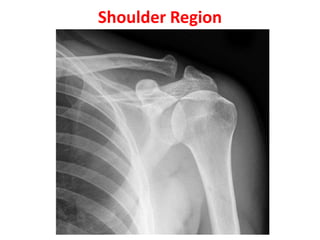
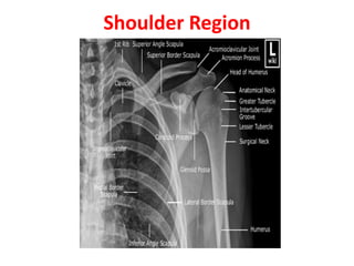
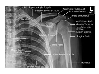
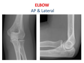
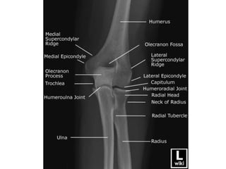
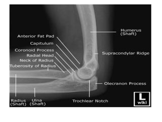
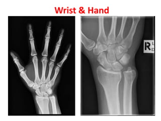
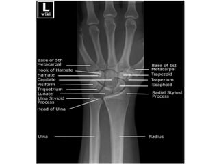
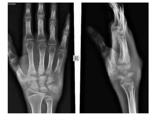

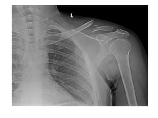
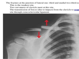
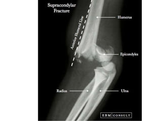
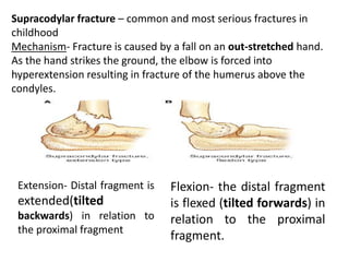
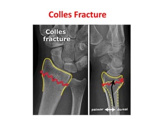
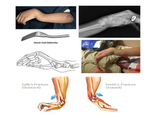
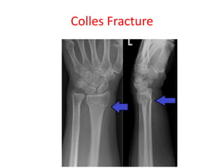
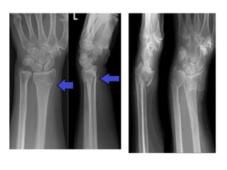
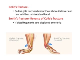
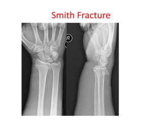
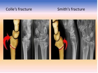
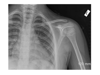
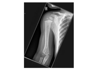
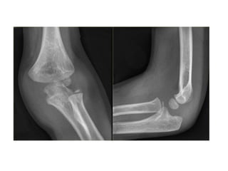

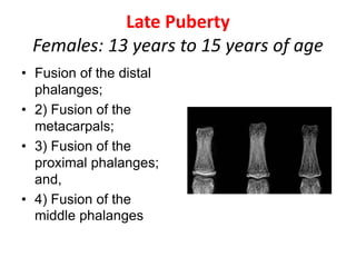
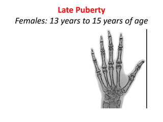
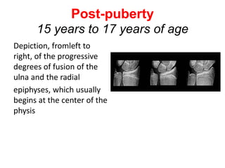
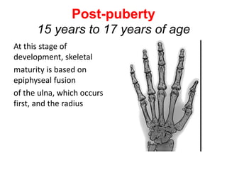
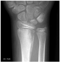


1) The document discusses radiology of the upper limb, including the shoulder, elbow, hand, and wrist regions. It covers common fractures like supracondylar fractures in children and Colles' fractures in the wrist. 2) Details are provided on age estimation based on appearance and fusion of epiphyseal centers in long bones, with timelines given for the shoulder, elbow, hand, and wrist regions. 3) Assessment of bone age is described for different developmental stages from infancy to post-puberty based on features of the epiphyses.


