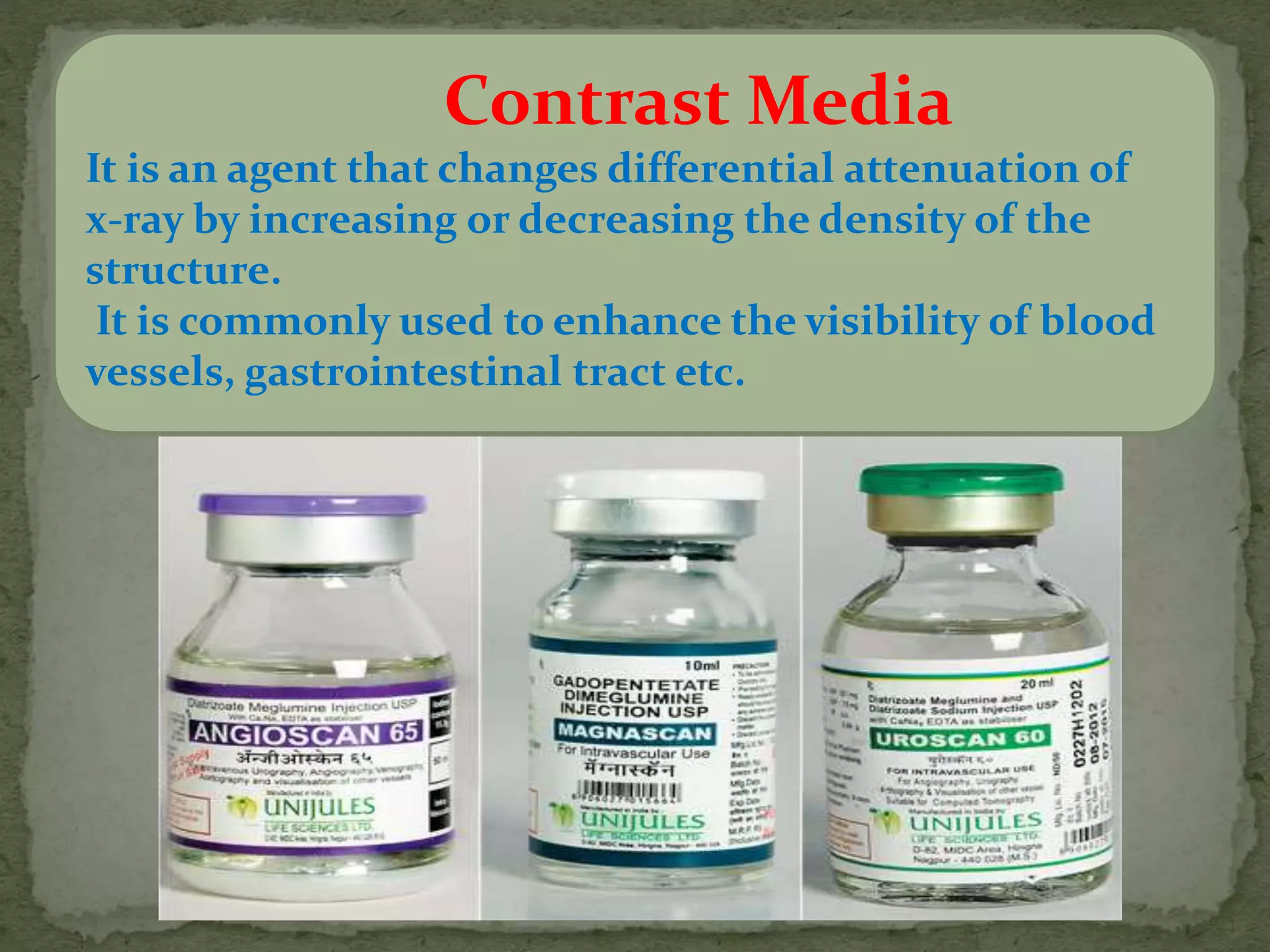
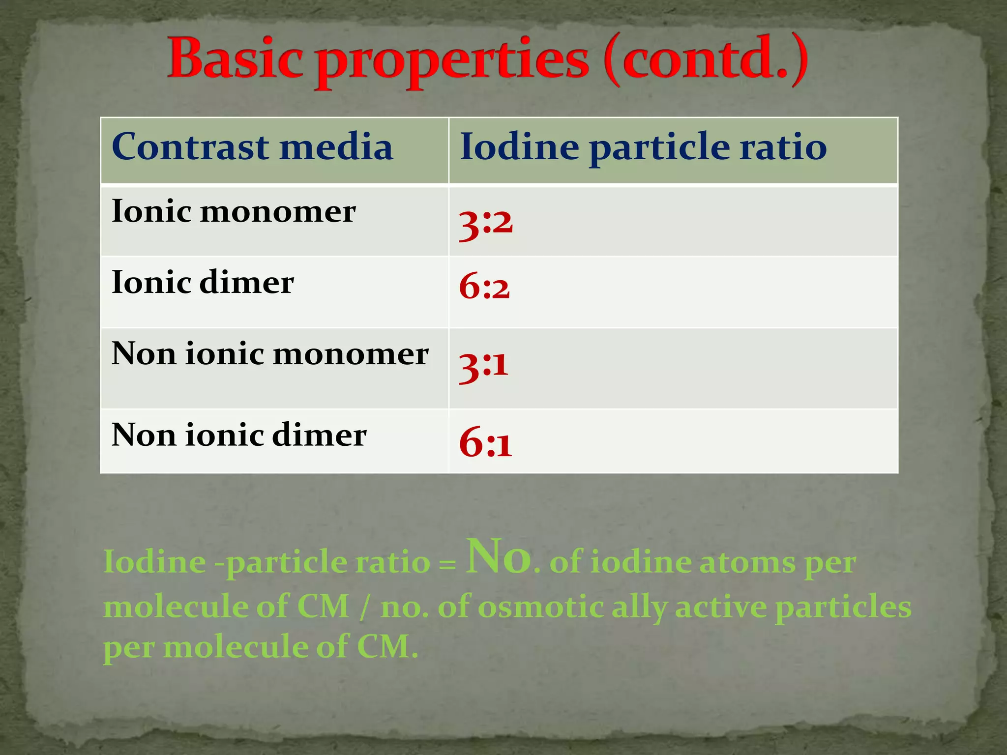
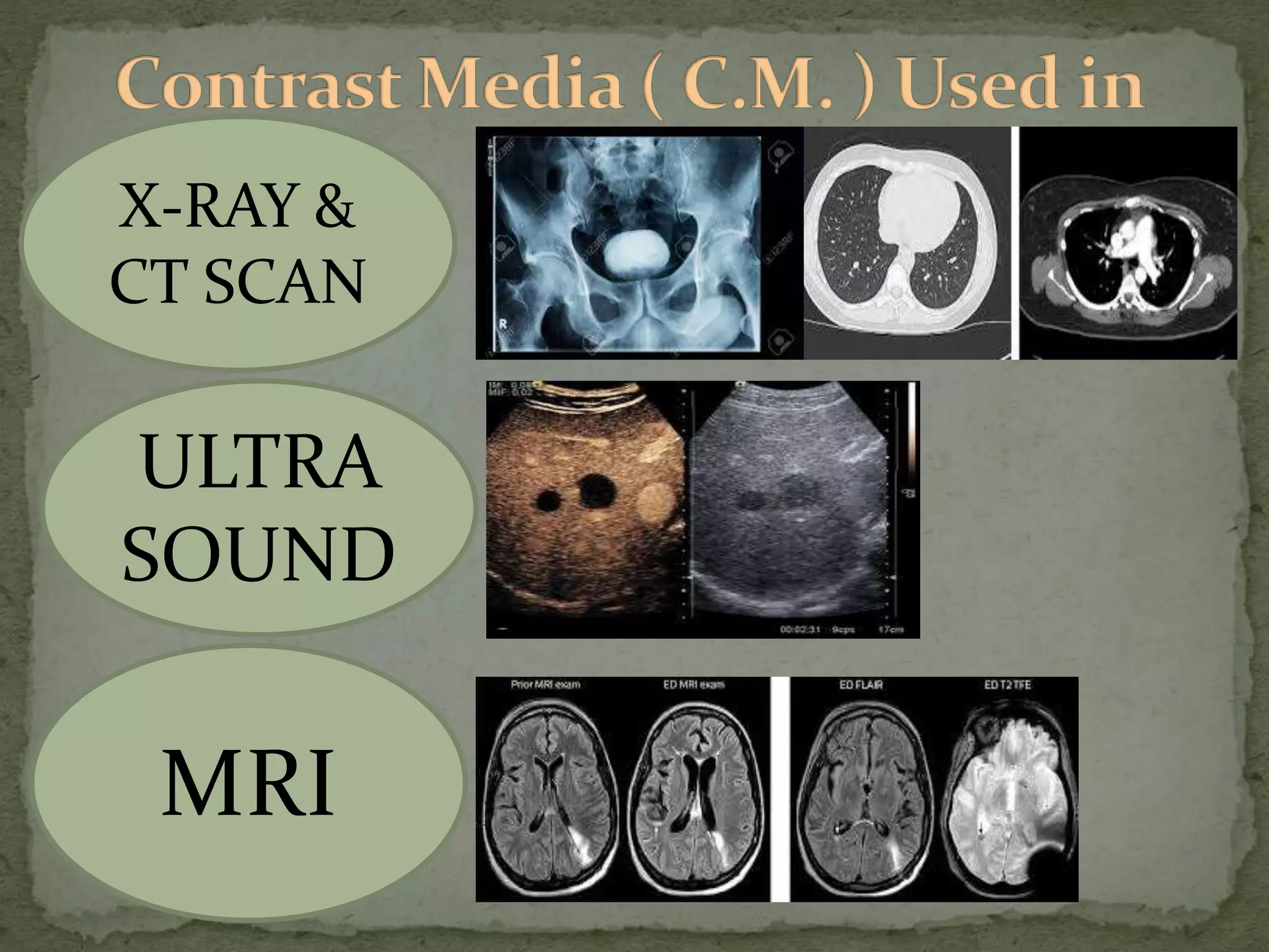
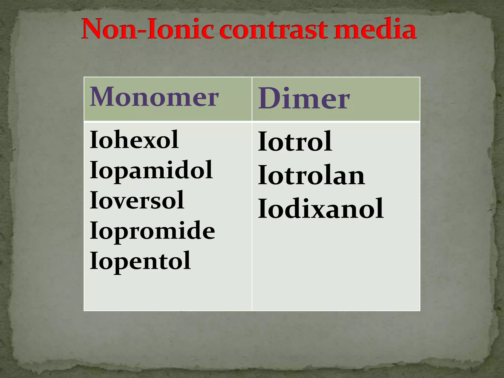
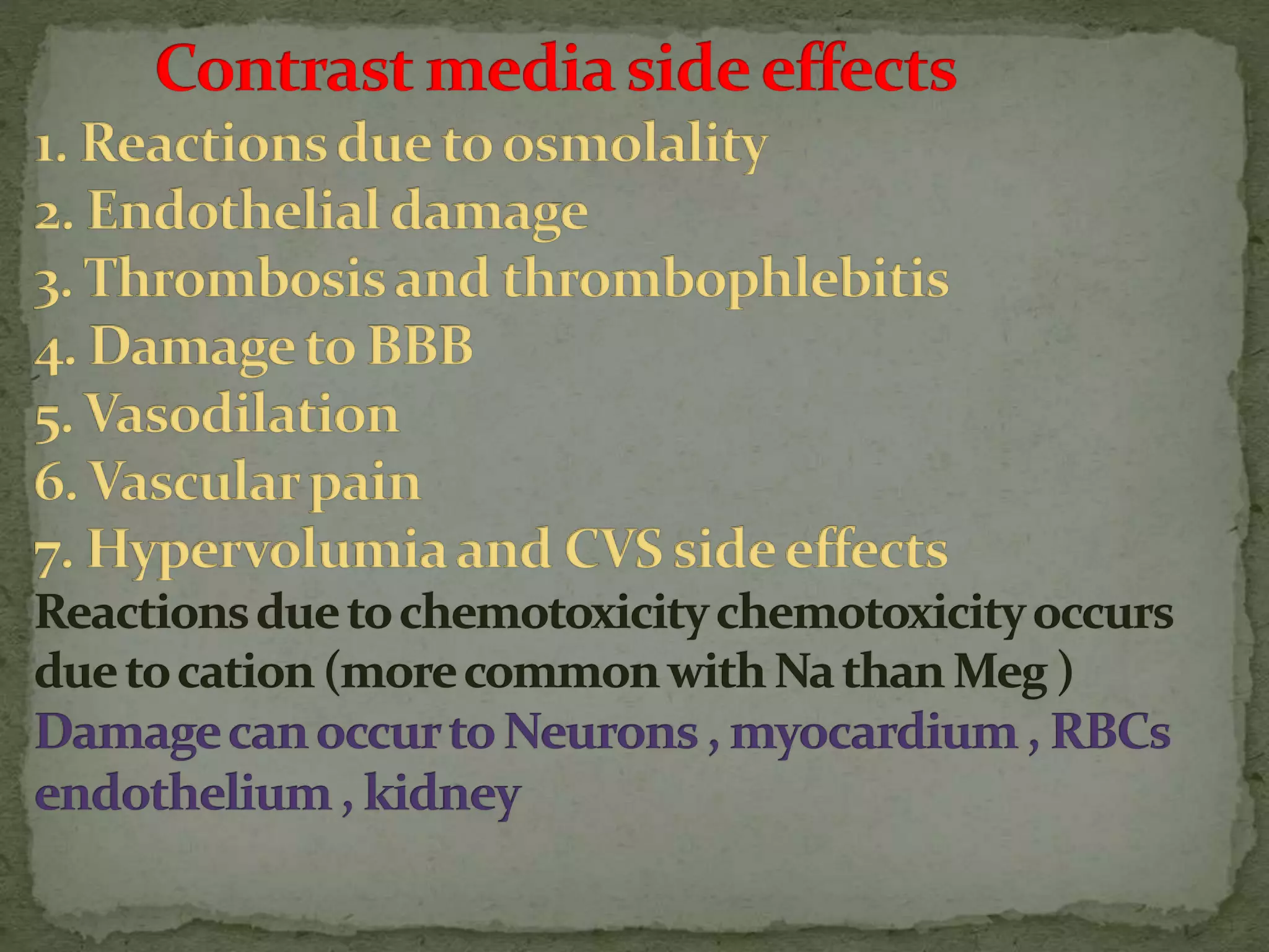
Contrast Media Presentation Uploaded by SONU SHARMA
Student of B.Sc Radiography & Imaging Technology 3rd Year
at Pt.B.D.Sharma University of Health & Science ,
P.G.I.M.S. Rohtak,Haryana,India.
Contrast media are substances used to enhance the contrast of structures or fluids within the body during medical imaging. There are two main types - positive contrast agents that are high in atomic number like iodine and barium, and negative contrast agents that are low in atomic number like air. Positive contrast agents can be water soluble like iodine compounds used for intravenous injections, or water insoluble like barium sulphate used to study the gastrointestinal tract. While generally safe, contrast media do carry risks of minor or major reactions, with ionic contrast media having higher reaction probabilities than non-ionic varieties.
1. Contrast agents are substances that have different atomic numbers or electron densities than surrounding tissues, allowing visualization of internal organs on imaging.
2. Early contrast agents included barium sulfate, iodinated compounds, and air or CO2. Modern agents are classified as ionic or non-ionic monomers and dimers with varying osmolalities and viscosities.
3. Contrast agents are used with multiple imaging modalities like CT, MRI, ultrasound, and fluoroscopy. They are administered orally, intravenously, or directly into structures. The choice depends on the physical properties and safety of the specific agent and the anatomy being imaged.
This document provides an overview of contrast media including its introduction, mechanism of action, basic chemistry and properties, classifications, commonly used preparations, pharmacodynamics, interactions with body systems, potential side effects and their management, and prevention strategies. Contrast media helps improve visualization of tissues during medical imaging by increasing beam attenuation. Iodine is commonly used as the contrast agent due to its high atomic number. Both ionic and non-ionic varieties exist with different properties. Potential side effects range from minor to severe reactions.
MRI pulse sequences are programmed sets of changing magnetic gradients used to generate images. There are several types of sequences, including spin echo, gradient echo, and inversion recovery sequences. Sequences are defined by parameters like time to echo, time to repetition, and flip angle. Functional techniques like diffusion-weighted imaging, perfusion imaging, and fMRI are used to evaluate brain physiology rather than just anatomy. Echo planar imaging allows for very fast image acquisition, while other sequences like spin echo provide different types of tissue contrast.
This document defines and provides examples of common CT artifacts, including their causes and potential solutions. It discusses ring artifact caused by a miscalibrated detector element; noise (photon starvation) caused by low photon counts; metal artifacts from multiple factors like beam hardening; and beam hardening artifact from polychromatic X-rays. Motion artifacts from patient movement are also addressed. Solutions include recalibrating detectors, increasing mA or kV, and using techniques like faster pitch or cardiac gating.
Contrast media are agents used to enhance the visibility of structures in medical imaging. There are several types including positive contrast media which make structures appear brighter on scans, and negative contrast media which make structures seem darker. Common contrast agents contain iodine and can be ionic monomers, ionic dimers, non-ionic monomers, or non-ionic dimers. While contrast imaging provides important medical information, the agents sometimes cause side effects from mild reactions like nausea to more severe issues like pulmonary edema. Care must be taken with patients having risk factors for complications.
The document discusses various contrast media used in radiology including iodinated contrast media, barium sulfate, gadolinium, and ultrasound contrast agents. It provides classifications of contrast media based on their atomic number, water solubility, and excretion route. It describes the differences between high-osmolar and low-osmolar iodinated contrast media and their safety profiles. MRI contrast agents discussed include gadolinium chelates and their indications. The document also covers ultrasound contrast microbubbles and their encapsulation, as well as barium sulfate mixtures used for gastrointestinal imaging.
1) The document discusses the historical development of radiographic contrast media from its earliest uses in the late 19th century to the development of tri-iodinated benzene derivatives and modern low-osmolar non-ionic contrast agents.
2) It covers the ideal characteristics, classification, and basic chemistry of iodinated contrast media including differences between ionic and non-ionic monomers and dimers.
3) The risks of contrast media administration are discussed including predictable chemo-toxic reactions affecting tissues and organ systems as well as unpredictable anaphylactoid reactions.
This document provides an overview of radiographic contrast media. It discusses how contrast media enhance images by increasing the absorption of x-rays in certain tissues. It describes the ideal properties of contrast media and classifications such as iodinated versus non-iodinated, ionic versus non-ionic, monomer versus dimer. Examples are given for different types of contrast media including barium sulfate, iodinated monomers and dimers, oil-soluble agents, and MRI contrast agents containing gadolinium. The document covers the history, properties, advantages, disadvantages and examples of various contrast media used in radiology.
MRI artifacts can occur due to hardware issues, software problems, physiological phenomena or physical limitations of the MRI device. Common artifacts include chemical shift artifacts seen at fat-water interfaces, aliasing artifacts due to an undersized field of view, black boundary artifacts at tissue borders, and motion artifacts from patient movement. Understanding the sources and appearances of artifacts is important for technicians to maintain image quality and avoid confusing artifacts with pathology.
This document provides an overview of basic pulse sequences in MRI. It discusses introductory concepts like pulse sequences and parameters such as TR, TE, and TI. The two main pulse sequences covered are spin echo and gradient echo. Spin echo uses 90 and 180 degree RF pulses to generate an echo and compensate for T2* decay, while gradient echo lacks the 180 degree pulse. Fast spin echo is also summarized as a faster version of spin echo. Other sequences like inversion recovery, FLAIR, and steady state are briefly introduced along with their purposes and timing parameters.
Susmita Shrestha presented on contrast media in radiology. The document provided a historical overview of contrast media development beginning in the late 19th century. It described the basic properties and types of contrast media used for X-ray, CT, MRI, and ultrasound imaging. Contrast media allow tissues and structures to be more clearly visualized and include barium, iodine, gadolinium, and microbubbles depending on the imaging modality. The optimal contrast medium maximizes visibility while minimizing toxicity.
Computed tomography (CT) uses computer-processed X-rays to create cross-sectional images of the body. CT works by rotating an X-ray tube and detectors around the patient, acquiring multiple transmission measurements at different angles to reconstruct a 3D image. Image reconstruction involves algorithms like back projection and filtered back projection that use the transmission data to calculate the attenuation coefficients of different tissues and generate tomographic images representing slices of the body. CT numbers, measured in Hounsfield units, provide standardized values related to tissue density and visibility.
Beam hardening artifact occurs when an X-ray beam passes through multiple materials of varying densities within a scan volume. This causes the beam to become harder as lower energy photons are preferentially absorbed, leading to streaks or shading in the reconstructed CT image. Photon starvation is another cause of streak artifacts, occurring when there is insufficient photon flux passing through areas of higher attenuation, such as across the shoulders. Adaptive filtering and modulating tube current based on attenuation can help reduce these artifacts. Ring artifacts from defective detector elements in older CT scanners appear as rings in the reconstructed images.
This document describes a barium swallow procedure used to examine the esophagus and detect esophageal diseases. It discusses the anatomy of the pharynx and esophagus, the contrast agent used, and provides details on the different phases of swallowing. It outlines the technique for performing a barium swallow study, including evaluation of the pharynx and esophagus. Key findings on radiographic images are described. The document emphasizes analyzing swallowing studies by looking for asymmetry, stasis, cricopharyngeal dysfunction, and aspiration.
Anatomia y Posicionamiento de las extremidades superiores. Deseo aclarar que el video no me pertenece de ninguna manera. Se esta compartiendo publicamente con el fin de ayudar a los futuros tecnologos a obtener conocimiento para su revalida.
MRI contrast agents are compounds used to improve visibility of internal structures in MRI scans. The most commonly used agents contain gadolinium which shortens relaxation times of nearby protons, altering tissue contrast. Contrast agents must be able to interact with proton magnetic moments in tissues to change relaxation rates between tissues differentially. While generally safe, gadolinium-based agents rarely cause allergic reactions or nephrogenic systemic fibrosis in patients with kidney disease. Oral contrasts are also used to enhance gastrointestinal tract imaging.
This document discusses various components of an MRI system including magnets, RF coils, gradient coils, and safety considerations. It describes the different types of magnets used in MRI like permanent, resistive, and superconducting magnets. It explains the purpose and types of RF coils and gradient coils used to generate the magnetic field gradients needed for spatial encoding in MRI. Safety aspects such as screening for metallic objects, specific absorption rate limits, and absolute contraindications for MRI are also summarized.
The document summarizes the history and development of computed tomography (CT) scanning technology. It describes the key events and innovations such as the development of the first CT scanner by Godfrey Hounsfield in 1972 (1), the introduction of whole body scanning in 1975 (2), and Hounsfield and Cormack being awarded the Nobel Prize in 1979 (3). Subsequent generations of CT scanners incorporated improvements like faster scanning speeds, multiple detectors, and eliminating moving parts to enable ultra-fast scanning.
This document provides information on small bowel enema/enteroclysis procedure. It discusses the indications for the procedure including partial small bowel obstruction and Crohn's disease. It outlines the preparation process and describes how to position the Bilbao Dotter tube through the nose into the duodenum. The document discusses performing the procedure with single or double contrast and imaging techniques. Potential findings and complications are also summarized.
Barium follow through and small bowel enema sahara mahatosahara mahato
This document discusses barium follow through and small bowel enema examinations. It begins by explaining that barium is used as a contrast medium to visualize the small bowel on radiographs. It then describes the anatomy and divisions of the small bowel. The document outlines the procedures for barium follow through and small bowel enema, including patient preparation, technique, and potential findings. Complications are also briefly mentioned. In summary, it provides an overview of using barium and radiography to examine the small intestine.
1. This document provides information on various radiological procedures including enteroclysis, ERCP, ascending urethrogram, barium swallow, single contrast enema, T-tube cholangiography, and barium enema. It describes the indications, contraindications, required equipment, contrast agents, techniques, and potential complications for each procedure.
2. Five high risk factors for reactions to ionic contrast media are discussed. Ionic contrast agents contain both positively and negatively charged ions which can increase the risk of allergic-like reactions compared to non-ionic contrast.
3. Key details are provided for performing a single or double contrast barium enema examination including the indications, contraindications,
The document discusses discography, a minimally invasive diagnostic imaging test where contrast material is injected into one or more spinal discs under x-ray guidance to help determine if a specific disc is the source of back pain. It covers the normal anatomy of discs, indications for the procedure, contraindications, techniques, potential findings, and risks. Discography provides information that can help evaluate patients with chronic back pain.
This document discusses techniques for visualizing soft tissues in radiography. Soft tissues have less differential attenuation compared to bones, making contrast reduced. Special techniques are needed to improve contrast and demonstrate soft tissues clearly. These include adjusting the kVp and adding filters to change image contrast. Using a normal or low kVp can help visualize certain soft tissues like adenoid and effusions more clearly. High kVp is useful for exams like BA enemas where thicker tissues are involved. Digital technology also helps improve soft tissue visibility compared to conventional radiography. Proper technique selection is important to optimize contrast and sharpness while reducing artifacts.
This document discusses quality control procedures for CT scanners, including checking image quality metrics like resolution, noise, and CT number accuracy using phantoms. Regular quality control is recommended to establish baselines, identify potential problems early, and reduce downtime. Various tests are described to check parameters like resolution, noise, CT numbers, distance measurement accuracy, slice thickness, table movement, and laser alignment using specialized phantoms and protocols.
Dr. Mustafa Zuhair Mahmoud has degrees from several universities including a B.Sc from SUST in Khartoum, Sudan, an M.Sc from AAU in Khartoum and JUREI in Philadelphia, and a Ph.D from Ludes in Lugano, Swiss and SUST in Khartoum. The document provides a quick anatomical review of the digestive system including the alimentary canal and accessory glands. It then discusses digestive system radiography including indications for imaging the upper and lower GI tract, contrast media, and patient preparation for procedures like barium swallow, barium meal, barium follow through, and barium enema.
CONTRAST MEDIA in diagnostic radiographyRitupanta1
Contrast media are diagnostic agents used in radiology to enhance images. There are different types including barium sulfate, which is used to examine the gastrointestinal tract, and iodinated contrast media, which contains iodine and is preferred due to its high contrast density. Iodinated contrast media can be ionic or non-ionic, and classified further based on whether they contain ionic monomers, dimers, or non-ionic monomers and dimers. The classification depends on the iodine to particle ratio in solution.
TITLE -Radiopaque contrast media/PHARMACEUTICAL CHEMISTRYSUSHANT OJHA
Contrast media are used to enhance images from medical imaging scans. Positive contrast media like barium and iodine absorb more x-rays, appearing denser on images. Negative contrast media like gases absorb fewer x-rays, appearing darker. An ideal contrast medium would be non-toxic, concentrated in the desired area, and rapidly eliminated. Iodine is commonly used for CT scans while barium is used for imaging the gastrointestinal tract. Contrast media allow tissues to be differentiated for easier diagnosis.
Here are the key steps in administering intravenous contrast media safely:
- Obtain informed consent
- Check for allergies and reactions to previous contrast administrations
- Consider risk factors like renal impairment, diabetes, or cardiac conditions
- Use low-osmolar contrast for high-risk patients
- Monitor vital signs during and after injection
- Have resuscitation equipment and medications available in case of reaction
Contrast media and medical imaging part 1Gopal Panda
Medical imaging uses contrast agents to improve visualization of internal organs and tissues. Contrast agents work by absorbing or altering electromagnetic waves or ultrasound, enhancing the contrast between tissues in images. India has a large and varied radiology market to serve its large population, but relatively few radiologists per capita. The major modalities used are X-ray, ultrasound, CT, and MRI. Contrast agents improve visibility of structures for these modalities. The most common types are iodine-based agents for X-ray and gadolinium for MRI; ultrasound uses microbubbles. Iodine-based agents are classified based on iodine concentration and osmolarity.
Radioisotopes -B for nuclear Engineering Course.pptxDrSafiurRahman
1) Radioisotopes are atoms of an element that have the same number of protons but a different number of neutrons. They can be stable or unstable. Unstable isotopes decay by emitting particles like alpha or beta particles or gamma rays.
2) The fundamental unit of radioactivity is the Curie, defined as 3.7x10¹⁰ disintegrations per second. Some common units used are millicurie and microcurie.
3) Radioisotopes are used safely in medicine if their emissions have low energy, they have a short physical half-life, and a short biological half-life. Common medical isotopes include carbon-14, sodium-24, hydrogen-3, and tech
Radiographic contrast media were developed starting in the late 19th century to increase differences in tissue attenuation and improve organ visualization on radiographs. The first effective contrast agents used barium and iodine compounds. Modern contrast media are tri-iodinated benzene derivatives that are either ionic or non-ionic, and monomeric or dimeric in structure. They carry risks of both idiosyncratic and non-idiosyncratic adverse reactions that depend on their osmolality, viscosity, and other properties. Appropriate use and precautions seek to minimize risks while maximizing diagnostic value.
The document discusses the structure of atoms including subatomic particles like protons, neutrons and electrons. It describes atomic number and mass number, isotopes, radioactive decay, and different types of radiation (alpha, beta, gamma). It explains how radiation can be detected and some uses and biological effects of radiation including cancer risks from ionizing radiation. The concept of half-life is introduced with examples of how radioactive materials decay over time in a predictable pattern.
This document provides an overview of contrast media used in radiology. It discusses the history of contrast media beginning with their discovery in 1896. It then covers the basics of contrast media including their physiology, modes of administration, classifications for different imaging modalities, and examples of agents used for X-ray/CT, ultrasound, and MR imaging. Specific contrast agents are described in detail including their properties, uses, and side effects. The document emphasizes the importance of using lower osmolar iodinated contrast media to reduce risks when possible.
basics of contrastmedia-171027192651.pdfAIDA BORLAZA
This document provides an overview of contrast media used in medical imaging. It discusses the history of contrast media beginning with their discovery in 1896. It then covers the physiology of contrast agents, how they are administered and classified. Specific contrast media are described for X-ray/CT including barium sulfate and iodinated agents. Ultrasound contrast media consisting of gas-filled microbubbles are also outlined. The document provides details on the various generations of contrast agents developed for optimal imaging and safety.
This document discusses contrast media used in radiology. It introduces positive and negative contrast media, which increase or decrease density during imaging. Positive contrast agents contain iodine, bromine or barium, while negative agents include air, carbon dioxide and oxygen. Contrast media is classified as ionic or non-ionic, with ionic further divided into high- and low-osmolar types based on iodine concentration. Non-ionic agents have lower osmolality and are less likely to cause negative reactions in patients. The document outlines advantages like improved visualization but also disadvantages like possible aspiration if inhaled.
1. Diagnostic agents include radiopaque agents which are used to detect abnormalities through x-rays. Radiopaque agents absorb x-rays, appearing lighter on images.
2. Iodinated organic compounds are commonly used radiopaque agents as iodine provides high contrast while being less toxic than inorganic iodine.
3. Iodinated radiopaque agents are classified based on osmolarity, with high osmolar agents being avoided due to side effects. Low and iso-osmolar agents are preferred.
Contrast agents are used to highlight areas of the body during imaging procedures. They work by enhancing the density of tissues so they appear differently than surrounding areas. There are two main types - negative contrast agents which appear darker, and positive contrast agents which appear brighter. Barium sulfate is commonly used orally or rectally for GI studies, while iodine compounds are used for angiography and urography. Ideal contrast agents are water soluble, chemically stable, non-toxic, and selectively excreted by the kidneys. High osmolar ionic dimeric and monomeric agents can cause more adverse effects than low osmolar non-ionic variants. Newer non-ionic dimeric and monom
This presentation talks about the basics of radioactivity and how it can be applied in our day to day lives. It also lists properties of radiation and some important radioisotopes. Ideal for revision or studying IGCSE physics.
The International Agency for Research on Cancer projects that by 2030 almost 21.4 million new cancer cases will be diagnosed annually, a sharp rise from the estimated 12.7 million new cases in 2008. The agency predicts that cancer rates will continue rising as populations increase and expand in developing nations.
This document discusses different types of contrast agents used in medical imaging. It describes:
1. Positive contrast agents like iodine and barium that appear white on images as they attenuate x-rays more than soft tissue. Negative contrast agents like air and CO2 appear dark as they attenuate x-rays less.
2. Iodinated contrast agents are most commonly used. They are classified as high-osmolar ionic monomers, low-osmolar ionic dimers and non-ionic monomers/dimers based on their molecular structure and osmolarity.
3. Important factors for contrast agents include water solubility, stability, biocompatibility, and renal excretion.
This document discusses the attenuation of x-rays as they pass through matter. It defines key terms like quantity, quality, intensity, attenuation, linear attenuation coefficient, half value layer, and mass attenuation coefficient. It describes how the energy of x-rays, the density and atomic number of the absorbing material, and whether radiation is monoenergetic or polyenergetic affect attenuation. Higher energy x-rays or absorbers with lower density/atomic number experience less attenuation. Attenuation follows an exponential curve on semi-log graphs. Photoelectric interactions dominate at low energies while Compton scattering increases at higher energies. These principles underlie the contrast seen in x-ray images.
This slide includes physical, biological properties of proton and its advantage over the photon. It also provides information from beam production to treatment planning system of proton therapy, its potential applications, cost effectiveness and demerits.
This document discusses different types of microscopy and specimen preparation techniques. It describes how compound microscopes work using objective and ocular lenses to magnify images. Electron microscopes are able to achieve higher resolutions than light microscopes due to the shorter wavelength of electrons. Various staining techniques are covered, including Gram staining to distinguish between gram-positive and gram-negative bacteria and acid-fast staining for mycobacteria. Special staining methods are needed to visualize structures like capsules, endospores and flagella.
Contrast media are agents used to improve visualization of internal structures in medical imaging. They work by increasing the contrast between tissues. Contrast media can be classified as positive or negative based on whether they appear bright or dark on images. The main types of contrast media used are barium sulfate for x-rays and iodinated compounds for CT and angiography. Ultrasound contrast agents contain microbubbles that enhance backscatter and improve tissue contrast on ultrasound images. They allow better evaluation of organ perfusion and detection of abnormalities. Contrast media are important tools that improve diagnostic accuracy in medical imaging.
Contrast media are agents used to improve visualization of internal structures that otherwise cannot be seen clearly. There are two main types - positive contrast agents that are radiopaque, and negative contrast agents that are not. Iodinated contrast media are most commonly used today. They can be ionic monomers, ionic dimers, non-ionic monomers or non-ionic dimers. The ideal contrast medium has high solubility, stability, low toxicity and viscosity, and is excreted selectively like by the kidneys. Ultrasound contrast agents contain microbubbles that enhance echogenicity and improve tissue contrast. They act by resonating within the ultrasound beam. Newer agents use more stable low solubility gases and encapsulation to increase
Similar to Radiological procedure CONTRAST MEDIA (20)
Test bank clinical nursing skills a concept based approach 4e pearson educati...rightmanforbloodline
Test bank clinical nursing skills a concept based approach 4e pearson education
Dr. Sherman Lai, MD — Guelph's Dedicated Medical ProfessionalSherman Lai Guelph
Guelph native Dr. Sherman Lai, MD, is a committed medical practitioner renowned for his thorough medical knowledge and caring patient care. Dr. Lai guarantees that every patient receives the best possible medical care and assistance that is customized to meet their specific needs. She has years of experience and is dedicated to providing individualized health solutions.
Emotional and Behavioural Problems in Children - Counselling and Family Thera...PsychoTech Services
A proprietary approach developed by bringing together the best of learning theories from Psychology, design principles from the world of visualization, and pedagogical methods from over a decade of training experience, that enables you to: Learn better, faster!
Benefits:
Linga mudra generates excessive heat within the body and is very useful for dealing with colds.
It also helps in boosting the immune system and makes the body more resistant to colds and similar infections.
The benefits of penis posture also extend to the respiratory system and it can help loosen the phlegm accumulated from the throat.
This posture also helps in weight loss.
Discomfort experienced in an air conditioned room is relieved by this mudra.
Difficulty in breathing can be relieved by this mudra.
Congested nose can be relieved by this mudra immediately and one can get good sleep.
It controls the flow of the menstrual cycle. Performing the Linga mudra with the Sun Mudra gives better results – both 15 minutes each, one after the other.
When navel center is shifted from its original place, comes back to its place by this mudra.
Cancer treatment has advanced significantly over the years, offering patients various options tailored to their specific type of cancer and stage of disease. Understanding the different types of cancer treatments can help patients make informed decisions about their care. In this ppt, we have listed most common forms of cancer treatment available today.
The Ultimate Guide in Setting Up Market Research System in Health-TechGokul Rangarajan
How to effectively start market research in the health tech industry by defining objectives, crafting problem statements, selecting methods, identifying data collection sources, and setting clear timelines. This guide covers all the preliminary steps needed to lay a strong foundation for your research.
"Market Research it too text-booky, I am in the market for a decade, I am living research book" this is what the founder I met on the event claimed, few of my colleagues rolled their eyes. Its true that one cannot over look the real life experience, but one cannot out beat structured gold mine of market research.
Many 0 to 1 startup founders often overlook market research, but this critical step can make or break a venture, especially in health tech.
But Why do they skip it?
Limited resources—time, money, and manpower—are common culprits.
"In fact, a survey by CB Insights found that 42% of startups fail due to no market need, which is like building a spaceship to Mars only to realise you forgot the fuel."
Sudharsan Srinivasan
Operational Partner Pitchworks VC Studio
Overconfidence in their product’s success leads founders to assume it will naturally find its market, especially in health tech where patient needs, entire system issues and regulatory requirements are as complex as trying to perform brain surgery with a butter knife. Additionally, the pressure to launch quickly and the belief in their own intuition further contribute to this oversight. Yet, thorough market research in health tech could be the key to transforming a startup's vision into a life-saving reality, instead of a medical mishap waiting to happen.
Example of Market Research working
Innovaccer, founded by Abhinav Shashank in 2014, focuses on improving healthcare delivery through data-driven insights and interoperability solutions. Before launching their platform, Innovaccer conducted extensive market research to understand the challenges faced by healthcare organizations and the potential for innovation in healthcare IT.
Identifying Pain Points: Innovaccer surveyed healthcare providers to understand their difficulties with data integration, care coordination, and patient engagement. They found widespread frustration with siloed systems and inefficient workflows.
Competitive Analysis: Analyzed competitors offering similar solutions in healthcare analytics and interoperability. Identified gaps in comprehensive data aggregation, real-time analytics, and actionable insights.
Regulatory Compliance: Ensured their platform complied with HIPAA and other healthcare data privacy regulations. This compliance was crucial to gaining trust from healthcare providers wary of data security issues.
Customer Validation: Conducted pilot programs with several healthcare organizations to validate the platform's effectiveness in improving care outcomes and operational efficiency. Gathered feedback to refine features and user interface.
Test bank calculating drug dosages a patient safe approach to nursing and mat...rightmanforbloodline
Test bank calculating drug dosages a patient safe approach to nursing and math 2nd edition by castillo werner mccullough
Nursing management of the patient with Tonsillitis PPTblessyjannu21
Prepared by Prof. Blessy Thomas MSc Nursing, FNCON, SPN. The tonsils are two small glands that sit on either side of the throat.
In young children, they help to fight germs and act as a barrier against infection.
Tonsils act as filters, trapping germs that could otherwise enter the airways and cause infection.
They also make antibodies to fight infection.
But sometimes, they get overwhelmed by bacteria or viruses.
This can make them swollen and inflamed.
Tonsillitis is an infection of the tonsils, two masses of tissue at the back of the throat.
Tonsillitis is inflammation of the tonsils, two oval-shaped pads of tissue at the back of the throat — one tonsil on each side.
Tonsillitis is common, especially in children.
It can happen once in a while or come back again and again in a short period.Nursing management of Tonsillitis is important.
A comprehensive understanding of the operations for management of Tonsillitis and areas requiring special attention would be important.
2. Contrast Media
It is an agent that changes differential attenuation of
x-ray by increasing or decreasing the density of the
structure.
It is commonly used to enhance the visibility of blood
vessels, gastrointestinal tract etc.
3.
4.
5.
6.
7.
8. Contrast media Iodine particle ratio
Ionic monomer 3:2
Ionic dimer 6:2
Non ionic monomer 3:1
Non ionic dimer 6:1
Iodine -particle ratio = No. of iodine atoms per
molecule of CM / no. of osmotic ally active particles
per molecule of CM.
10. Negative C.M. Positive C.M.
Which make image clearly
visible and enhance contrast by
decreasing Atomic No. and
Atomic Weight.
Organs become more
radiolucent.
X-rays penetrate more
easily.
Ex: Air,CO2
Which make image clearly
visible and enhance conteast by
increasing Atomic No. and
Atomic weight.
Substance absorbs x rays
Organ become radiopaque
Most common media I, Ba
Example :
Barium Sulphate ( Ba So4 )
Oily CM
Iodinate CM (Water Soluble )
14. Solubility Less Better
Tolerance Less Better
BBB effect Crosses Not
Vascular effects More Less
Viscosity Low High
Diuretic Less Strong
Opacification Better Less
Bronchospasm No Yes
Parameter Sodium Meglumine
41. Mild Moderate Severe
->O2 10 L/m
by face mask.
->Metered
dose inhaler
(Albuterol 2-3
inhalation )
-> Epinephrine
0.1-0.3 ml SC
->Repeat
for 10-15 min.
->Aminophylline
5mg/kg I.V. Slowly
over 10-20 min.
Epinephrine
I.V.
42.
43. Mild Severe
->Release any
abdominal
compression
->Elevate legs
->O2 10L/m
->Isotonic I.V.
fluids administered
rapidly
Bradycardia Techycardia
->Atropine
0.6-1 mg I.V.
slowly
->Repeat
after 2-5
min.
->Max dose
3mg.
->Epinephrine
1-3 ml I.V.
->Dopamine
->Max dose
10ml
44. Mild Severe
-> Turn patient to one side to
avoid aspiration
->Be sure airway should be
open and clean
-> Pure O2 10 L/m
->Diazepam – 5mg I.V. slowly
->Prevent aspiration
-> O2 inhalation 10 L/m
Hypertensive Crisis
-> O2 10L/m
->Tab. Nitroglycerine- 0.4 mg
sub-lingually
If No Response
-> Capsule Nifedipine 10mg Sub-ling.
->Monitor B.P. closely
-> Phentorlamine 5 mg I.V
( Pheochromocytome)
->Furosemide 40 mg I.V.