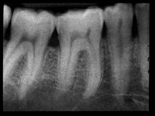
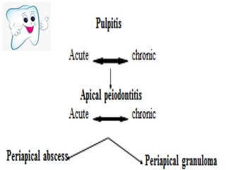
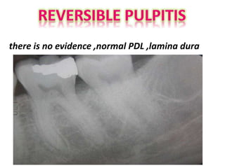
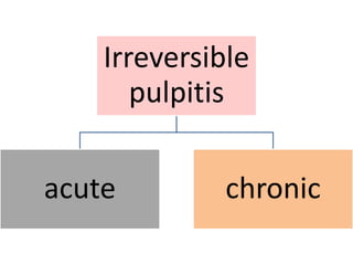
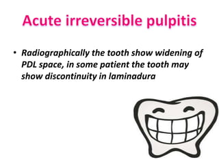
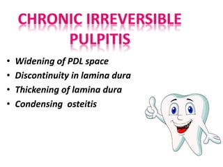
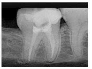
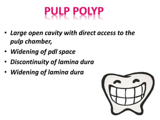
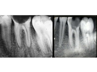
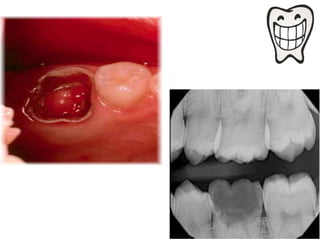
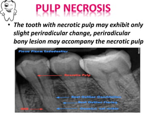
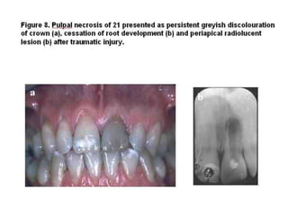
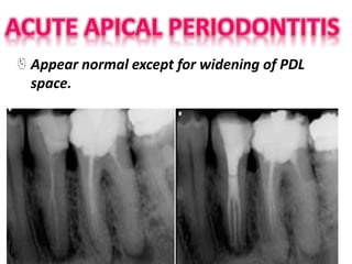
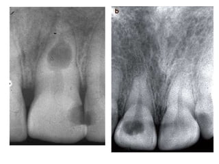
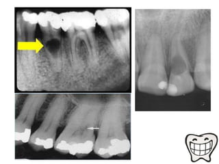
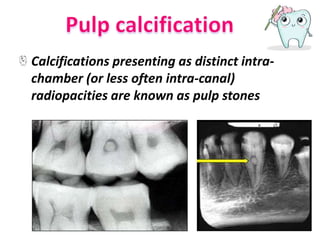
• Tooth pulp : the area of tooth that contain
blood vessels and efferent nerves and divided
into:
1. Pulp chamber
2. Root canal
The normal pulp appear as radiolucent area
in the center of the tooth (radiolucency of
pulp because it contain no minerals within it).
•Pulpitis
Reversible pulpitis
Irreversible pulptis(acute and chronic)
•Pulp necrosis
•Pulp polyps
•Periapical abscess
•Periapical granuloma
•Pulp calcification
•Internal resorption
•dens invaginatus
•taurodontism
there is no evidence ,normal PDL ,lamina dura
Irreversible
pulpitis
acute chronic
• Radiographically the tooth show widening of
PDL space, in some patient the tooth may
show discontinuity in laminadura
• Widening of PDL space
• Discontinuity in lamina dura
• Thickening of lamina dura
• Condensing osteitis
• Large open cavity with direct access to the
pulp chamber,
• Widening of pdl space
• Discontinuity of lamina dura
• Widening of lamina dura
• The tooth with necrotic pulp may exhibit only
slight periradicular change, periradicular
bony lesion may accompany the necrotic pulp
Appear normal except for widening of PDL
space.
Radiographical features:
In initial stages,periapical granuloma show widening
of the
periodontal ligament space of the tooth.
Fully developed lesions usually produce a well
defined
radiolucent area of varying size which appears to be
in continuity with the root apex.
Long standing periapical granuloma may show
varying
degrees of root resorption.
• Radiographical features:
Radiographical changes seen are limited to
only slight thickening of the periodontal
ligament space in apex region of the involved
tooth.
While in chronic periapical abscess,
radiograph often reveal small radiolucent
area at the root apex of the involved tooth
with poorly defined margins.
Apical Periodontal Cyst
• RADIOGRAPHIC FEATURES:
• Radiolucency – round/ ovoid with a narrow
opaque margin which is continuous with
lamina dura.
• In long standing cyst bone resorption of
affected teeth & occasional resorption of adj.
teeth may be seen.
• It presents a well defined,spherical
shaped,radiolucent area in dentine which
usually continuous with the pulp
• Enlargement of root canal
• Original root canal outline is distored
Calcifications presenting as distinct intra-
chamber (or less often intra-canal)
radiopacities are known as pulp stones
The shape of the invagination varying from a
narrow and undilated fissure to a
tear-shaped loop pointing towards the main
body of the pulp
Radio pulp diseases
Radio pulp diseases
Radio pulp diseases
Radio pulp diseases
Radio pulp diseases
Radio pulp diseases

Radio pulp diseases

  • 2.
    • Tooth pulp: the area of tooth that contain blood vessels and efferent nerves and divided into: 1. Pulp chamber 2. Root canal The normal pulp appear as radiolucent area in the center of the tooth (radiolucency of pulp because it contain no minerals within it).
  • 4.
    •Pulpitis Reversible pulpitis Irreversible pulptis(acuteand chronic) •Pulp necrosis •Pulp polyps •Periapical abscess •Periapical granuloma •Pulp calcification •Internal resorption •dens invaginatus •taurodontism
  • 6.
    there is noevidence ,normal PDL ,lamina dura
  • 7.
  • 8.
    • Radiographically thetooth show widening of PDL space, in some patient the tooth may show discontinuity in laminadura
  • 9.
    • Widening ofPDL space • Discontinuity in lamina dura • Thickening of lamina dura • Condensing osteitis
  • 11.
    • Large opencavity with direct access to the pulp chamber, • Widening of pdl space • Discontinuity of lamina dura • Widening of lamina dura
  • 14.
    • The toothwith necrotic pulp may exhibit only slight periradicular change, periradicular bony lesion may accompany the necrotic pulp
  • 16.
    Appear normal exceptfor widening of PDL space.
  • 17.
    Radiographical features: In initialstages,periapical granuloma show widening of the periodontal ligament space of the tooth. Fully developed lesions usually produce a well defined radiolucent area of varying size which appears to be in continuity with the root apex. Long standing periapical granuloma may show varying degrees of root resorption.
  • 19.
    • Radiographical features: Radiographicalchanges seen are limited to only slight thickening of the periodontal ligament space in apex region of the involved tooth. While in chronic periapical abscess, radiograph often reveal small radiolucent area at the root apex of the involved tooth with poorly defined margins.
  • 21.
    Apical Periodontal Cyst •RADIOGRAPHIC FEATURES: • Radiolucency – round/ ovoid with a narrow opaque margin which is continuous with lamina dura. • In long standing cyst bone resorption of affected teeth & occasional resorption of adj. teeth may be seen.
  • 24.
    • It presentsa well defined,spherical shaped,radiolucent area in dentine which usually continuous with the pulp • Enlargement of root canal • Original root canal outline is distored
  • 27.
    Calcifications presenting asdistinct intra- chamber (or less often intra-canal) radiopacities are known as pulp stones
  • 28.
    The shape ofthe invagination varying from a narrow and undilated fissure to a tear-shaped loop pointing towards the main body of the pulp