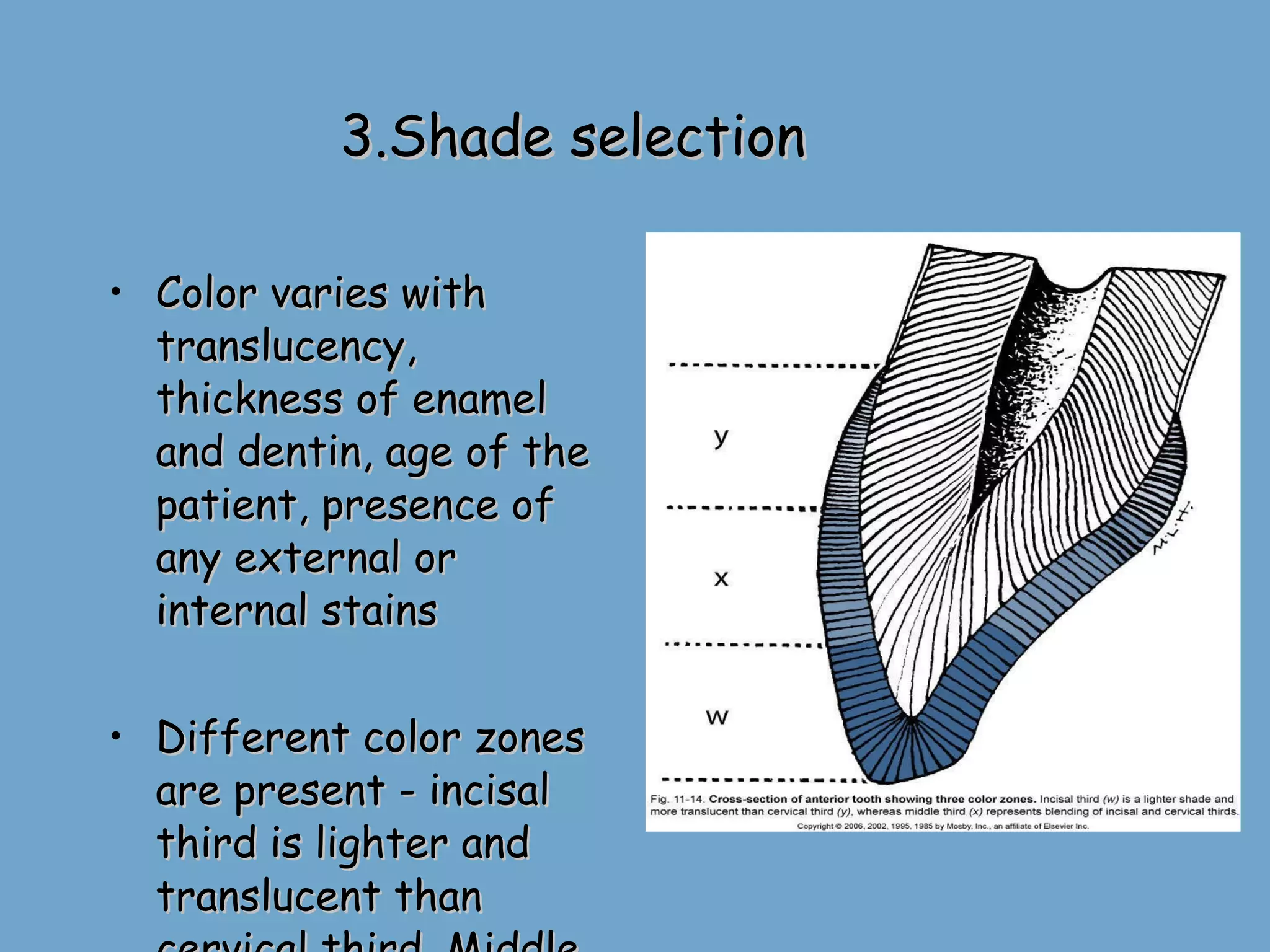
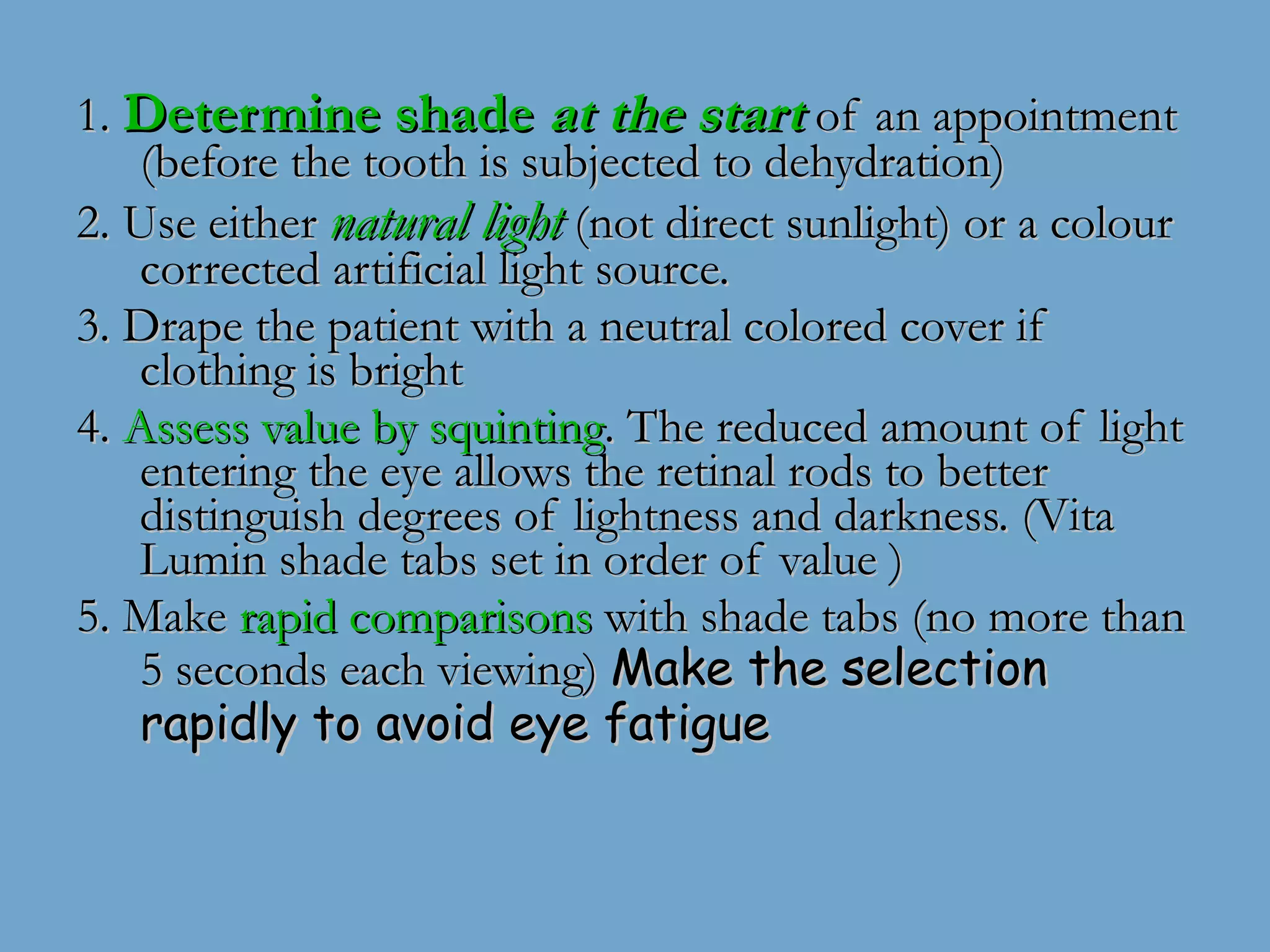
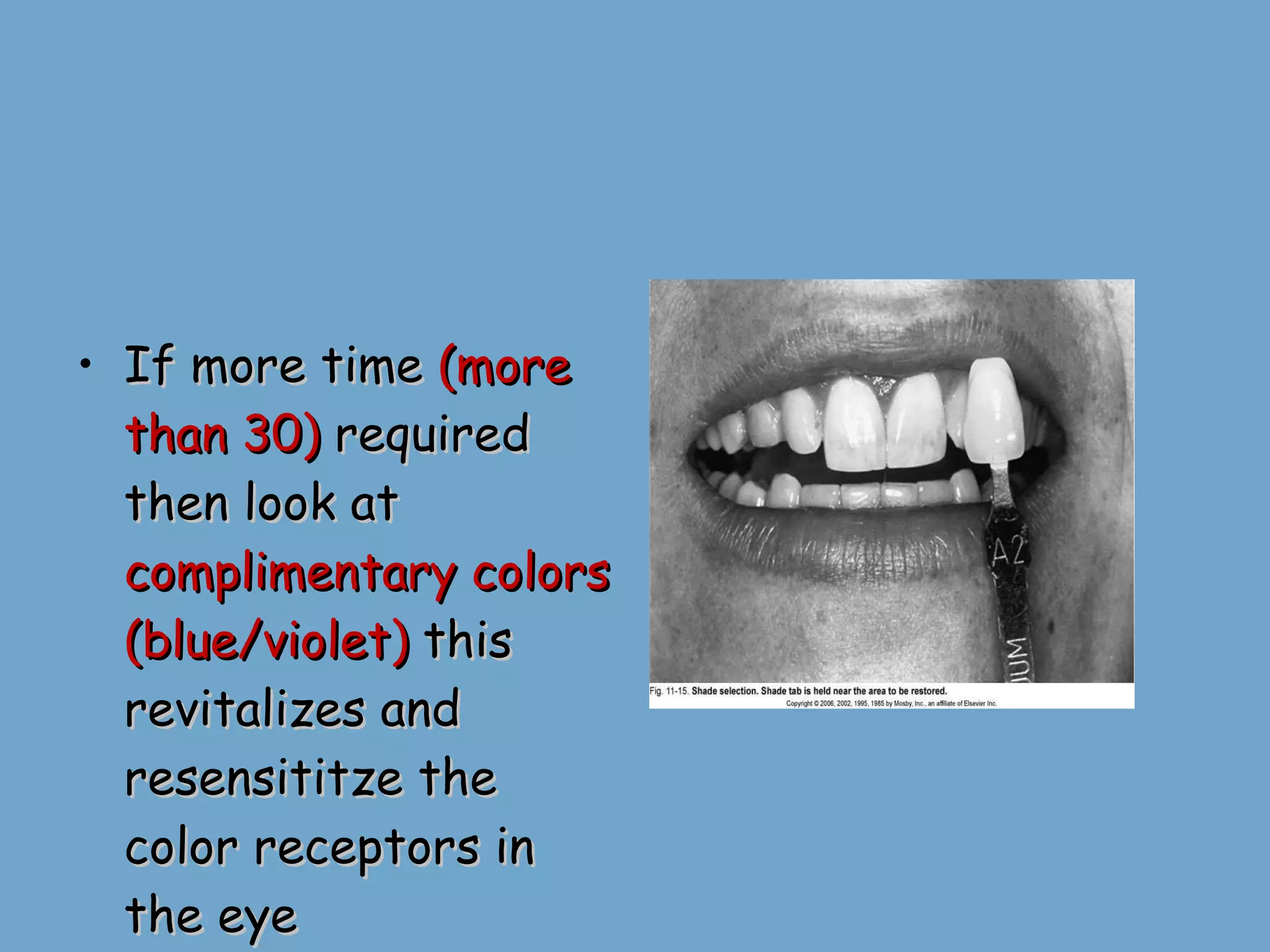
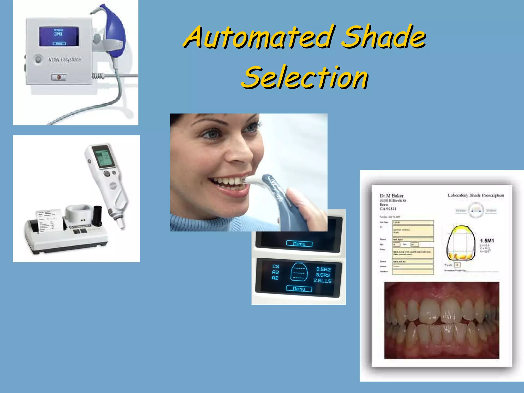
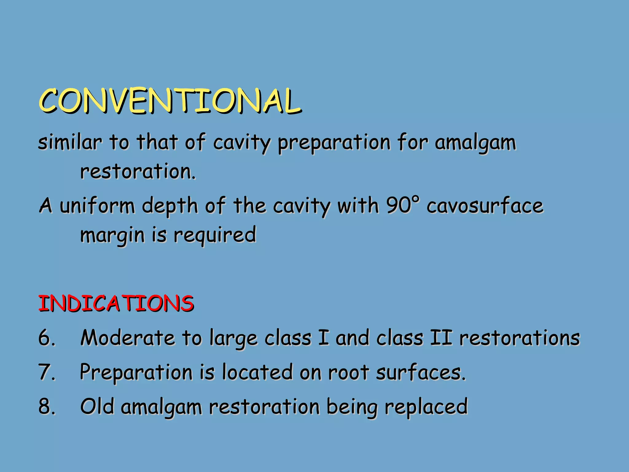
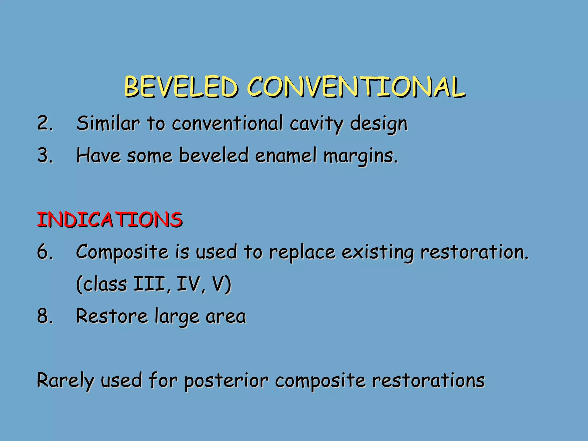
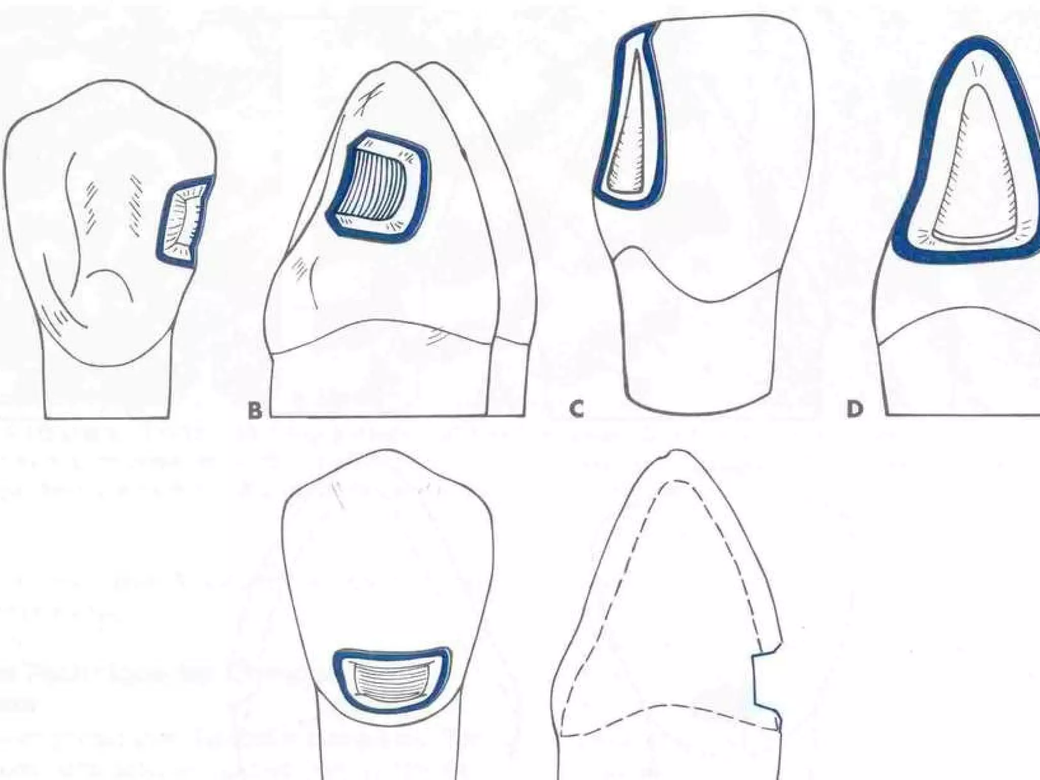
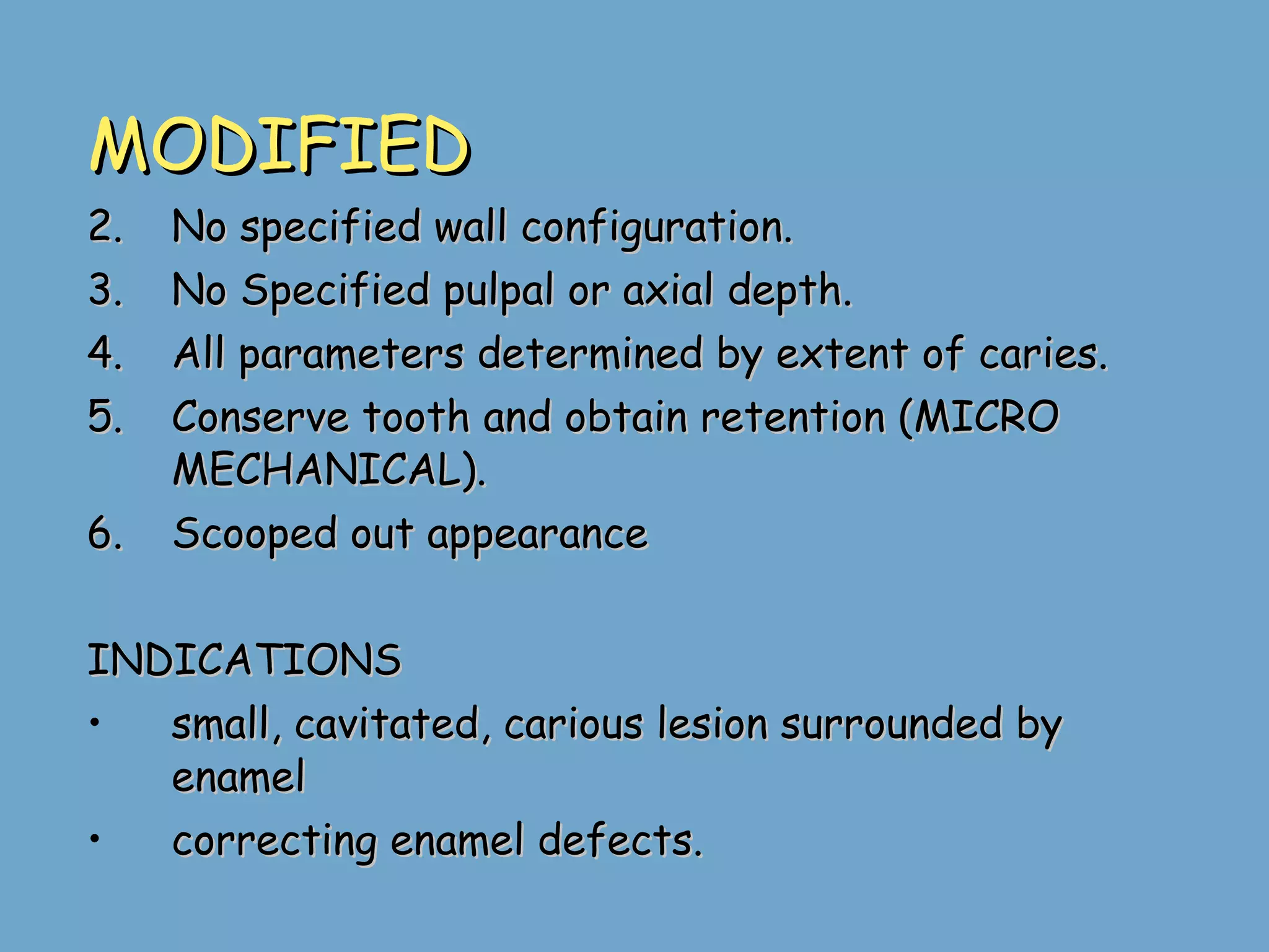
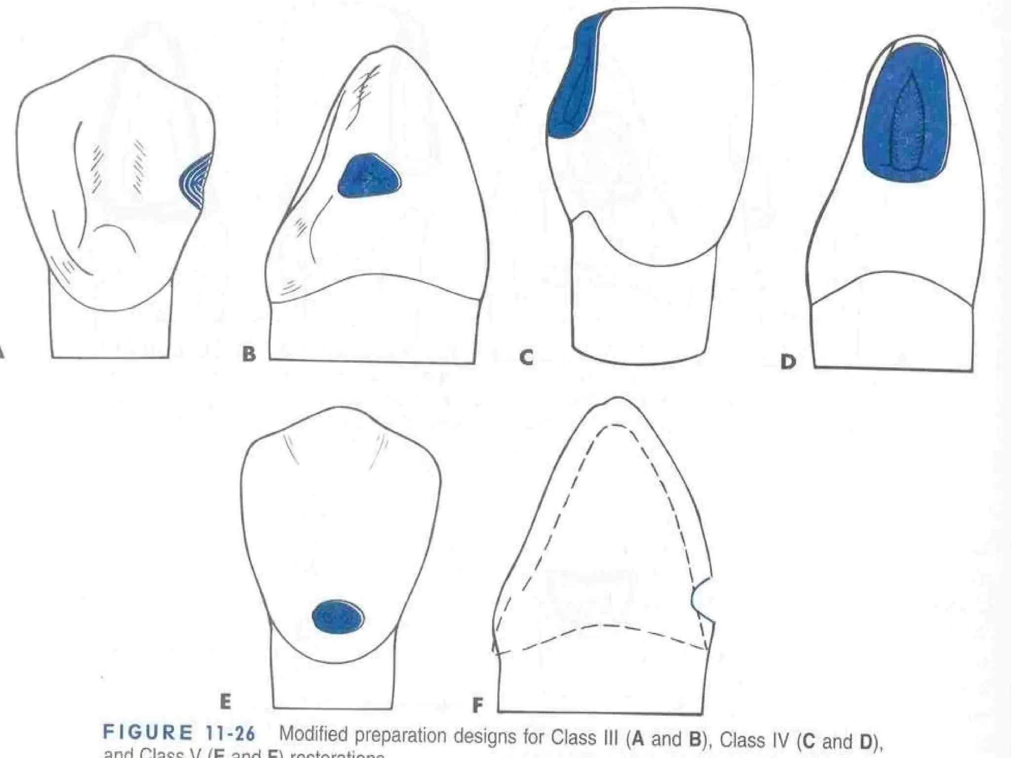
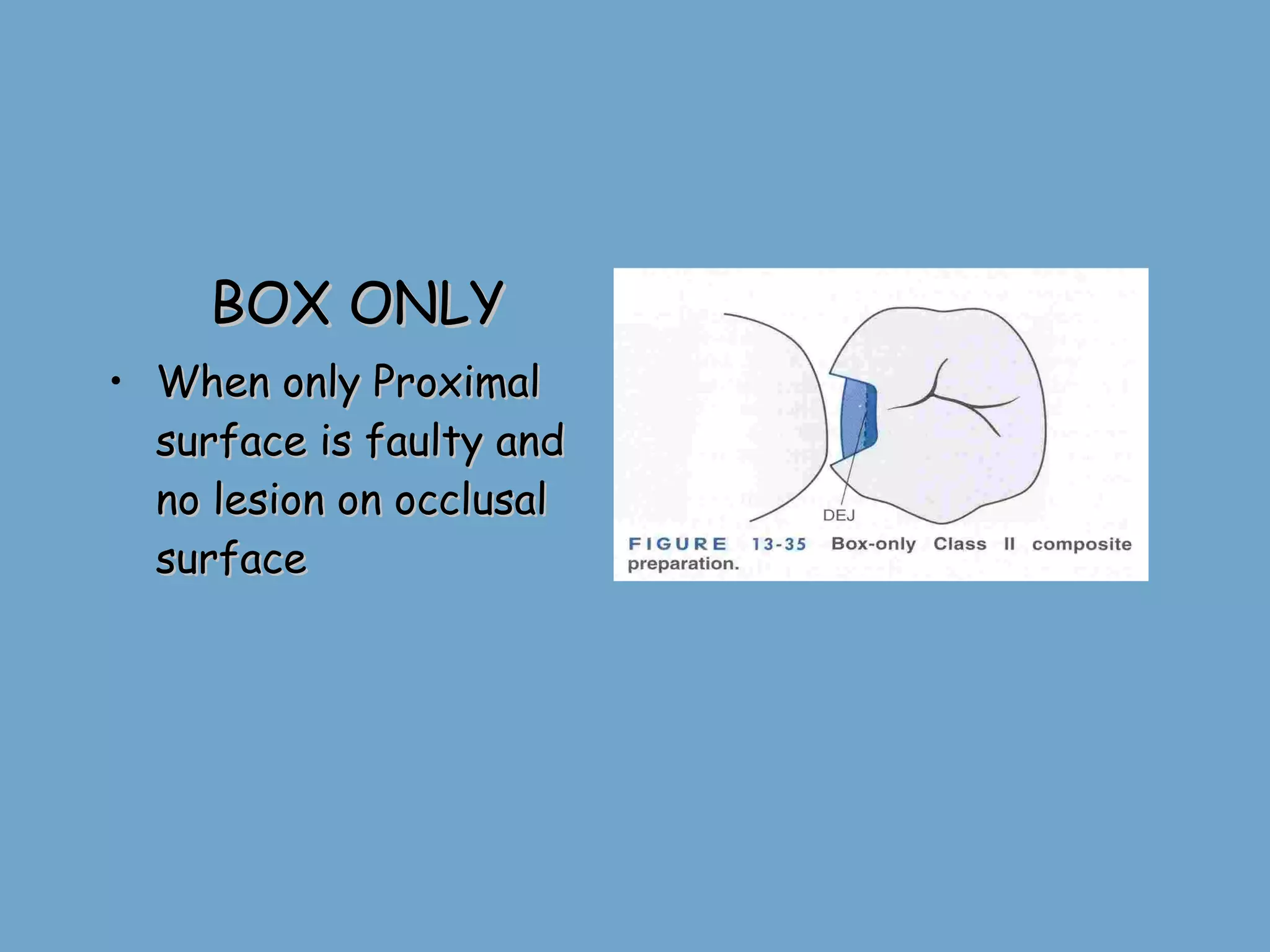
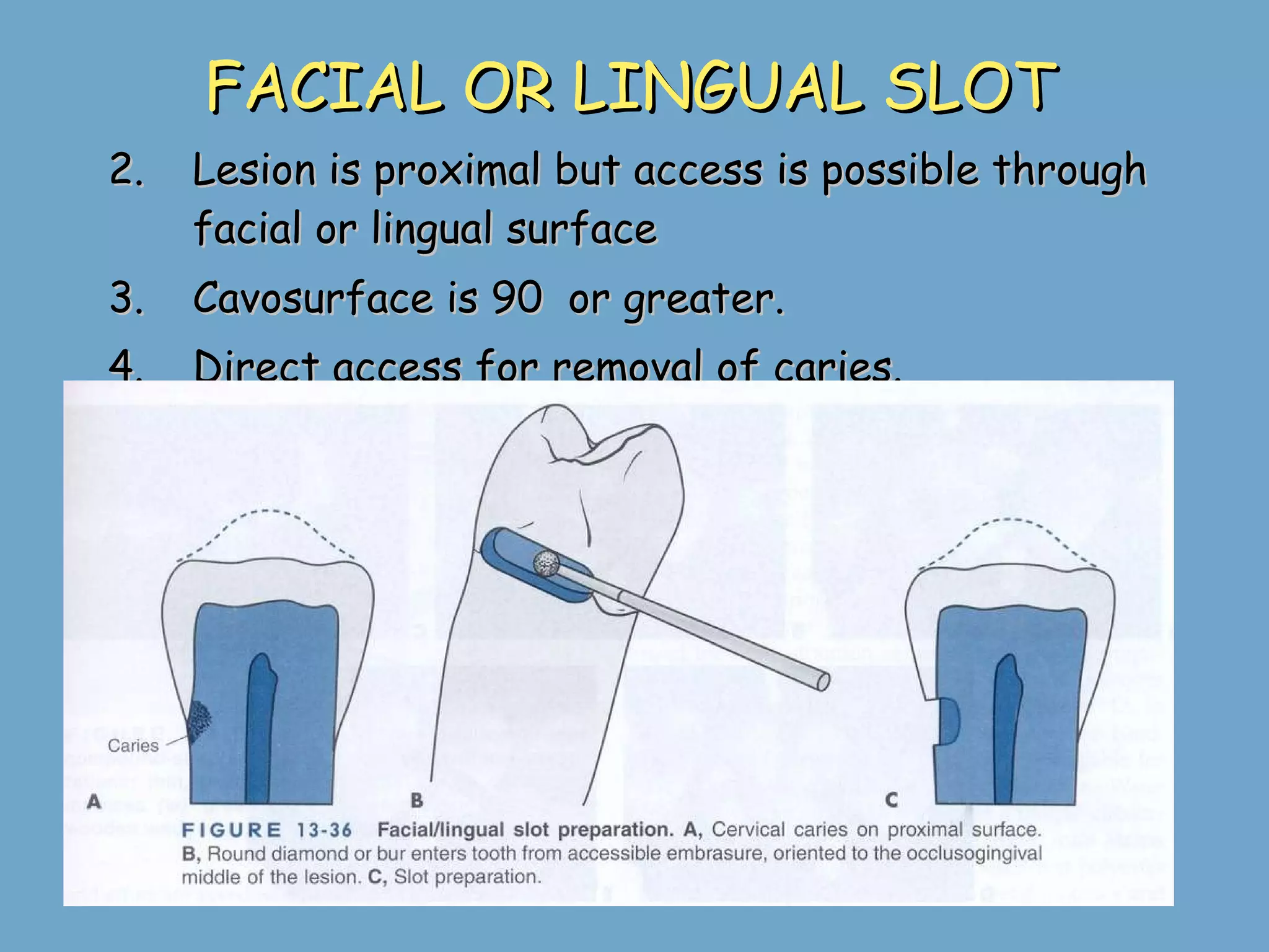
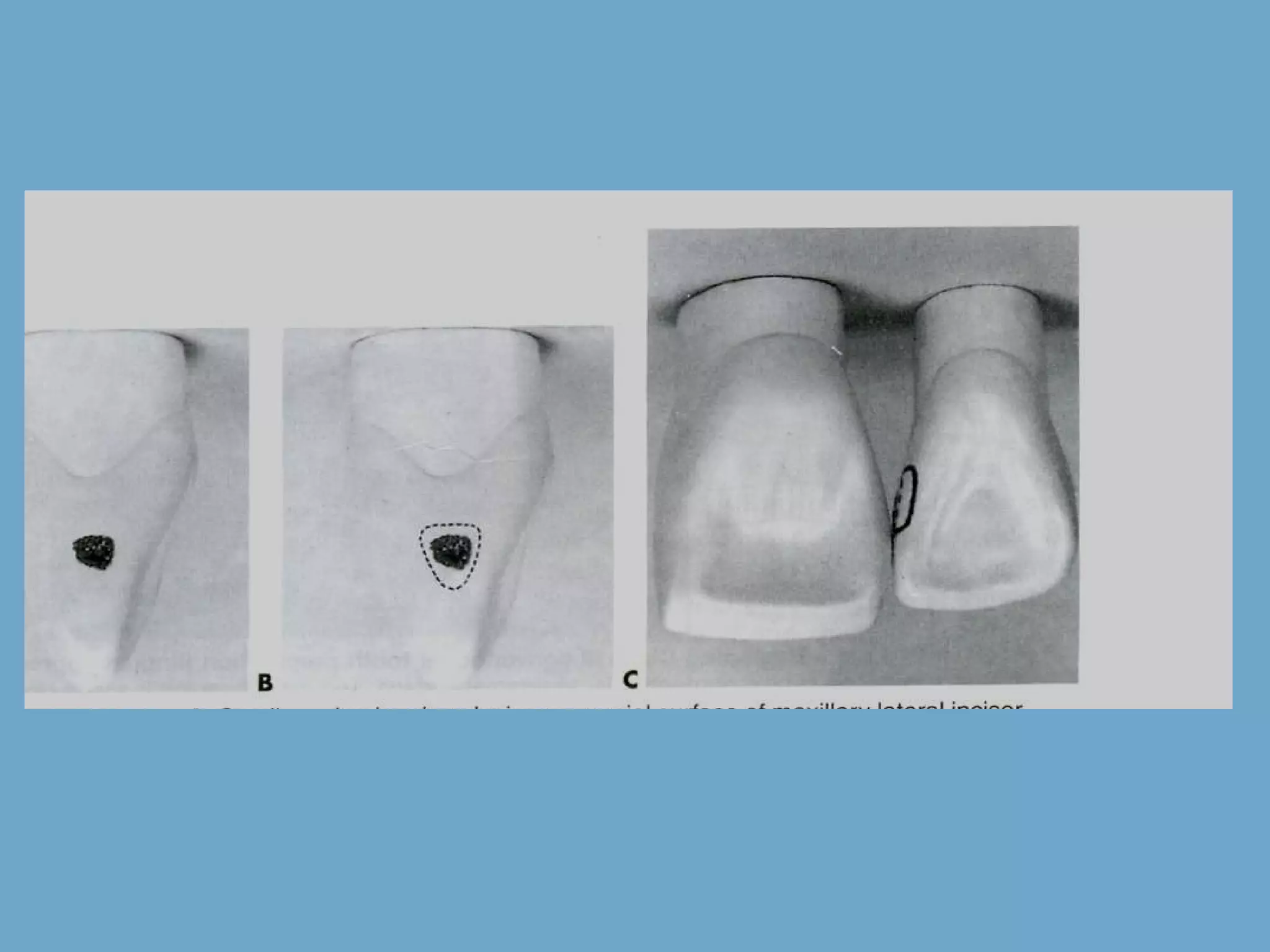
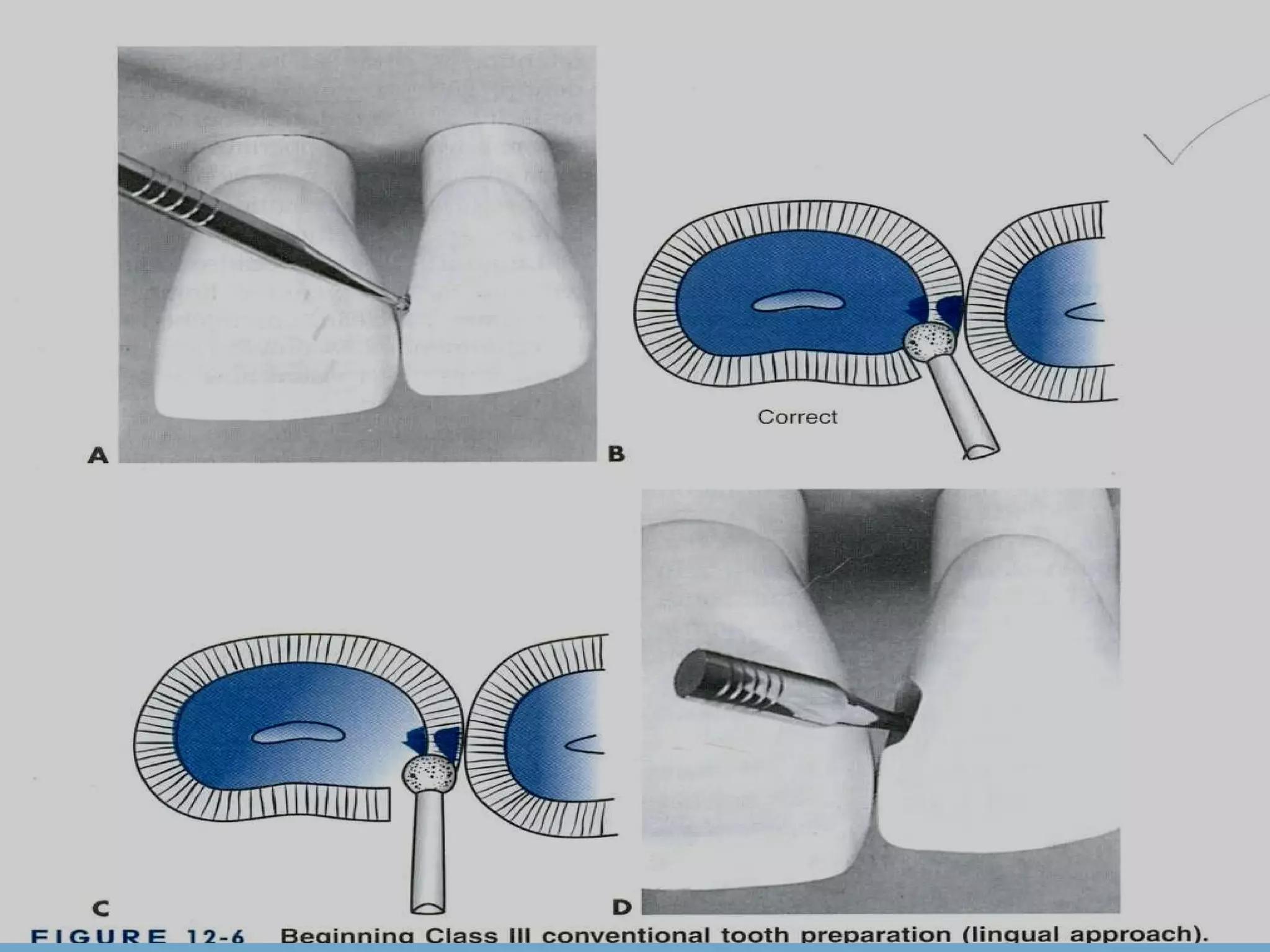
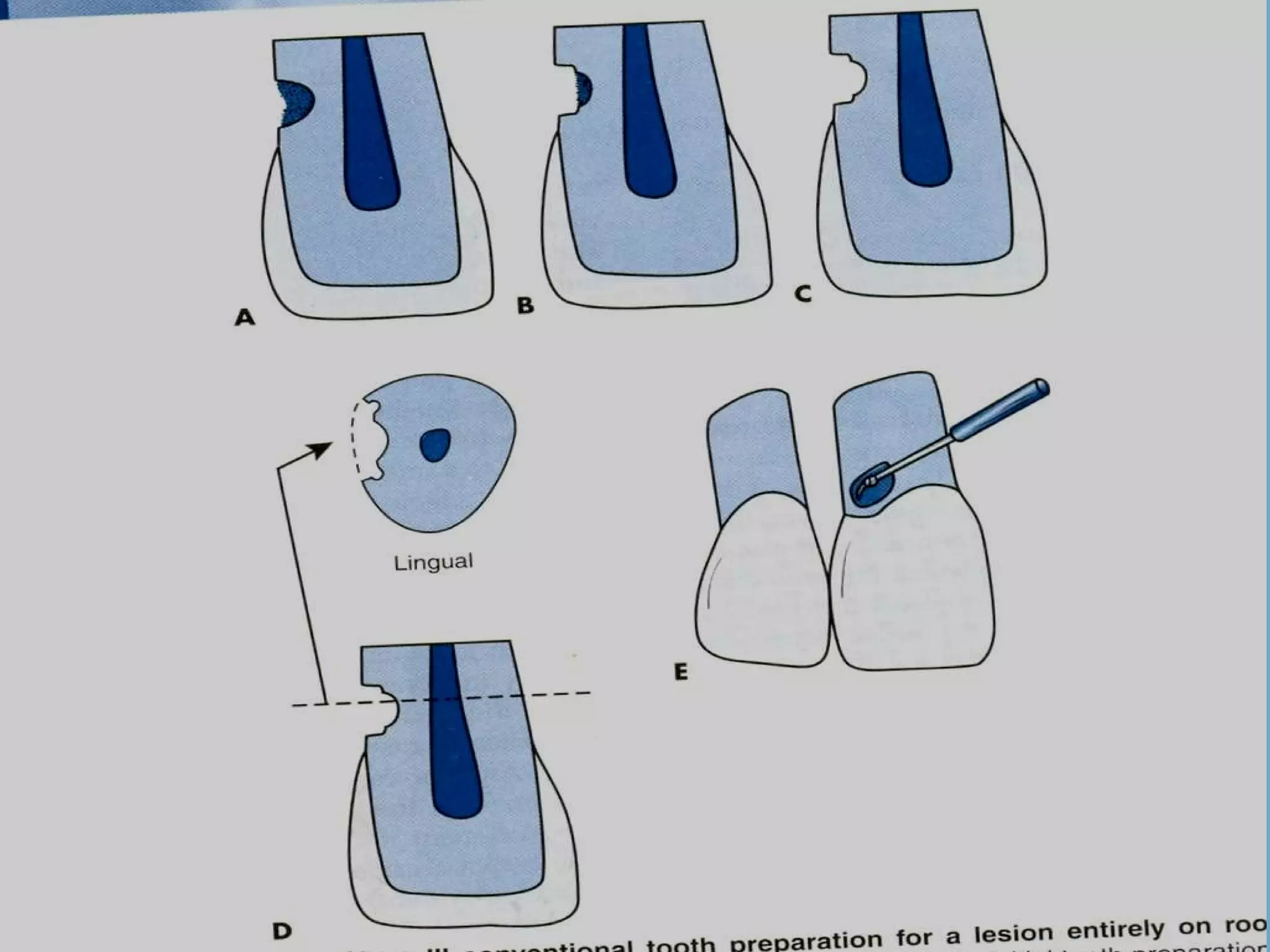
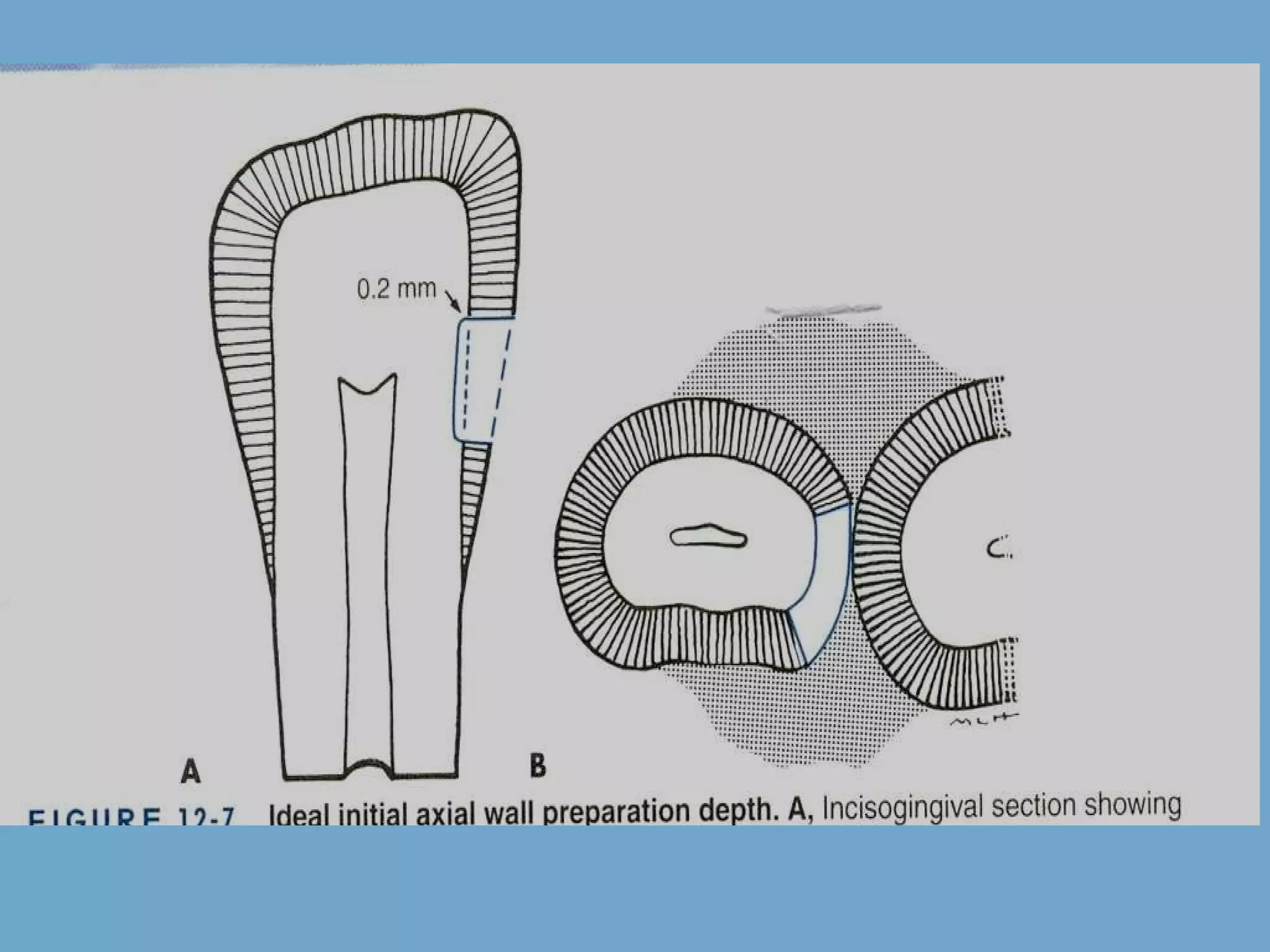
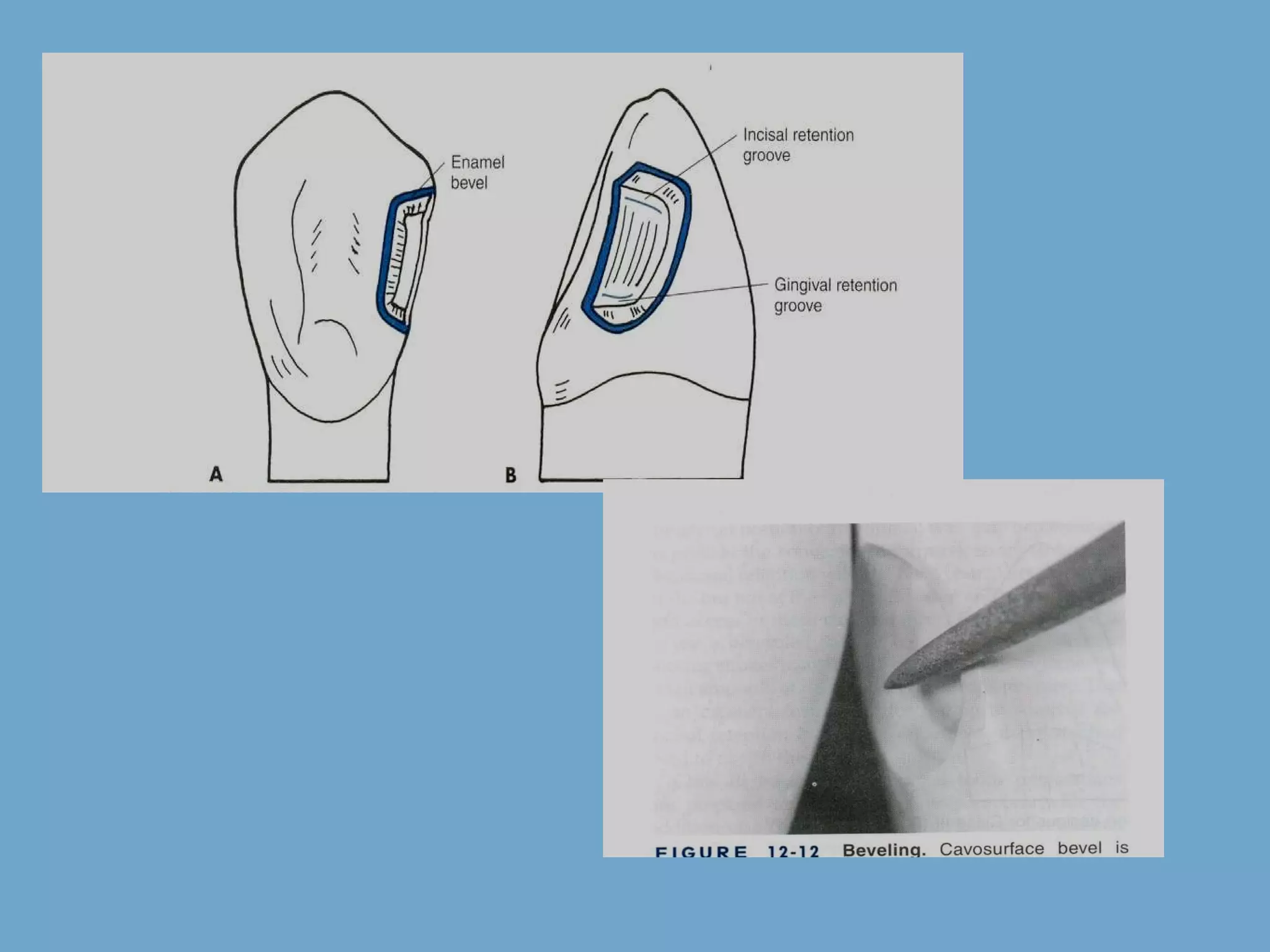
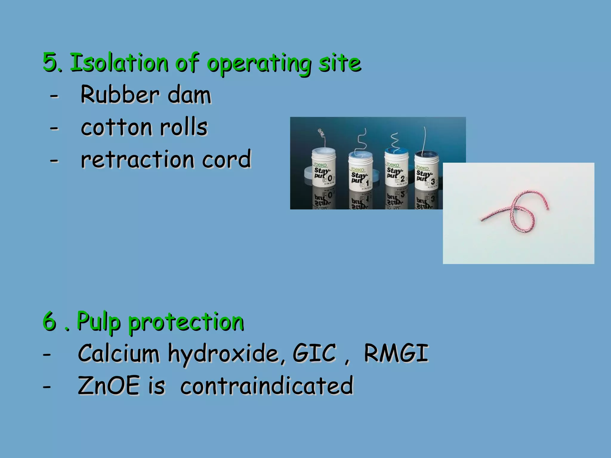
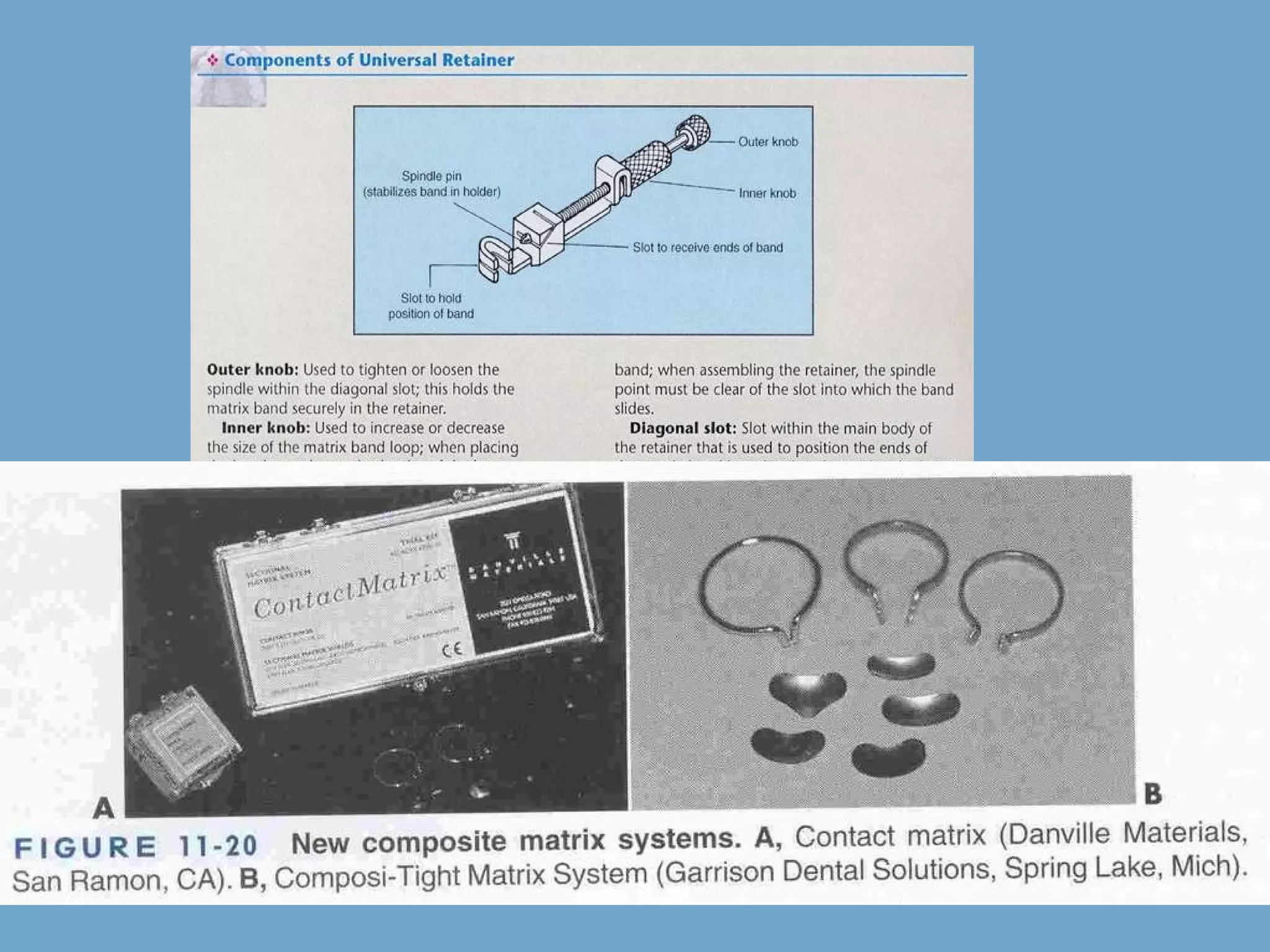
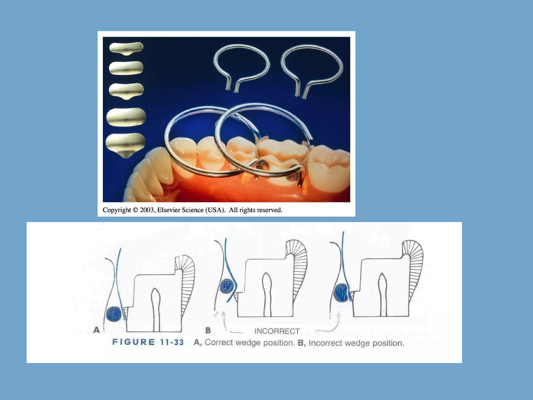
Clinical technique of composite restoration presented by: Faisal Alanazi
Clinical technique of composite restoration Initial clinical procedures, Tooth preparation for composite Restorative technique for composite Repairing composite restorations Fahad will complete C and D
Clinical technique  Initial clinical procedures, 1.  Local anesthesia   - patient is more relaxed - reduced salivation 2.  Preparation of operating site – clean the operating site with slurry of pumice to remove any  debris, plaque  , pellicle, and superficial stains .   Calculus removal  Prophy pastes containing flavoring agents, or fluorides act as contaminants and  should be  avoided  to prevent a possible conflict with the  acid-etch  technique.
3.Shade selection Color varies with translucency, thickness of enamel and dentin, age of the patient, presence of any external or internal stains  Different color zones are present - incisal third is lighter and  translucent than cervical third. Middle third is blend of two


Vita Lumin: A= reddish brown B = reddish yellow C = grey shades D = reddish grey B1 A1 B2 D2 A2 C1 C2 D4 A3 D3 B3 A3.5 B4 C3 A4 C4
3D Master
1.  Determine shade  at the start  of an appointment (before the tooth is subjected to dehydration) 2. Use either  natural light  (not direct sunlight) or a colour corrected artificial light source. 3. Drape the patient with a neutral colored cover if clothing is bright  4.  Assess value by squinting . The reduced amount of light entering the eye allows the retinal rods to better distinguish degrees of lightness and darkness. (Vita Lumin shade tabs set in order of value ) 5. Make  rapid comparisons  with shade tabs (no more than 5 seconds each viewing)  Make the selection rapidly to avoid eye fatigue
If more time  (more than 30)  required then look at  complimentary colors (blue/violet)  this revitalizes and resensititze the color receptors in the eye
6. Choose the dominant hue and chroma within the value range chosen. The canines - useful guide to assessing hue. 7. Compare selected tabs under different conditions eg wet vs dry, different lip positions, artificial and natural light from different angles. 8. Look carefully for colour characterisation such as stained imbrication lines, white spots, neck colouration, incisal edge translucency
Automated Shade Selection
B.Tooth preparation for composite 4.Cavity preparation
Tooth preparation principles and criteria External Outline form Extent is determined by size, shape, and location of defect . should include all Caries, any fault, defective, old friable tooth structure. Removal of discolored tooth structure as required for esthetics. Create prepared enamel margin of 90° or greater by giving bevel wherever required. Create 90° cavosurface on root surfaces Pulpally, no uniform depth is needed Depth should be sufficient to identify and remove caries or existing restoration.
RETENTION Micromechanical retention by etching of enamel and dentin. Mechanical undercuts when margins terminate in cementum.
Advantages of beveling. Increase in surface area because stronger enamel to resin bond  Ends of enamel rods  are etched. Esthetic blending due to cavosurface bevel.
 
Cavity designs for composite cavity preparation Conventional  Beveled conventional Modified Box shape Facial/lingual slot
CONVENTIONAL similar to that of cavity preparation for amalgam restoration. A uniform depth of the cavity with 90° cavosurface margin is required  INDICATIONS Moderate to large class I and class II restorations  Preparation is located on root surfaces. Old amalgam restoration being replaced
BEVELED CONVENTIONAL Similar to conventional cavity design Have some beveled enamel margins.  INDICATIONS Composite is used to replace existing restoration. (class III, IV, V) Restore large area Rarely used for posterior composite restorations
 
Combined design
MODIFIED No specified wall configuration. No Specified pulpal or axial depth. All parameters determined by extent of caries. Conserve tooth and obtain retention (MICRO MECHANICAL).  Scooped out appearance INDICATIONS small, cavitated, carious lesion surrounded by  enamel  correcting enamel defects.
 
BOX ONLY When only Proximal surface is faulty and no lesion on occlusal surface
FACIAL OR LINGUAL SLOT Lesion is proximal but access is possible through facial or lingual surface Cavosurface is 90  or greater. Direct access for removal of caries.


5. Isolation of operating site -  Rubber dam -  cotton rolls -  retraction cord 6 . Pulp protection Calcium hydroxide, GIC ,  RMGI ZnOE is  contraindicated
7. Matrix placement Two types of matrices are available - Polyester matrix - metal matrix  Various matrix retainer which can be used are Tofflemire retainer Compound supported metal matrix Sectional matrix system- palodent contact matrix
 
 
Polyester matrix -  used especially CLASS III, CLASSIV ,CLASS V cavities Advantage  -  they allow the light to pass Disadvantage  - they are not rigid and get deform during placement of rigid material and contact cannot be properly restored Metal matrix -  Ultrathin metal matrices 001- .002 inch are used - Band should be precontoured outside the mouth
Thank you Theodore.M.Roberson,Harald.O.Heymann,Edward.J.SwiftJR.Sturdevant's  Art and Science of Operative Dentistry.Mosby publications;2002; 483-492 Reference

Composite preparation

  • 1.
    Clinical technique ofcomposite restoration presented by: Faisal Alanazi
  • 2.
    Clinical technique ofcomposite restoration Initial clinical procedures, Tooth preparation for composite Restorative technique for composite Repairing composite restorations Fahad will complete C and D
  • 3.
    Clinical technique Initial clinical procedures, 1. Local anesthesia - patient is more relaxed - reduced salivation 2. Preparation of operating site – clean the operating site with slurry of pumice to remove any debris, plaque , pellicle, and superficial stains . Calculus removal Prophy pastes containing flavoring agents, or fluorides act as contaminants and should be avoided to prevent a possible conflict with the acid-etch technique.
  • 4.
    3.Shade selection Colorvaries with translucency, thickness of enamel and dentin, age of the patient, presence of any external or internal stains Different color zones are present - incisal third is lighter and translucent than cervical third. Middle third is blend of two
  • 5.
  • 6.
  • 7.
  • 8.
    Vita Lumin: A=reddish brown B = reddish yellow C = grey shades D = reddish grey B1 A1 B2 D2 A2 C1 C2 D4 A3 D3 B3 A3.5 B4 C3 A4 C4
  • 9.
  • 10.
    1. Determineshade at the start of an appointment (before the tooth is subjected to dehydration) 2. Use either natural light (not direct sunlight) or a colour corrected artificial light source. 3. Drape the patient with a neutral colored cover if clothing is bright 4. Assess value by squinting . The reduced amount of light entering the eye allows the retinal rods to better distinguish degrees of lightness and darkness. (Vita Lumin shade tabs set in order of value ) 5. Make rapid comparisons with shade tabs (no more than 5 seconds each viewing) Make the selection rapidly to avoid eye fatigue
  • 11.
    If more time (more than 30) required then look at complimentary colors (blue/violet) this revitalizes and resensititze the color receptors in the eye
  • 12.
    6. Choose thedominant hue and chroma within the value range chosen. The canines - useful guide to assessing hue. 7. Compare selected tabs under different conditions eg wet vs dry, different lip positions, artificial and natural light from different angles. 8. Look carefully for colour characterisation such as stained imbrication lines, white spots, neck colouration, incisal edge translucency
  • 13.
  • 14.
    B.Tooth preparation forcomposite 4.Cavity preparation
  • 15.
    Tooth preparation principlesand criteria External Outline form Extent is determined by size, shape, and location of defect . should include all Caries, any fault, defective, old friable tooth structure. Removal of discolored tooth structure as required for esthetics. Create prepared enamel margin of 90° or greater by giving bevel wherever required. Create 90° cavosurface on root surfaces Pulpally, no uniform depth is needed Depth should be sufficient to identify and remove caries or existing restoration.
  • 16.
    RETENTION Micromechanical retentionby etching of enamel and dentin. Mechanical undercuts when margins terminate in cementum.
  • 17.
    Advantages of beveling.Increase in surface area because stronger enamel to resin bond Ends of enamel rods are etched. Esthetic blending due to cavosurface bevel.
  • 18.
  • 19.
    Cavity designs forcomposite cavity preparation Conventional Beveled conventional Modified Box shape Facial/lingual slot
  • 20.
    CONVENTIONAL similar tothat of cavity preparation for amalgam restoration. A uniform depth of the cavity with 90° cavosurface margin is required INDICATIONS Moderate to large class I and class II restorations Preparation is located on root surfaces. Old amalgam restoration being replaced
  • 21.
    BEVELED CONVENTIONAL Similarto conventional cavity design Have some beveled enamel margins. INDICATIONS Composite is used to replace existing restoration. (class III, IV, V) Restore large area Rarely used for posterior composite restorations
  • 22.
  • 23.
  • 24.
    MODIFIED No specifiedwall configuration. No Specified pulpal or axial depth. All parameters determined by extent of caries. Conserve tooth and obtain retention (MICRO MECHANICAL). Scooped out appearance INDICATIONS small, cavitated, carious lesion surrounded by enamel correcting enamel defects.
  • 25.
  • 26.
    BOX ONLY Whenonly Proximal surface is faulty and no lesion on occlusal surface
  • 27.
    FACIAL OR LINGUALSLOT Lesion is proximal but access is possible through facial or lingual surface Cavosurface is 90 or greater. Direct access for removal of caries.
  • 28.
  • 29.
  • 30.
  • 31.
  • 32.
  • 33.
    5. Isolation ofoperating site - Rubber dam - cotton rolls - retraction cord 6 . Pulp protection Calcium hydroxide, GIC , RMGI ZnOE is contraindicated
  • 34.
    7. Matrix placementTwo types of matrices are available - Polyester matrix - metal matrix Various matrix retainer which can be used are Tofflemire retainer Compound supported metal matrix Sectional matrix system- palodent contact matrix
  • 35.
  • 36.
  • 37.
    Polyester matrix - used especially CLASS III, CLASSIV ,CLASS V cavities Advantage - they allow the light to pass Disadvantage - they are not rigid and get deform during placement of rigid material and contact cannot be properly restored Metal matrix - Ultrathin metal matrices 001- .002 inch are used - Band should be precontoured outside the mouth
  • 38.
    Thank you Theodore.M.Roberson,Harald.O.Heymann,Edward.J.SwiftJR.Sturdevant's Art and Science of Operative Dentistry.Mosby publications;2002; 483-492 Reference