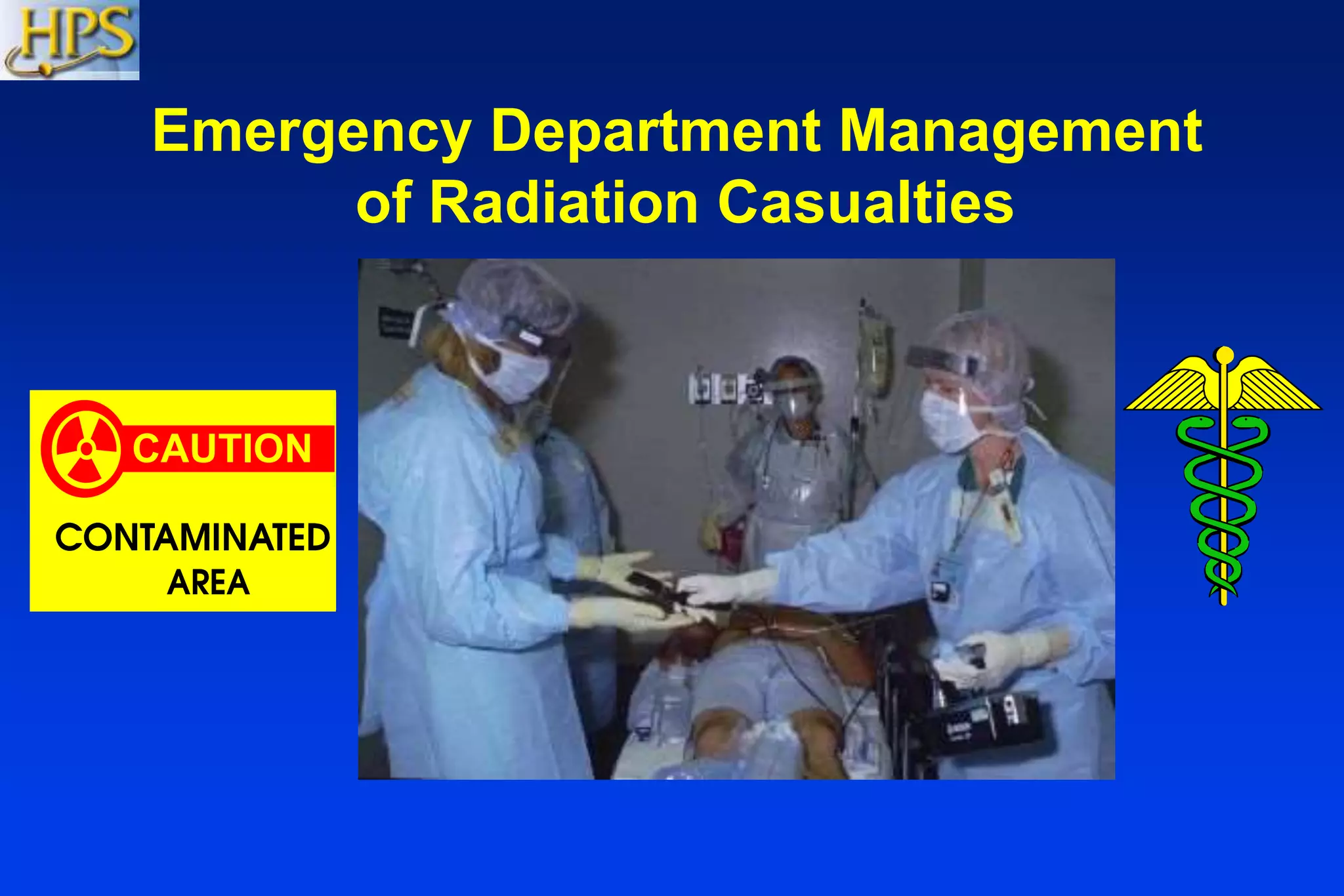
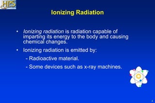
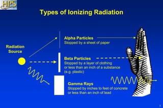
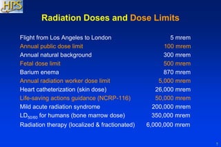
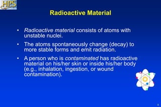
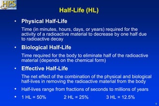
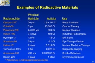
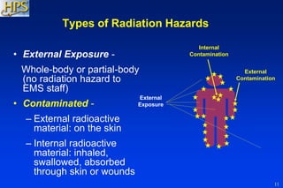
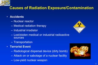
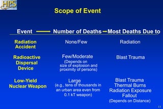
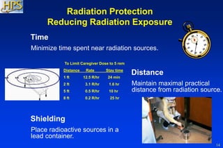
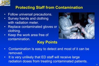
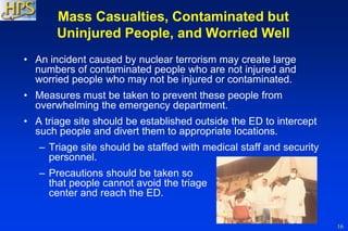
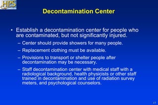
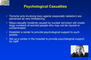
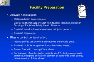
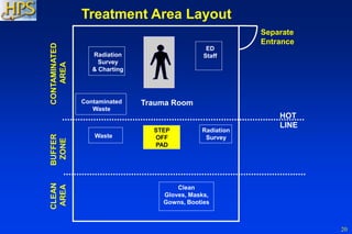
It has been concluded that the management of radiation accidents is a very challenging process and that nuclear medicine physicians have to be well organized in.
This document discusses emergency response and preparedness in a radiation department. It defines a radiation emergency and classifies emergencies by whether they affect equipment, an individual patient, or many patients. Potential sources of error leading to emergencies in radiotherapy, nuclear medicine, brachytherapy, and diagnostic radiology are described. Regulations regarding reporting and investigating emergencies are summarized. Steps for handling common emergency situations like source stucks are outlined. The responsibilities of licensees and radiation safety officers in emergency planning and response are also covered.
This document discusses ionizing radiation, its biological effects, and safety issues. It begins by defining ionizing radiation and its units of measurement. It then describes the mechanisms by which ionizing radiation can damage cells, particularly DNA, and potentially lead to genetic mutations and cancer initiation. Key factors that influence radiosensitivity, such as the cell cycle phase and tissue type, are also covered. The document discusses deterministic effects, which occur above threshold doses, and stochastic effects like cancer that occur probabilistically. Guidelines for radiation protection emphasize justification of exposures and optimizing procedures to minimize risks.
The document discusses recommendations from ICRP 60 & 103 regarding radiation protection. It begins with background on natural and artificial radiation sources and their effects. It then summarizes the evolution of ICRP recommendations over time, from early annual dose limits of 1000 mSv reduced gradually to current limits. Key concepts discussed include justification of practices, optimization of protection, and application of dose limits. Occupational, public, and medical exposure dose limits are provided. ICRP 103 introduced changes like new tissue weighting factors and computational phantoms.
Radiation protection, also known as radiological protection, is defined by the International Atomic Energy Agency (IAEA) as "The protection of people from harmful effects of exposure to ionizing radiation, and the means for achieving this". Exposure can be from a source of radiation external to the human body or due to internal irradiation caused by the ingestion of radioactive contamination.
Ionizing radiation is widely used in industry and medicine, and can present a significant health hazard by causing microscopic damage to living tissue. There are two main categories of ionizing radiation health effects. At high exposures, it can cause "tissue" effects, also called "deterministic" effects due to the certainty of them happening, conventionally indicated by the unit gray and resulting in acute radiation syndrome. For low level exposures there can be statistically elevated risks of radiation-induced cancer, called "stochastic effects" due to the uncertainty of them happening, conventionally indicated by the unit sievert.
Fundamental to radiation protection is the avoidance or reduction of dose using the simple protective measures of time, distance and shielding. The duration of exposure should be limited to that necessary, the distance from the source of radiation should be maxi mised, and the source shielded wherever possible. To measure personal dose uptake in occupational or emergency exposure, for external radiation personal dosimeters are used, and for internal dose to due to ingestion of radioactive contamination, bioassay techniques are applied.
This document discusses the history and development of radiation protection. Some key points:
- The harmful effects of radiation were initially not well understood after X-rays were discovered in the late 19th century. Several early researchers and technicians suffered health effects.
- Over time, concepts like tolerance doses, maximum permissible doses, and the "as low as reasonably achievable" principle were developed to set safe radiation exposure limits.
- International organizations like the ICRP and IAEA were formed to make recommendations on radiation safety standards and regulation. National bodies like AERB regulate radiation protection in India.
- The principles of justification, optimization and dose limitation form the foundation of modern radiation protection practices and regulation. Exposure
This document discusses radiation protection for medical workers. It explains that radiologists, radiographers and other medical staff are subject to some radiation exposure through their work, so radiation protection aims to keep doses as low as possible to prevent health risks. It describes different types of radiation effects and dose limits for workers. Various methods of radiation monitoring are outlined, including use of dosimeters like film badges, pocket ionization chambers and thermoluminescent dosimeters to measure individual radiation exposures.
1. Photons can interact with matter through various processes including photoelectric effect, Compton scattering, pair production, and photodisintegration.
2. The dominant interaction depends on the photon energy and atomic number (Z) of the absorbing material. Low energy photons mainly undergo the photoelectric effect while high energy photons undergo Compton scattering and pair production.
3. Each interaction process results in the photon transferring some or all of its energy to electrons or matter. This energy deposition is important for applications in medical imaging and radiation therapy.
This document discusses various topics relating to radiation, including:
- Types of radiation such as particle radiation (alpha, beta, neutron) and electromagnetic radiation like X-rays.
- Properties of X-rays including their wavelength range and ability to ionize atoms.
- Sources of radiation including cosmic radiation from space, radon gas, radioactive substances, nuclear activities, and particle accelerators.
- Biological effects of radiation like genetic effects impacting future generations, and somatic effects impacting individuals. Effects can be deterministic where the severity increases with dose above a threshold, or stochastic like cancer induction where risk increases linearly with dose.
The objectives of radiation protection according to the ICRP and NCRP are to prevent serious radiation effects and reduce stochastic effects to acceptable levels while allowing beneficial practices involving radiation exposure. This is achieved through principles of justification, optimization and dose limitation. Justification requires that practices only be adopted if benefits outweigh radiation risks. Optimization aims to keep exposures as low as reasonably achievable. Dose limitation sets defined exposure limits for workers and the public.
Radiation is energy that is given off by particular materials and devices.
Radiation protection, also known as radiological protection, is defined by the International Atomic Energy Agency (IAEA) as "The protection of people from harmful effects of exposure to ionizing radiation, and the means for achieving this". Exposure can be from a source of radiation external to the human body or due to internal irradiation caused by the ingestion of radioactive contamination
This document discusses various radiation quantities and units used to characterize ionizing radiation. It describes key concepts such as activity, kerma, exposure, absorbed dose, equivalent dose, effective dose, annual limit intake (ALI), and derived air concentration (DAC). The International Commission on Radiation Protection (ICRP) and International Commission on Radiation Units (ICRU) help define these quantities and their relationships. Primary quantities like equivalent dose relate radiation risk, while operational quantities like exposure are used for measurements. Tissue weighting factors account for different tissue sensitivities in calculating effective dose from equivalent dose.
1) Radiation protection standards aim to restrict radiation risks while allowing beneficial uses of radiation. Exposure standards are set to limit stochastic effects probabilistically and prevent deterministic effects.
2) Radiation can cause both stochastic (random) effects like cancer that have no threshold and increase in risk with increasing dose, as well as non-stochastic (deterministic) effects like burns that have thresholds below which they will not occur.
3) Various quantities like absorbed dose, equivalent dose, and effective dose are used to quantify radiation exposure and the associated biological harm from different radiation types. Operational quantities allow direct measurement and monitoring.
The document discusses the history of radiation protection, including early pioneers who discovered radiation hazards and effects. It describes some key events like the establishment of the ICRP and AERB, and definitions of key radiation terms. It also outlines the biological effects of radiation exposure, distinguishing between deterministic and stochastic effects. The three principles of radiation protection - justification, optimization and dose limitation - are explained.
Radiation emergencies and preparedness in radiotherapyDeepjyoti saha
In a Radiotherapy Department where cancer patients are being treated with high energy photons,gamma rays,electrons; all the radiation workers should be alert regarding radiation accidents & how to face the situation.
This document discusses radiation protection and dosimetry concepts. It defines key terms like absorbed dose, equivalent dose, effective dose and their calculations. It describes stochastic and deterministic effects and the objectives of radiation protection to limit both. The ALARA principle and its application are explained. Various radiation measurement instruments like survey meters, dosimeters and their uses are outlined. The document also discusses radiation shielding calculations and definitions of controlled, supervised and uncontrolled areas.
This power-point presentation is very important for radiology resident radiologist and radiographers and technician. this includes principles, technique , biological effects of radiation and how to protect, whats should normal radiation dose with latest update. This slide also includes ALARA PRINCIPLE thanks.
This document discusses various radiation units used to quantify radiation exposure and its effects. It defines units of radioactivity like curie and becquerel, exposure units like roentgen, absorbed dose units like rad and gray, and equivalent and effective dose units like rem and sievert used to account for radiation type and organ sensitivity. It also discusses concepts like attenuation, kerma, absorbed dose, and weighting factors used to calculate equivalent and effective doses from radiation exposure.
Isodose lines represent points of equal absorbed radiation dose on a dose distribution map. They are depicted as curves on isodose charts showing the volumetric and planar variations in absorbed dose. Factors influencing isodose curves include beam quality, field size, source-to-skin distance, beam modifiers like wedges or bolus, and depth. Isodose curves are used in radiation therapy treatment planning to evaluate dose distributions and ensure tumor coverage while sparing surrounding healthy tissues. They provide critical information about the radiation dose profile essential for safe and effective treatment.
This document provides information on occupational radiation safety for radiologic technologists. It discusses the risks of ionizing radiation exposure and strategies to minimize that exposure through proper use of time, distance, and shielding techniques. Protective equipment discussed includes lead aprons, thyroid collars, gloves, glasses and face masks. The document emphasizes the importance of radiation safety given the increased risk of health issues like cataracts for those who work regularly with medical imaging that uses ionizing radiation.
This document discusses central axis depth doses in water for both SSD and SAD techniques. For SSD technique:
- Percentage depth dose (PDD) curves measure attenuation at different depths and are affected by beam quality, field size, and SSD.
- Buildup region occurs as secondary electrons deposit energy downstream, increasing dose with depth until maximum.
- Depth dose maximum (zmax) depends on beam energy and field size.
- PDD increases with larger field sizes due to increased scatter radiation.
- PDD increases with longer SSD due to the inverse square law of radiation intensity.
Rp003 biological effects of ionizing radiation 2lanka007
The document discusses the biological effects of ionizing radiation. It covers early observations of radiation effects from 1895 onwards. It describes direct and indirect cellular damage from radiation and outlines deterministic and stochastic effects. Deterministic effects have a threshold dose and include skin burns, cataracts and sterility. Stochastic effects have no threshold and include cancer and genetic mutations. Sensitive organs include the breast, lungs, bone and thyroid. Radiation exposure during pregnancy can cause lethal effects or malformations in the embryo/fetus.
Radiation safety in diagnostic nuclear medicineSGPGIMS
1. Radiation is a form of energy emitted by atoms in the form of electromagnetic waves or particles. Ionizing radiation can eject electrons from atoms and produce ions, while non-ionizing radiation excites electrons.
2. People are exposed to ionizing radiation from natural and man-made sources. Naturally occurring sources include terrestrial radiation, cosmic radiation, and internal radiation. Medical procedures such as CT scans, nuclear medicine exams, and fluoroscopy account for over 90% of man-made radiation exposure.
3. Radiation protection aims to take advantage of the benefits of radiation use while preventing deterministic effects and limiting stochastic effects to acceptable levels. Occupational dose limits are higher than public limits, and some populations like
This document discusses radiation therapy accidents that occurred in France between 2003-2007. It describes several cases where errors in planning, equipment use, or quality assurance led to overdoses and patient complications. Key lessons learned include ensuring safety of treatment machines and software, continuous training, independent dose verification, and adapting quality assurance for new techniques. National reforms in France since then have focused on strengthening quality assurance programs, reporting systems, safety inspections, and developing a culture of safety in radiotherapy.
Treatment Planning Ii Patient Data, Corrections, And Set Upfondas vakalis
The document discusses treatment planning for radiation therapy patients. It covers acquiring accurate patient data through CT, MRI, and ultrasound scans. It also discusses corrections that must be made for tissue inhomogeneities, contour irregularities, and patient positioning. Various methods are described for correcting for the effects of bone, lung tissue, air cavities, and other inhomogeneities on radiation dose distribution.
Beam modification devices are used in radiotherapy to modify the spatial distribution of radiation within the patient. The main types of beam modification are shielding to eliminate dose to some areas, compensation to allow for irregular surfaces and tissues, wedge filtration to modify isodose curves, and flattening filters to modify the natural beam profile. Beam modification devices can alter the dose distribution due to effects of primary radiation attenuation and scattering. Common beam modification devices include shielding blocks, compensators, wedges, and multileaf collimators.
1. Electronic Portal Imaging Devices (EPIDs) are imaging devices mounted on linear accelerators opposite the MV x-ray source.
2. EPIDs have a wide variety of applications including real-time patient setup verification during treatment and determining beam blocking shapes and leaf positions.
3. Commercially available EPIDs include scanning liquid-filled ion chamber devices, camera-based devices, and active matrix flat panel detectors. They provide localization quality images with doses less than 3 cGy.
This document discusses radiographic grids, which are devices placed between the patient and image receptor to absorb scatter radiation and improve image quality. It defines grids and their construction using lead strips and spacers. It describes different grid patterns, ratios, frequencies, and types. It also covers topics like primary transmission, grid conversion factor, contrast improvement, and causes of grid cut-off like decentering errors. The key purpose of grids is to absorb scattered radiation and improve radiographic contrast for diagnostic purposes, while minimizing additional patient dose. Grid selection involves balancing image quality with keeping patient exposure as low as reasonably achievable.
1. The quality of an x-ray beam is often described by its penetrating ability and can be specified by measuring parameters like the half-value layer (HVL) and peak kilovoltage (kVp).
2. Filters are used to selectively attenuate low energy rays and alter the beam's spectral distribution. Common filters include aluminum, copper, and flattening filters.
3. The HVL, kVp, and added filtration together characterize low energy x-ray beams, while megavoltage beams are typically specified by their peak energy.
This document discusses radiation protection and safety in radiotherapy. It covers the principles of radiation protection including justification, optimization and dose limitation. It describes biological effects of radiation, radiation quantities, radiation shielding, area monitoring, personnel monitoring, radiation safety programs and regulatory frameworks. Incidents and accidents in radiotherapy are discussed along with risk assessment methodologies. The goal of radiation protection is to prevent deterministic effects and limit stochastic effects by limiting radiation exposure.
Radiation protection course for radiologists L5Amin Amin
This document provides a summary of key principles of radiation protection from a lecture on the topic. It discusses the need for radiation protection given the risks of ionizing radiation exposure. It describes sources of background radiation including cosmic rays, terrestrial radiation, radionuclides in the body, and radon gas. It covers principles of radiation protection including justification, optimization, dose limits, and ALARA. It discusses dose limits for occupational, public, and medical exposures. The aims are to eliminate deterministic effects and reduce stochastic effects.
Occupational radiation safety in Radiotherapy, Timothy Peace Sohscmcvellore
This document discusses occupational radiation safety in radiotherapy. It outlines potential radiation hazards from teletherapy equipment like telecobalt units and linear accelerators, as well as brachytherapy sources. Case studies of accidents are presented to illustrate hazards that can occur from equipment malfunctions, improper safety procedures, and lack of regulatory oversight. The document recommends strict adherence to safety guidelines and regulatory standards to minimize risks and ensure occupational exposures are kept as low as reasonably achievable. Regular equipment maintenance, staff training, and quality assurance are emphasized.
PET - Radiation Safety Practices in a Radionuclide Produciton facility v2@Saudi_nmc
This document discusses radiation safety practices in a radionuclide production facility. It describes the two types of radiation effects - deterministic and stochastic - and notes that radiation protection aims to eliminate deterministic effects and reduce stochastic effects. It outlines the system of radiological protection established by ICRP, including justification of exposures, optimization of protection, and dose/risk limitations. The document also discusses specific radiation safety practices for staff protection like controls, classified areas, and protection of radiation workers.
This document provides information on medical radiation safety. It discusses natural and man-made sources of radiation exposure, units used to measure radiation doses, and key principles of radiation protection including minimizing time, distance, and shielding. The document also covers radiation risks and perceptions, dose limits for occupational exposure, and requirements for radioactive waste management programs.
X-rays are a form of ionizing radiation that produces charged particles when passing through matter. The goals of radiation protection are to protect persons from both short-term and long-term effects of radiation by adhering to an established radiation protection program. Effective radiation protection employs measures to safeguard patients, personnel, and the public from unnecessary radiation exposure.
Radiation protection principles aim to safeguard patients, personnel, and the public from unnecessary exposure to ionizing radiation during medical procedures. Key concepts include justifying procedures, applying ALARA principles to minimize dose, monitoring personnel dose, and protecting sensitive populations like pregnant workers and children. Radiation safety is an ongoing responsibility requiring adherence to protection programs and guidelines.
Radiation hazard and safety measure of radiation hazard.pptxDeathhunter009
The document discusses radiation safety and protection measures for medical imaging professionals. It covers key topics like:
1. The three principles of radiation protection are time, distance, and shielding which can be applied to patients and radiographers.
2. Medical radiation exposure should always be kept as low as reasonably achievable (ALARA) for patients and imaging personnel through dose-reduction methods and use of protective equipment.
3. Proper patient education and obtaining consent are important to ensure highest quality of care while communicating risks and potential benefits of imaging procedures honestly.
This document discusses various chemical and radiation hazards in industrial settings and methods for controlling exposures. It covers topics like chemical hazards from toxic materials and their sources in the environment. It also discusses ionizing radiation, types of radiation, units of measurement, and radiation protection. Non-ionizing radiation like ultraviolet, microwave, infrared and lasers are also explained. The roles and responsibilities of industrial hygienists in ensuring worker health and safety are summarized.
Dentists and dental health care workers may face potential occupational hazards due to exposure risks inherent in the profession . Dental practitioners are at the risk of exposure to blood-borne pathogens like HIV , HBV, HCV. STRESS can never be totally eliminated from dental practise , however it can be managed .
Acute Radiation Syndrome results from exposure to high doses of ionizing radiation which damages cellular DNA. It progresses through three stages - prodromal (GI symptoms within hours), latent (asymptomatic bone marrow suppression within days to weeks) and manifest (multi-system organ involvement within weeks). The severity of illness depends on radiation dose with doses over 1 Sievert likely causing acute radiation syndrome and over 3 Sieverts being potentially lethal without treatment. Management involves supportive care, antibiotics, blood products, and growth factors with prognosis guided by initial lymphocyte counts.
X-rays are a form of ionizing radiation that produces positively and negatively charged particles when passing through matter. The goals of radiation protection are to protect persons from both short-term and long-term effects of radiation by adhering to an established radiation protection program. Effective radiation protection measures are employed by radiation workers to safeguard patients, personnel, and the general public from unnecessary exposure to ionizing radiation.
Hygienic regulation of ionizing radiation as a base of radiation SafetyEneutron
This document discusses ionizing radiation as an environmental and occupational factor and outlines hygienic regulations for radiation safety. It describes various sources of radioactive contamination in the environment and characteristics of ionizing radiation sources used in medicine. The document outlines principles of radiation safety, including justification of practices, optimization of protection, and dose limits for different categories of exposed individuals. It also discusses characteristics and hygienic risks of different medical uses of radiation sources, as well as principles and measures for ensuring radiation safety in occupational and medical settings.
This training presentation provides an overview of radiation safety for medical professionals. It covers the different types of radiation, their biological effects, and occupational dose limits. Key aspects of radiation safety discussed include following the ALARA philosophy, NRC regulations, workers' rights, and safe practices for using radioactive materials including signage, shielding, and disposal. The goal is to equip participants with the tools and knowledge to safely work with radioactive materials in their daily practice.
The document provides information on emergency preparedness for industrial radiological accidents. It discusses the definition of a radiological accident, potential causes of accidents, types of accidents involving gamma exposure devices and x-ray devices. It emphasizes the importance of emergency planning and preparedness to effectively respond to accidents. Key components of emergency planning discussed include assessing hazards, acquiring emergency equipment, developing written procedures, and training. The document also outlines generic emergency response organizations and responsibilities at various levels. Specific procedures for responding to missing or stolen radioactive sources are presented.
New microsoft office power point presentation b arun kumarDavid Hunter
This document discusses radiation protection, including natural and manmade sources of radiation, units of measurement for radiation exposure and dose, the different types of ionizing radiation and their effects on cells. It describes determinisitic and stochastic health effects and emphasizes the importance of optimizing radiation protection to keep doses as low as reasonably achievable. Key aspects of a radiation protection program include establishing barriers against radiation, monitoring radiation exposure with personnel dosimeters, and regulatory bodies that set radiation safety standards.
Radiation Protection Course For Orthopedic Specialists: Lecture 3 of 4: Basic...Amin Amin
The document discusses the basics of radiation protection for orthopedic specialists, including the principles of justification, optimization and dose limitation for patients, staff and the public. It covers natural and artificial sources of radiation, dose limits, and the importance of controlling areas where radiation is used through procedures, signage and monitoring to restrict exposure.
This document provides an overview of radiation awareness and safety. It defines radiation as energy that can penetrate materials and cause ionization. There are two types: photons and particles. Radiation is not visible or detectable by our senses. Natural sources include cosmic rays, materials in our environment and bodies. Radiation protection aims to prevent deterministic effects and limit stochastic effects. The principles of justification, optimization and dose limits are explained in relation to patients, public and radiation workers. Various methods of protection include time, distance, shielding, protective equipment and monitoring with devices like film badges and TLD badges. The annual fatality rates from accidents are lower in radiation industries than most other occupations.
Similar to Radiation accident management powerpoint (20)
A Visual Guide to 1 Samuel | A Tale of Two HeartsSteve Thomason
These slides walk through the story of 1 Samuel. Samuel is the last judge of Israel. The people reject God and want a king. Saul is anointed as the first king, but he is not a good king. David, the shepherd boy is anointed and Saul is envious of him. David shows honor while Saul continues to self destruct.
This presentation was provided by Racquel Jemison, Ph.D., Christina MacLaughlin, Ph.D., and Paulomi Majumder. Ph.D., all of the American Chemical Society, for the second session of NISO's 2024 Training Series "DEIA in the Scholarly Landscape." Session Two: 'Expanding Pathways to Publishing Careers,' was held June 13, 2024.
How Barcodes Can Be Leveraged Within Odoo 17Celine George
In this presentation, we will explore how barcodes can be leveraged within Odoo 17 to streamline our manufacturing processes. We will cover the configuration steps, how to utilize barcodes in different manufacturing scenarios, and the overall benefits of implementing this technology.
Chapter wise All Notes of First year Basic Civil Engineering.pptxDenish Jangid
Chapter wise All Notes of First year Basic Civil Engineering
Syllabus
Chapter-1
Introduction to objective, scope and outcome the subject
Chapter 2
Introduction: Scope and Specialization of Civil Engineering, Role of civil Engineer in Society, Impact of infrastructural development on economy of country.
Chapter 3
Surveying: Object Principles & Types of Surveying; Site Plans, Plans & Maps; Scales & Unit of different Measurements.
Linear Measurements: Instruments used. Linear Measurement by Tape, Ranging out Survey Lines and overcoming Obstructions; Measurements on sloping ground; Tape corrections, conventional symbols. Angular Measurements: Instruments used; Introduction to Compass Surveying, Bearings and Longitude & Latitude of a Line, Introduction to total station.
Levelling: Instrument used Object of levelling, Methods of levelling in brief, and Contour maps.
Chapter 4
Buildings: Selection of site for Buildings, Layout of Building Plan, Types of buildings, Plinth area, carpet area, floor space index, Introduction to building byelaws, concept of sun light & ventilation. Components of Buildings & their functions, Basic concept of R.C.C., Introduction to types of foundation
Chapter 5
Transportation: Introduction to Transportation Engineering; Traffic and Road Safety: Types and Characteristics of Various Modes of Transportation; Various Road Traffic Signs, Causes of Accidents and Road Safety Measures.
Chapter 6
Environmental Engineering: Environmental Pollution, Environmental Acts and Regulations, Functional Concepts of Ecology, Basics of Species, Biodiversity, Ecosystem, Hydrological Cycle; Chemical Cycles: Carbon, Nitrogen & Phosphorus; Energy Flow in Ecosystems.
Water Pollution: Water Quality standards, Introduction to Treatment & Disposal of Waste Water. Reuse and Saving of Water, Rain Water Harvesting. Solid Waste Management: Classification of Solid Waste, Collection, Transportation and Disposal of Solid. Recycling of Solid Waste: Energy Recovery, Sanitary Landfill, On-Site Sanitation. Air & Noise Pollution: Primary and Secondary air pollutants, Harmful effects of Air Pollution, Control of Air Pollution. . Noise Pollution Harmful Effects of noise pollution, control of noise pollution, Global warming & Climate Change, Ozone depletion, Greenhouse effect
Text Books:
1. Palancharmy, Basic Civil Engineering, McGraw Hill publishers.
2. Satheesh Gopi, Basic Civil Engineering, Pearson Publishers.
3. Ketki Rangwala Dalal, Essentials of Civil Engineering, Charotar Publishing House.
4. BCP, Surveying volume 1
🔥🔥🔥🔥🔥🔥🔥🔥🔥
إضغ بين إيديكم من أقوى الملازم التي صممتها
ملزمة تشريح الجهاز الهيكلي (نظري 3)
💀💀💀💀💀💀💀💀💀💀
تتميز هذهِ الملزمة بعِدة مُميزات :
1- مُترجمة ترجمة تُناسب جميع المستويات
2- تحتوي على 78 رسم توضيحي لكل كلمة موجودة بالملزمة (لكل كلمة !!!!)
#فهم_ماكو_درخ
3- دقة الكتابة والصور عالية جداً جداً جداً
4- هُنالك بعض المعلومات تم توضيحها بشكل تفصيلي جداً (تُعتبر لدى الطالب أو الطالبة بإنها معلومات مُبهمة ومع ذلك تم توضيح هذهِ المعلومات المُبهمة بشكل تفصيلي جداً
5- الملزمة تشرح نفسها ب نفسها بس تكلك تعال اقراني
6- تحتوي الملزمة في اول سلايد على خارطة تتضمن جميع تفرُعات معلومات الجهاز الهيكلي المذكورة في هذهِ الملزمة
واخيراً هذهِ الملزمة حلالٌ عليكم وإتمنى منكم إن تدعولي بالخير والصحة والعافية فقط
كل التوفيق زملائي وزميلاتي ، زميلكم محمد الذهبي 💊💊
🔥🔥🔥🔥🔥🔥🔥🔥🔥
Elevate Your Nonprofit's Online Presence_ A Guide to Effective SEO Strategies...TechSoup
Whether you're new to SEO or looking to refine your existing strategies, this webinar will provide you with actionable insights and practical tips to elevate your nonprofit's online presence.
Andreas Schleicher presents PISA 2022 Volume III - Creative Thinking - 18 Jun...EduSkills OECD
Andreas Schleicher, Director of Education and Skills at the OECD presents at the launch of PISA 2022 Volume III - Creative Minds, Creative Schools on 18 June 2024.
THE SACRIFICE HOW PRO-PALESTINE PROTESTS STUDENTS ARE SACRIFICING TO CHANGE T...indexPub
The recent surge in pro-Palestine student activism has prompted significant responses from universities, ranging from negotiations and divestment commitments to increased transparency about investments in companies supporting the war on Gaza. This activism has led to the cessation of student encampments but also highlighted the substantial sacrifices made by students, including academic disruptions and personal risks. The primary drivers of these protests are poor university administration, lack of transparency, and inadequate communication between officials and students. This study examines the profound emotional, psychological, and professional impacts on students engaged in pro-Palestine protests, focusing on Generation Z's (Gen-Z) activism dynamics. This paper explores the significant sacrifices made by these students and even the professors supporting the pro-Palestine movement, with a focus on recent global movements. Through an in-depth analysis of printed and electronic media, the study examines the impacts of these sacrifices on the academic and personal lives of those involved. The paper highlights examples from various universities, demonstrating student activism's long-term and short-term effects, including disciplinary actions, social backlash, and career implications. The researchers also explore the broader implications of student sacrifices. The findings reveal that these sacrifices are driven by a profound commitment to justice and human rights, and are influenced by the increasing availability of information, peer interactions, and personal convictions. The study also discusses the broader implications of this activism, comparing it to historical precedents and assessing its potential to influence policy and public opinion. The emotional and psychological toll on student activists is significant, but their sense of purpose and community support mitigates some of these challenges. However, the researchers call for acknowledging the broader Impact of these sacrifices on the future global movement of FreePalestine.
2. 1
This presentation, "Emergency Department Management of
Radiation Casualties,” was prepared as a public service by the
Health Physics Society for hospital staff training.
The presentation includes talking points on the Notes pages,
which can be viewed if you go to the File Menu and "Save As"
a PowerPoint file to your computer.
The talking points are provided with each slide to assist the
presenter in answering questions. It is not expected that all the
information in the talking points will be presented during the
training.
The presentation can be edited to fit the needs of the user. The
authors request that that appropriate attribution be given for
this material and would like to know who is presenting it and to
what groups. That information and comments may be sent to
Jerrold T. Bushberg, PhD, UC Davis Health System, at
jtbushberg@ucdavis.edu.
Version 2.9
3. 2
Scope of Training
• Characteristics of ionizing radiation and
radioactive materials
• Differentiation between radiation exposure
and radioactive material contamination
• Staff radiation protection procedures
and practices
• Facility preparation
4. 3
Scope of Training (Cont.)
• Patient assessment and management of
radioactive material contamination and
radiation injuries
• Health effects of acute and chronic radiation
exposure
• Psychosocial considerations
• Facility recovery
• Resources
5. 4
Ionizing Radiation
• Ionizing radiation is radiation capable of
imparting its energy to the body and causing
chemical changes.
• Ionizing radiation is emitted by:
- Radioactive material.
- Some devices such as x-ray machines.
6. 5
Types of Ionizing Radiation
Alpha Particles
Stopped by a sheet of paper
Beta Particles
Stopped by a layer of clothing
or less than an inch of a substance
(e.g. plastic)
Gamma Rays
Stopped by inches to feet of concrete
or less than an inch of lead
Radiation
Source
7. 6
Measure of
Amount of
radioactive material
Ionization in air
Absorbed energy
per mass
Absorbed dose
weighted by type of
radiation
Radiation Units
For most types of radiation 1 R 1 rad 1 rem
Quantity
Activity
Exposure
Absorbed
Dose
Dose
Equivalent
Unit
curie (Ci)
roentgen (R)
rad
rem
8. 7
Radiation Doses and Dose Limits
Flight from Los Angeles to London 5 mrem
Annual public dose limit 100 mrem
Annual natural background 300 mrem
Fetal dose limit 500 mrem
Barium enema 870 mrem
Annual radiation worker dose limit 5,000 mrem
Heart catheterization (skin dose) 26,000 mrem
Life-saving actions guidance (NCRP-116) 50,000 mrem
Mild acute radiation syndrome 200,000 mrem
LD50/60 for humans (bone marrow dose) 350,000 mrem
Radiation therapy (localized & fractionated) 6,000,000 mrem
9. 8
Radioactive Material
• Radioactive material consists of atoms with
unstable nuclei.
• The atoms spontaneously change (decay) to
more stable forms and emit radiation.
• A person who is contaminated has radioactive
material on his/her skin or inside his/her body
(e.g., inhalation, ingestion, or wound
contamination).
10. 9
Half-Life (HL)
• Physical Half-Life
Time (in minutes, hours, days, or years) required for the
activity of a radioactive material to decrease by one half due
to radioactive decay
• Biological Half-Life
Time required for the body to eliminate half of the radioactive
material (depends on the chemical form)
• Effective Half-Life
The net effect of the combination of the physical and biological
half-lives in removing the radioactive material from the body
• Half-lives range from fractions of seconds to millions of years
• 1 HL = 50% 2 HL = 25% 3 HL = 12.5%
11. 10
Physical
Radionuclide Half-Life Activity Use
Cesium-137* 30 yrs 1.5 x 10⁶ Ci Blood Irradiator
Cobalt-60 5 yrs 15,000 Ci Cancer Therapy
Plutonium-239 24,000 yrs 600 Ci Nuclear Weapon
Iridium-192 74 days 100 Ci Industrial Radiography
Hydrogen-3 12 yrs 12 Ci Exit Signs
Strontium-90 29 yrs 0.1 Ci Eye Therapy Device
Iodine-131 8 days 0.015 Ci Nuclear Medicine Therapy
Technetium-99m 6 hrs 0.025 Ci Diagnostic Imaging
Americium-241 432 yrs 0.000005 Ci Smoke Detectors
Radon-222 4 days 1 pCi/l Environmental Level
* Potential use in radiological dispersion device
Examples of Radioactive Materials
12. 11
Types of Radiation Hazards
• External Exposure -
Whole-body or partial-body
(no radiation hazard to
EMS staff)
• Contaminated -
– External radioactive
material: on the skin
– Internal radioactive
material: inhaled,
swallowed, absorbed
through skin or wounds
External
Exposure
Internal
Contamination
External
Contamination
13. 12
Causes of Radiation Exposure/Contamination
• Accidents
– Nuclear reactor
– Medical radiation therapy
– Industrial irradiator
– Lost/stolen medical or industrial radioactive
sources
– Transportation
• Terrorist Event
– Radiological dispersal device (dirty bomb)
– Attack on or sabotage of a nuclear facility
– Low-yield nuclear weapon
14. 13
Scope of Event
Event Number of Deaths Most Deaths Due to
Radiation
Accident
None/Few Radiation
Radioactive
Dispersal
Device
Few/Moderate
(Depends on
size of explosion and
proximity of persons)
Blast Trauma
Low-Yield
Nuclear Weapon
Large
(e.g., tens of thousands in
an urban area even from
0.1 kT weapon)
Radiation Exposure
Blast Trauma
Thermal Burns
Fallout
(Depends on Distance)
15. 14
Time
Minimize time spent near radiation sources.
Radiation Protection
Reducing Radiation Exposure
Distance
Maintain maximal practical
distance from radiation source.
Shielding
Place radioactive sources in a
lead container.
To Limit Caregiver Dose to 5 rem
Distance Rate Stay time
1 ft 12.5 R/hr 24 min
2 ft 3.1 R/hr 1.6 hr
5 ft 0.5 R/hr 10 hr
8 ft 0.2 R/hr 25 hr
16. 15
Key Points
• Contamination is easy to detect and most of it can be
removed.
• It is very unlikely that ED staff will receive large
radiation doses from treating contaminated patients.
Protecting Staff from Contamination
• Follow universal precautions.
• Survey hands and clothing
with radiation meter.
• Replace contaminated gloves or
clothing.
• Keep the work area free of
contamination.
17. 16
Mass Casualties, Contaminated but
Uninjured People, and Worried Well
• An incident caused by nuclear terrorism may create large
numbers of contaminated people who are not injured and
worried people who may not be injured or contaminated.
• Measures must be taken to prevent these people from
overwhelming the emergency department.
• A triage site should be established outside the ED to intercept
such people and divert them to appropriate locations.
– Triage site should be staffed with medical staff and security
personnel.
– Precautions should be taken so
that people cannot avoid the triage
center and reach the ED.
18. 17
Decontamination Center
• Establish a decontamination center for people who
are contaminated, but not significantly injured.
– Center should provide showers for many people.
– Replacement clothing must be available.
– Provisions to transport or shelter people after
decontamination may be necessary.
– Staff decontamination center with medical staff with a
radiological background, health physicists or other staff
trained in decontamination and use of radiation survey
meters, and psychological counselors.
19. 18
Psychological Casualties
• Terrorist acts involving toxic agents (especially radiation) are
perceived as very threatening.
• Mass-casualty incidents caused by nuclear terrorism will create
large numbers of worried people who may not be injured or
contaminated.
• Establish a center to provide psychological support to such
people.
• Set up a center in the hospital to provide psychological support
for staff.
20. 19
Facility Preparation
• Activate hospital plan:
– Obtain radiation survey meters.
– Call for additional support: Staff from Nuclear Medicine, Radiation
Oncology, Radiation Safety (Health Physics).
– Establish area for decontamination of uninjured persons.
– Establish triage area.
• Plan to control contamination:
– Instruct staff to use universal precautions and double glove.
– Establish multiple receptacles for contaminated waste.
– Protect floor with covering if time allows.
– For transport of contaminated patients into ED, designate separate
entrance, designate one side of corridor, or transfer to clean gurney
before entering, if time allows.
22. 21
Detecting and Measuring Radiation
• Instruments
– Locate contamination - GM Survey Meter (Geiger counter)
– Measure exposure rate - Ion Chamber
• Personal Dosimeters - Measure doses to staff
– Radiation Badge - Film/TLD
– Self-reading dosimeter
(analog and digital)
23. 22
Patient Management - Priorities
Triage
• Medical triage is the highest priority.
• Radiation exposure and contamination
are secondary considerations.
• Degree of decontamination is dictated
by number of and capacity to treat
other injured patients.
24. 23
Patient Management - Triage
Triage based on:
• Injuries
• Signs and symptoms - nausea,
vomiting, fatigue, diarrhea
• History - Where were you when
the bomb exploded?
• Contamination survey
25. 24
Patient Management - Decontamination
• Carefully remove and bag patient’s clothing and
personal belongings (typically removes 95 percent
of contamination).
• Survey patient and, if practical, collect samples.
• Handle foreign objects with care until proven
nonradioactive with survey meter.
• Decontamination priorities:
– Decontaminate wounds first, then intact skin.
– Start with highest levels of contamination.
• Change outer gloves frequently to minimize spread
of contamination.
26. 25
Patient Management - Decontamination (Cont.)
• Protect noncontaminated wounds with waterproof dressings.
• Contaminated wounds:
– Irrigate and gently scrub with surgical sponge.
– Extend wound debridement for removal of contamination only
in extreme cases and upon expert advice.
• Avoid overly aggressive decontamination.
• Change dressings frequently.
• Decontaminate intact skin and hair by washing with soap & water.
• Remove stubborn contamination on hair by
cutting with scissors or electric clippers.
• Promote sweating.
• Use survey meter to monitor progress of
decontamination.
27. 26
Patient Management - Decontamination (Cont.)
• Cease decontamination of skin and wounds:
– When the area is less than twice background, or
– When there is no significant reduction between decon
efforts, and
– Before intact skin becomes abraded.
• Contaminated thermal burns
– Gently rinse. Washing may increase severity of injury.
– Additional contamination will be removed when dressings
are changed.
• Do not delay surgery or other necessary medical
procedures or exams . . . residual contamination can
be controlled.
28. 27
• Radionuclide-specific
• Most effective when administered early
• May need to act on preliminary information
• NCRP Report No. 65, Management of Persons
Accidentally Contaminated with Radionuclides
Treatment of Internal Contamination
Radionuclide Treatment Route
Cesium-137 Prussian blue Oral
Iodine-125/131 Potassium iodide Oral
Strontium-90 Aluminum phosphate Oral
Americium-241/ Ca- and Zn-DTPA IV infusion,
Plutonium-239/ nebulizer
Cobalt-60
29. 28
Patient Management - Patient Transfer
Transport injured, contaminated
patient into or from the ED:
• Cover clean gurney with two
sheets.
• Lift patient onto clean gurney.
• Wrap sheets over patient.
• Roll gurney into ED or out of
treatment room.
30. 29
Facility Recovery
• Remove waste from the emergency department and
triage area.
• Survey facility for contamination.
• Decontaminate as necessary:
– Normal cleaning routines (mop, strip waxed floors) typically
very effective.
– Periodically reassess contamination levels.
– Replace furniture, floor tiles, etc., that cannot
be adequately decontaminated.
• Decontamination Goal: Less than twice normal
background . . . higher levels may be acceptable.
31. 30
• Occurs only in patients who have received very high
radiation doses (greater than approximately 100 rem)
to most of the body
• Dose ~15 rem
– no symptoms, possible chromosomal aberrations
• Dose ~50 rem
– no symptoms, minor decreases in white cells and platelets
Radiation Sickness
Acute Radiation Syndrome
32. 31
• Prodromal Stage
– Symptoms may include nausea, vomiting, diarrhea, and fatigue.
– Higher doses produce more rapid onset and greater severity.
• Latent Period (Interval)
– Patient appears to recover.
– Decreases with increasing dose.
• Manifest Illness Stage
– Hematopoietic
– Gastrointestinal
– CNS
Acute Radiation Syndrome (Cont.)
For Doses > 100 rem
Time of Onset
Severity of Effect
33. 32
• Dose ~100 rem
– ~10 percent exhibit nausea and vomiting within 48 hrs
– mildly depressed blood counts
• Dose ~350 rem
– ~90 percent exhibit nausea/vomiting within 12 hrs, 10 percent exhibit diarrhea
within 8 hrs
– severe bone marrow depression
– ~50 percent mortality without supportive care
• Dose ~500 rem
– ~50 percent mortality with supportive care
• Dose ~1,000 rem
– 90-100 percent mortality despite supportive care
Acute Radiation Syndrome (Cont.)
Hematopoietic Component - latent period from weeks to days
34. 33
• Dose > 1,000 rem - damage to GI system
– severe nausea, vomiting, and diarrhea (within minutes)
– short latent period (days to hours)
– usually fatal in weeks to days
• Dose > 3,000 rem - damage to CNS
– vomiting, diarrhea, confusion, and severe hypotension
within minutes
– collapse of cardiovascular system and CNS
– fatal within 24 to 72 hours
Acute Radiation Syndrome (Cont.)
Gastrointestinal and CNS Components
35. 34
• Estimating the severity of radiation injury is difficult.
– Signs and symptoms (N,V,D,F): Rapid onset and greater severity
indicate higher doses. Can be psychosomatic.
– CBC with absolute lymphocyte count
– Chromosomal analysis of lymphocytes (requires special lab)
• Treat symptomatically. Prevention and management
of infection is the primary objective.
– Hematopoietic growth factors, e.g., GM-CSF, G-CSF (24-48 hours)
– Irradiated blood products
– Antibiotics/reverse isolation
– Electrolytes
• Seek the guidance of experts.
– Radiation Emergency Assistance Center/Training Site (REAC/TS)
– Medical Radiobiology Advisory Team (MRAT)
Treatment of Large External Exposures
36. 35
• Skin - No visible injuries < 100 rem
– Main erythema, epilation >500 rem
– Moist desquamation >1,800 rem
– Ulceration/Necrosis >2,400 rem
• Cataracts
– Acute exposure >200 rem
– Chronic exposure >600 rem
• Permanent Sterility
– Female >250 rem
– Male >350 rem
Localized Radiation Effects - Organ System
Threshold Effects
37. 36
Special Considerations
• High radiation dose and trauma interact
synergistically to increase mortality.
• Close wounds on patients with doses > 100 rem.
• Wound care, burn care, and surgery should be
done in the first 48 hours or delayed for 2 to 3
months (> 100 rem).
24-48 Hours ~3 Months
Emergency
Surgery
Hematopoietic Recovery
No Surgery
After adequate
hematopoietic recovery
Surgery
Permitted
38. 37
Chronic Health Effects from Radiation
• Radiation is a weak carcinogen at low doses.
• There are no unique effects (type, latency, pathology).
• Natural incidence of cancer is ~40 percent;
mortality ~25 percent.
• Risk of fatal cancer is estimated as ~5 percent per 100
rem.
• A dose of 5 rem increases the risk of fatal cancer
by ~0.25 percent.
• A dose of 25 rem increases the risk of fatal cancer
by ~1.25 percent.
39. 38
What Are the Risks to Future Children?
Hereditary Effects
• Magnitude of hereditary risk per rem is ~10 percent that
of fatal cancer risk.
• Risk to caregivers who would likely receive low doses is
very small; 5 rem increases the risk of severe hereditary
effects by ~0.02 percent.
• Risk of severe hereditary effects to a patient population
receiving high doses is estimated as ~0.4 percent per
100 rem.
40. 39
Fetal Irradiation
No significant risk of adverse
developmental effects below 10 rem
• Little chance of malformation
• Most probable effect, if any, is
death of embryo
• Reduced lethal effects
• Teratogenic effects
• Growth retardation
• Impaired mental ability
• Growth retardation with higher
doses
• Increased childhood cancer
risk (~0.6 percent per 10 rem)
<2
2-7
7-40
All
Pre-implantation
Organogenesis
Fetal
Weeks After
Fertilization
Period of
Development Effects
41. 40
Key Points
• Medical stabilization is the highest priority.
• Train/drill to ensure competence and confidence.
• Preplan to ensure adequate supplies and survey
instruments are available.
• Universal precautions and decontaminating patients
minimize exposure and contamination risk.
• Early symptoms and their intensity are an indication
of the severity of the radiation injury.
• The first 24 hours are the worst; then you will likely
have many additional resources.
42. 41
Resources
• Radiation Emergency Assistance Center/Training Site (REAC/TS),
865-576-1005, www.orise.orau.gov/reacts
• Medical Radiobiology Advisory Team (MRAT) Armed Forces Radiobiology
Research Institute (AFRRI), 301-295-0530, www.afrri.usuhs.mil
– Medical Management of Radiological Casualties Handbook, 2003; and
Terrorism with Ionizing Radiation Pocket Guide
• Web sites:
– http://remm.nlm.gov/ - Radiation Event Medical Management by Department
of Health & Human Services
– http://emergency.cdc.gov/radiation/ - Response to Radiation Emergencies by
the Centers for Disease Control and Prevention
– www.acr.org - “Disaster Preparedness for Radiology Professionals” by the
American College of Radiology, (search for “disaster” on website)
– www1.va.gov/emshg - Medical Treatment of Radiological Casualties
43. 42
Resources
• Books:
– Gusev I, Guskova A, Mettler F, eds. Medical management of radiation accidents, 2nd
ed. Boca Raton, FL: CRC Press; 2001.
– Mettler F, Upton A. Medical effects of ionizing radiation, 2nd ed. Philadelphia:
Saunders; 1995.
– The Medical Basis for Radiation-Accident Preparedness; REAC/TS Conference,
2002.
– National Council on Radiation Protection and Measurements. Management of
persons accidentally contaminated with radionuclides. Bethesda, MD: NCRP; NCRP
Report No. 65.
– National Council on Radiation Protection and Measurements. Management of
terrorist events involving radioactive material. Bethesda, MD: NCRP; NCRP Report
No. 138.
• Articles:
– Mettler F, Voelz G. Major radiation exposure - What to expect and how to respond.
New England Journal of Medicine 346:1554-1561; 2002.
– Waselenko J, et.al. Medical management of the acute radiation syndrome:
Recommendations of the strategic national stockpile radiation working group. Annals
of Internal Medicine 140:1037-1051; 2004.
– Gerber GB, Thomas RG, eds. Guidebook for the treatment of accidental internal
radionuclide contamination of workers. Radiation Protection Dosimetry. 41:1; 1992.
44. 43
Acknowledgments
Prepared by the Medical Response Subcommittee of the National
Health Physics Society Homeland Security Committee.
Jerrold T. Bushberg, PhD, Chair
Kenneth L. Miller, MS
Marcia Hartman, MS
Robert Derlet, MD
Victoria Ritter, RN, MBA
Edwin M. Leidholdt, Jr., PhD
Consultants
Fred A. Mettler, Jr., MD
Niel Wald, MD
William E. Dickerson, MD
Appreciation to Linda Kroger, MS, who assisted in this effort.
45. 44
Health Physics Society* Version 2.9
Disclaimer: The information contained herein was current as of May 9, 2009, and is
intended for educational purposes only. The authors and the Health Physics Society
(HPS) do not assume any responsibility for the accuracy of the information presented
herein. The authors and the HPS are not liable for any legal claims or damages that
arise from acts or omissions that occur based on its use.
*The Health Physics Society is a non profit scientific professional organization whose
mission is to promote the practice of radiation safety. Since its formation in 1956, the
Society has grown to approximately 6,000 scientists, physicians, engineers, lawyers,
and other professionals representing academia, industry, government, national
laboratories, the department of defense, and other organizations. Society activities
include encouraging research in radiation science, developing standards, and
disseminating radiation safety information. Society members are involved in
understanding, evaluating, and controlling the potential risks from radiation relative to
the benefits. Official position statements are prepared and adopted in accordance with
standard policies and procedures of the Society. The Society may be contacted at:
1313 Dolley Madison Blvd., Suite 402, McLean, VA 22101; phone: 703-790-1745; FAX:
703-790-2672; email: HPS@BurkInc.com.