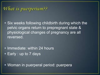
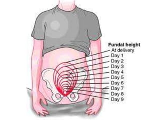
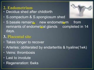
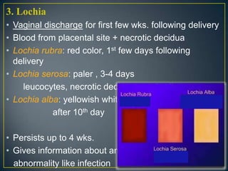
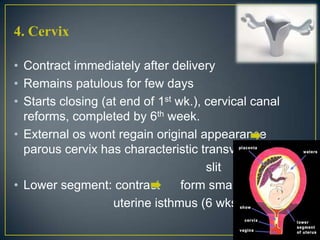
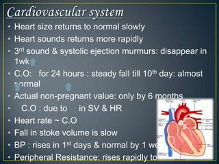
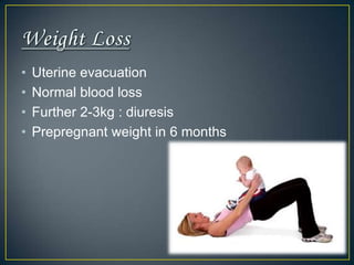
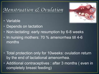
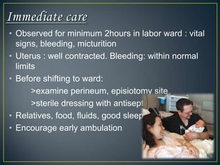
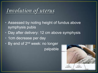
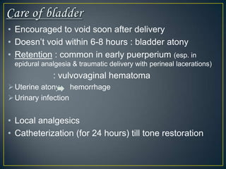
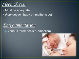
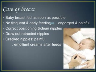
The normal puerperium period lasts 6 weeks following childbirth. During this time, the pelvic organs and body return to their pre-pregnant state. Physiological changes include the involution of the uterus, shedding of the endometrium and lochia discharge, closing of the cervix, and normalization of other organs and systems. Care during this period involves monitoring for complications, encouraging rest and early ambulation, perineal care, lactation support, nutrition, immunizations, contraception counseling, and postnatal checks to ensure full recovery.
INTRODUCTION
DEFINITION
TYPES
CAUSES
MANAGEMENT-Management of 3rd stage bleeding
Actual management
MANAGEMENT OF 3RD STAGE BLEEDING
Steps of management
1. Placental site bleeding-
To palpate the fundus and massage the uterus to make it hard. The massage is to be done by placing four fingers behind the uterus and thumb in front.
To start crystalloid solution (NS or RL) with oxytocin (1L with 20 units) at 60 drops per minute and to arrange for blood transfusion if necessary.
Oxytocin 10 unit IM or methergine 0.2 mg is given intravenously.
To catheterize the bladder.
To give antibiotics (Ampicillin 2gm and Metronidazole 500mg IV)
2. Management of traumatic bleed
The uterovaginal canal is to be explored under general anesthesia after the placenta is expelled and haemostatic sutures are placed on the offending sites.
STEPS OF MANUAL REMOVAL OF PLACENTA
The patient is placed in lithotomy position. With all aseptic measures, the bladder is catheterized.
One hand is introduced into the uterus in cone shaped manner following the cord. While introducing the hand, the labia are separated by the fingers at the other hand.
Counter pressure on the uterine fundus is applied by the hand placed over the abdomens. The abdominal hand should steady the fundus and guide the movement of the fingers inside the uterine cavity till the placenta is completely separated.
it contains a presentation on injuries that occur during baby birth
summary:
Maternal injuries following childbirth process are quite common.
VULVA
PERINEUM
RISK FACTORS FOR THIRD DEGREE PERINEL TEAR
REPAIR OF COMPLETE PERINEAL TEAR
VAGINA
CERVIX
PELVIC HEMATOMA
DIAGNOSIS OF RUPTURE UTERUS
Definition-
The destructive operations are designed to diminish the bulk of the fetus so as to facilitate easy delivery through the birth canal
types
Craniotomy
Eviceration
Decapitation
Cleidotomy
CRANIOTOMY
Definition
It is an operation to make a perforation on the fetal head to evacuate the contents followed by extraction of the fetus
DECAPITATION
Definition
It is a destructive operation whereby the fetal head is severed from the trunk and the delivery is completed with the extraction of the trunk and that of the decapitated head per vaginam
CLEIDOTOMY
Definition
The operation consist of reduction in the bulk of the shoulder girdle by division of one or both the clavicles
Indications
Dead fetus with shoulder dystocia
Procedure
The clavicles are divided by the embryotomy scissors or long straight scissors introduced under the guidance of left two fingers placed inside the vagina
Puerperium is the period following childbirth during which the body tissues, specially the pelvic organs revert back approximately to the pre-pregnant state both anatomically and physiologically. puerperium begins as soon as the placenta is expelled and lasts for approximately 6 weeks when the uterus becomes regressed almost to the non-pregnant size.
Intrapartum Care was developed for doctors and advanced midwives who care for women who deliver in district hospitals. It contains theory chapters and skills workshops adapted from the labour chapters of Maternal Care. Monitoring the mother, fetus, and progress of labour, the second and third stages of labour, managing pain, the puerperium and family planning
Intrapartum Care: Monitoring and management of the first stage of labourSaide OER Africa
Intrapartum Care was developed for doctors and advanced midwives who care for women who deliver in district hospitals. It contains theory chapters and skills workshops adapted from the labour chapters of Maternal Care. monitoring the mother, fetus, and progress of labour, the second and third stages of labour, managing pain, the puerperium and family planning
The puerperium is the period of time following childbirth, during which a woman's body returns to its pre-pregnancy state. This period typically lasts around 6-8 weeks, and during this time, the woman may experience physical and emotional changes. Lactation is the process of producing and secreting milk from the mammary glands, and it typically begins during the puerperium. The hormones released during pregnancy, specifically, prolactin and oxytocin, help to stimulate lactation and the production of milk. While lactation is a natural process, it can be challenging for some women and may require support and guidance.
what is the Puerperium and Postpartum Period
what's the normal?
what's the abnormal Puerperium?
and what are the most common complications and how to manage it?
Define the puerperium.
List the physical changes which occur during the puerperium.
Manage the normal puerperium.
Assess a patient at the 6-week postnatal visit.
Diagnose and manage the various causes of puerperal pyrexia.
Recognise the puerperal psychiatric disorders.
Diagnose and manage secondary postpartum haemorrhage.
Teach the patient the concept of ‘the mother as a monitor’.
3. Six weeks following childbirth during which the
pelvic organs return to prepregnant state &
physiological changes of pregnancy are all
reversed.
Immediate: within 24 hours
Early : up to 7 days
Woman in puerperal period: puerpera
5. 1. Involution of the uterus
• process by which postpartum uterus returns
to prepregnant state
• End of 3rd stage of labor: fundus just below
umbilicus
• By 2 weeks: descends into abdomen not palpable
through abdomen
• Weight: 1000g (immediately after delivery) 500g
by 1 wk. 300g by end of 2nd wk.
• Returns to prepregnant state 4 wks. after delivery
• Achieved by decrease in size of muscle fibers
• by autolysis of cytoplasm by proteolytic enzymes
6.
7. • After pains : infrequent, spasmodic pain in
lower abdomen for up to 3-4 days : common
due to uterine contraction
: during breast feeding (oxytocin)
Due to blood clots uterus contract
attempt to remove clot
More in primipara
If severe: antispasmodics
8. 2. Endometrium
• Decidua shed after childbirth
• S.compactum & S.spongiosum shed
• S.basale remains new endometrium from
remnants of endometrial glands completed in 14
days.
3. Placental site
• Takes longer to recover
• Arteries: obliterated by endarteritis & hyaline(1wk)
• Veins: thrombosis
• Last to involute
• Regeneration: 6wks
9. 3. Lochia
• Vaginal discharge for first few wks. following delivery
• Blood from placental site + necrotic decidua
• Lochia rubra: red color, 1st few days following
delivery
• Lochia serosa: paler , 3-4 days
leucocytes, necrotic decidua + red cells
• Lochia alba: yellowish white,
after 10th day
• Persists up to 4 wks.
• Gives information about any
abnormality like infection
10. 4. Cervix
• Contract immediately after delivery
• Remains patulous for few days
• Starts closing (at end of 1st wk.), cervical canal
reforms, completed by 6th week.
• External os wont regain original appearance
parous cervix has characteristic transverse
slit
• Lower segment: contract form small
uterine isthmus (6 wks.)
11. Other Pelvic Organs
Vagina: smooth & stretchable for first few days
size reduced & rugose within 4 wks.
Hymen: represented by caruncle myrtiformes
(small tags of tissue)
Relaxation of vaginal outlet & pelvic floor
12. Urinary system
Bladder: sustains trauma mucosal edema
& submucosal hemorrhages disappear in
few days
Prolonged& difficult delivery injury to bladder
innervation insensitive bladder capacity &
insensitive to intravesical fluid pressure
Over distension
Incomplete emptying
Residual urine / retention
Exaggerated in epidural & spinal anesthesia
After 1st week, recedes during the following weeks
13. Dilated ureters & pevicalyceal system:
normal in 8 wks
Renal plasma flow, GFR & creatinine clearance :
normal by end of puerperium
Increased extracellular water( in normal pregnancy)
becomes normal with postpartum diuresis
(2nd-5th days)
Urea & creatinine elimination : remain high for 2
wks
14. Delivery intestinal paresis constipation
Pain in perineum also contributes (episiotomy &
lacerations)
Early ambulation, high fiber diet, increased fluid
intake
15. • Rapid fall of plasma volume in 1st day
• Slowly to non-pregnant volume: end of 1st week.
• Red cell mass : normal within 24hours
• Hb concentration: minimum on 4th day & normal by
6 weeks
• ESR: in 1st week & normal by 4 weeks.
• Increased leukocytosis (during & after labor) :
subsides after 1 week
• Platelets: in no. with adhesiveness
• Fibrinogen: in 1st week.
• Coagulation factors: increased & persists in
16. • Heart size returns to normal slowly
• Heart sounds returns more rapidly
• 3rd sound & systolic ejection murmurs: disappear in
1wk
• C.O: for 24 hours : steady fall till 10th day: almost
normal
• Actual non-pregnant value: only by 6 months
• C.O : due to in SV & HR
• Heart rate ~ C.O
• Fall in stoke volume is slow
• BP : rises in 1st days & normal by 1 week
• Peripheral Resistance: rises rapidly to
17. • Uterine evacuation
• Normal blood loss
• Further 2-3kg : diuresis
• Prepregnant weight in 6 months
18. • Variable
• Depends on lactation
• Non-lactating: early resumption by 6-8 weeks
• In nursing mothers: 70 % amenorrhea till 4-6
months
• Total protection only for 10weeks: ovulation return
by the end of lactational amenorrhea.
• Additional contraceptives : after 3 months ( even in
completely breast feeding)
20. • Observed for minimum 2hours in labor ward : vital
signs, bleeding, micturition
• Uterus : well contracted. Bleeding: within normal
limits
• Before shifting to ward:
>examine perineum, episiotomy site
>sterile dressing with antiseptic
• Relatives, food, fluids, good sleep
• Encourage early ambulation
21. • Assessed by noting height of fundus above
symphysis pubis
• Day after delivery: 12 cm above symphysis
• 1cm decrease per day
• By end of 2nd week: no longer
palpable
22. • Encouraged to void soon after delivery
• Doesn’t void within 6-8 hours : bladder atony
• Retention : common in early puerperium (esp. in
epidural analgesia & traumatic delivery with perineal lacerations)
: vulvovaginal hematoma
Uterine atony hemorrhage
Urinary infection
• Local analgesics
• Catheterization (for 24 hours) till tone restoration
24. • Immediately start normal diet
• Plenty of fluids & milk daily : lactation
• Calorie & protein intake
• Additional daily requirement: 500kcal & 25g
proteins
• Fresh fruits & green leafy veg : vitamins
• Iron: continued for 3 months / whole lactating
period
25. • Regular antiseptic cleaning of episiotomy wound
• Wash with warm water & use sterile pads
• Analgesics
• Severe pain: hematoma
• Infection : antibiotics
• Episiotomy wound: healed by 3 weeks
26. • Must be adequate,
• Rooming in : baby on mother’s cot
Early ambulation
• Venous thrombosis & embolism
27. • Baby breast fed as soon as possible
• No frequent & early feeding engorged & painful
• Correct positioning &clean nipples
• Draw out retracted nipples
• Cracked nipples: painful
: emollient creams after feeds
28. • Episiotomy & perineal lacerations, breast
engorgement & after pains ( uterine contraction )
• Caesarean section: pain at incision site, post spinal
headache
• Analgesics
29. • Common in early puerperium : postpartum blues
• Reassurance & support
Hospital stay
Vaginal delivery: after 48hrs
Caesarian section : 4-5 days
31. 3. Medications
Adviced to avoid medications as far as possible
Take only on medical advice
4. Infant advice
Adequate follow up
Rooming in
BCG vaccine prior to going home
Immunisation schedule
Advice mother to feed on demand
32. 4. Postnatal exercises
Early in puerperium
Move limbs, deep breathing
exercises, abdominal muscle tightening
exercises everyday
Repeated 3-4 times a day
Perineal muscle exercises: improve vaginal muscle
tone, prevent vaginal laxity & stress incontinence
Back muscle toning
35. • Scheduled at 6 weeks
• Maternal problems discussed
• Breast-feeding
• Lochia & menses
• Clinical examination, abdomen, breasts
• Local examination : episiotomy & discharge
• Pelvic examination
• PIH, GDM
• Any medical problems
• Advice on resuming job, coitus,
other activities
36. • Breast feeding alone wont suffice as contraception
• Various options given, partner included
• IUCD inserted if acceptable
• Other options which wont reduce breast milk amount
Progesterone only pill
Injectable progestogens
(depot medroxy progesterone acetate)
37. Inform about possible side-effects
Should avoid estrogen-pregesterone
combination: affect quantity & quality of milk
Permanent sterilizations: postpartum/ interval
sterilization/
38. • Seen by pediatrician
• Checked for any problems
• Weight , feeding problems
• Reinforce immunization schedule
39. Restoration of maternal health to prepregnant state
Promotion of breast feeding
Correction of any problems arising due to delivery
Advice on baby care & immunization
Contraceptive advice