Downloaded 166 times

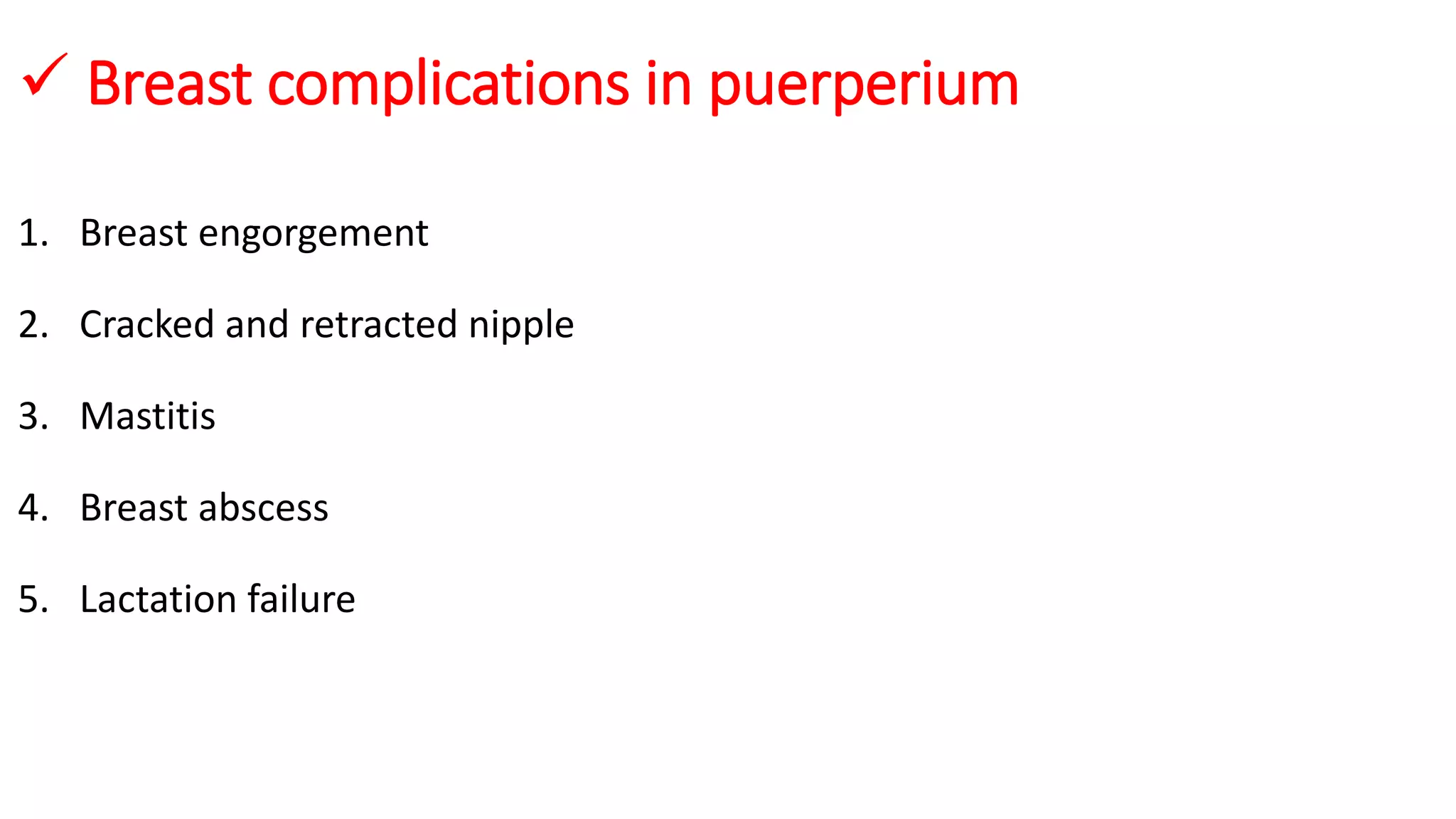
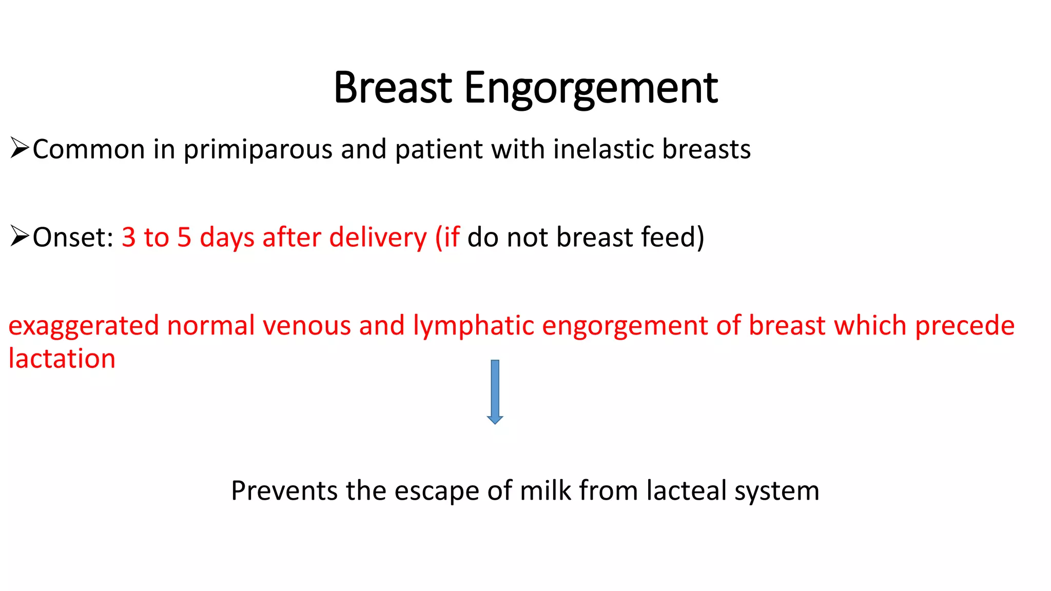


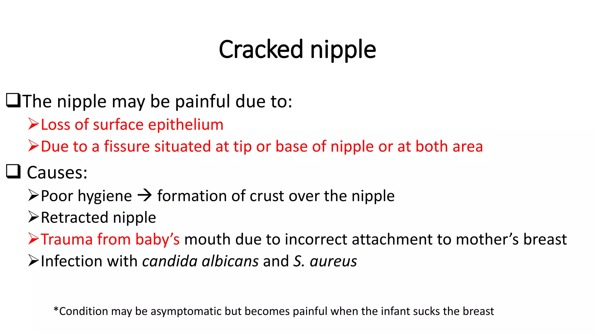


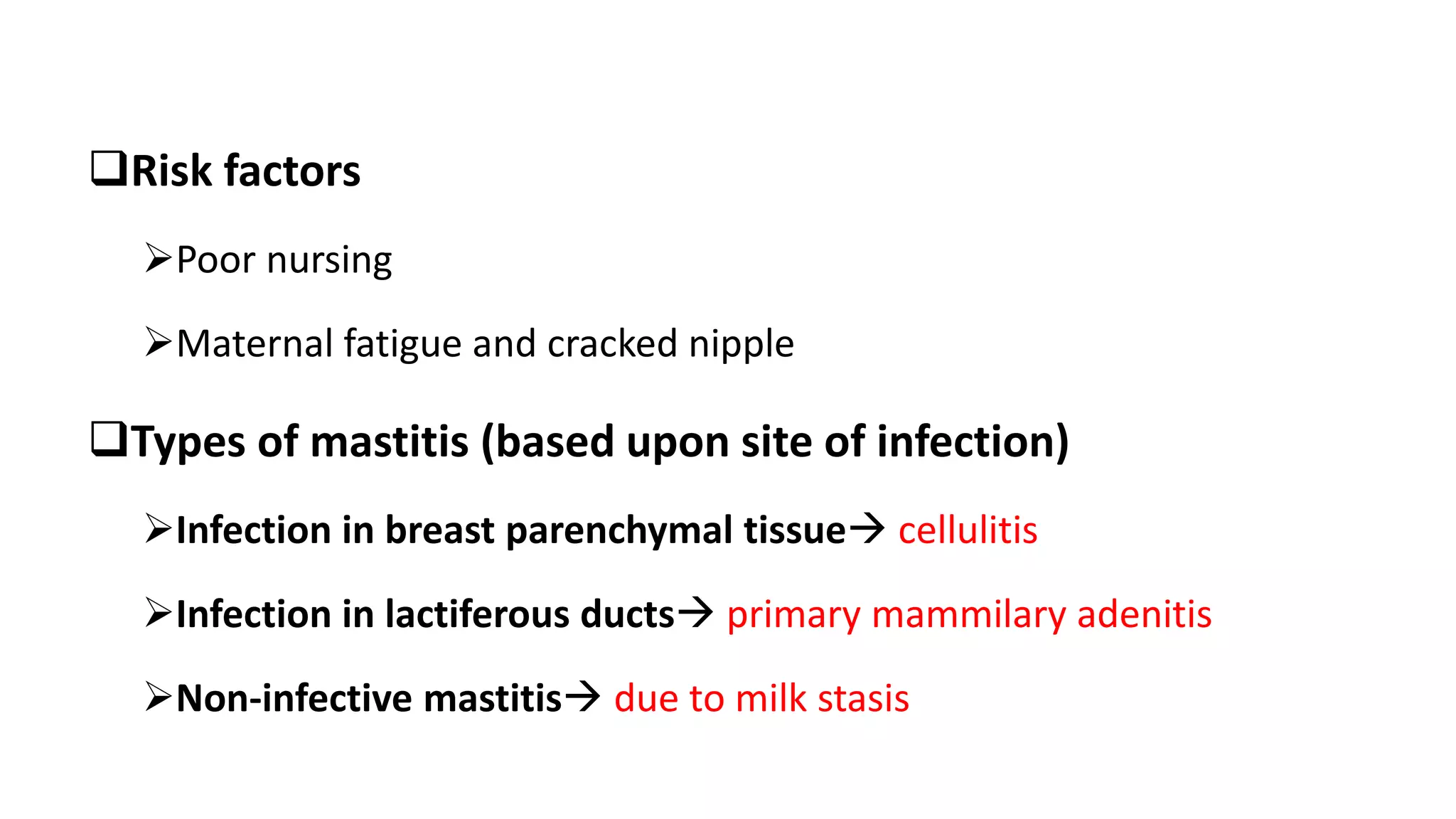


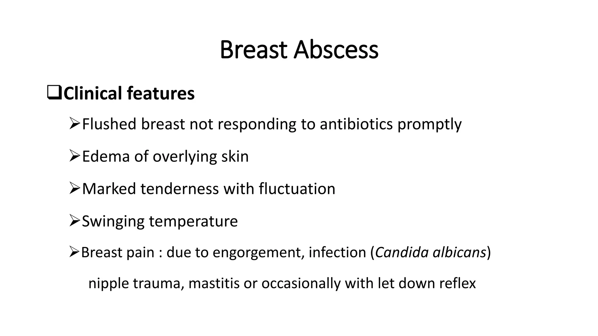
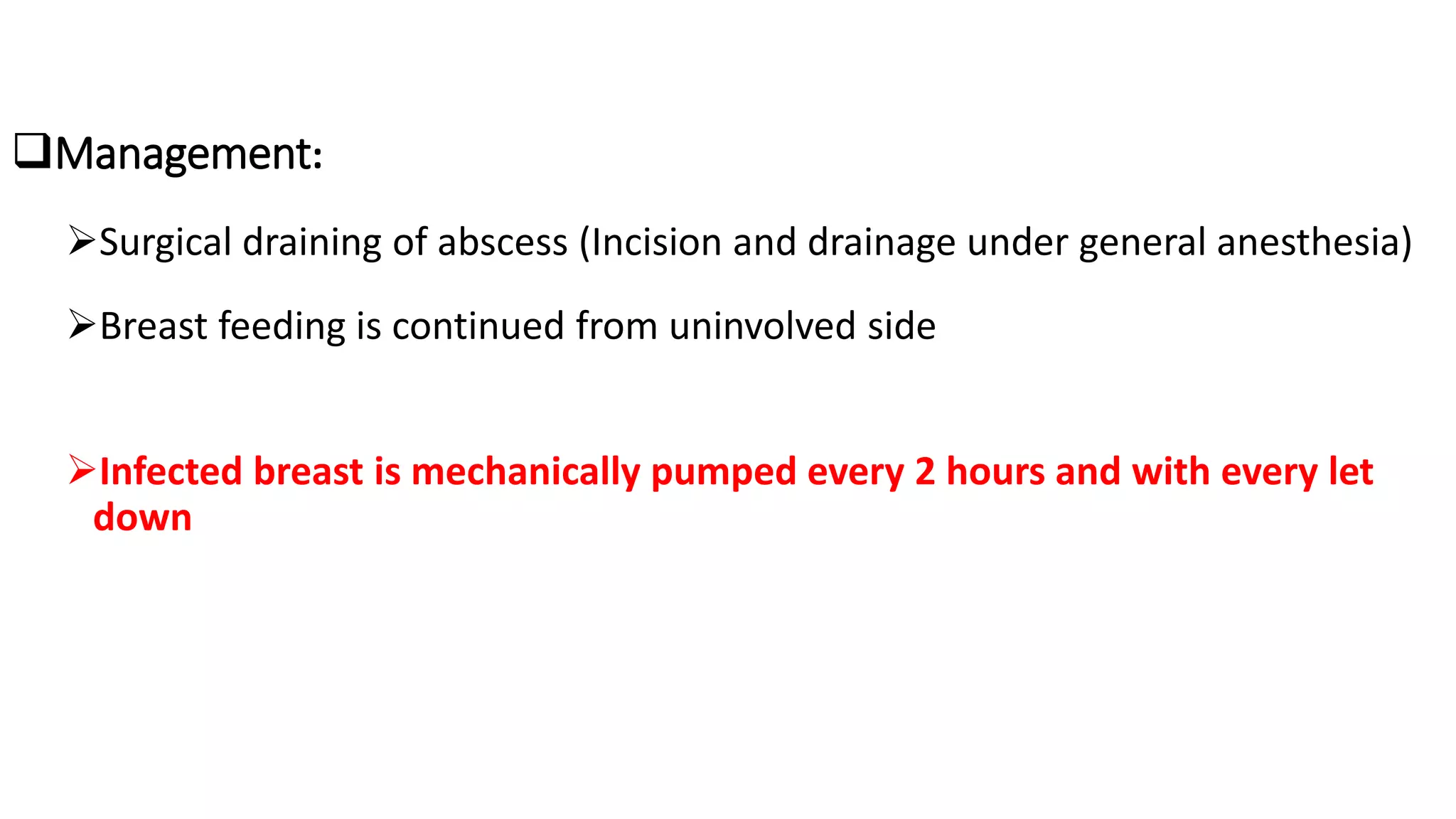


The document discusses various breast complications that may arise after delivery, including breast engorgement, cracked nipples, mastitis, breast abscess, and lactation failure, along with their prevention and management strategies. Symptoms, treatment options, and recommendations for maintaining breastfeeding are also outlined. Emphasis is placed on proper breastfeeding techniques, hygiene, and timely medical intervention for complications.