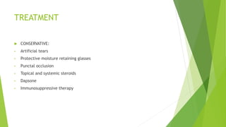
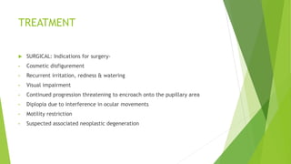
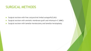
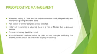
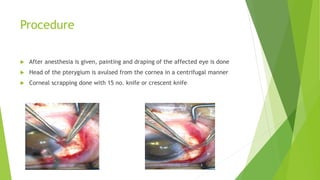
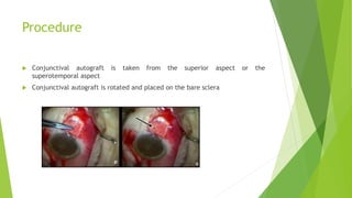
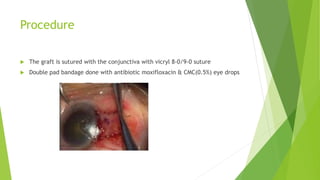
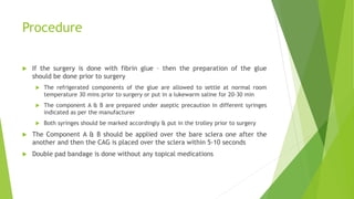
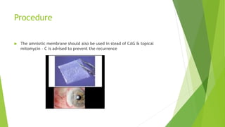
PTERYGIUM
Dr Jeel Garala
Ophthalmology Dept
CU Shah Medical college
DEFINITION
 PTERYGIUM is a triangular encroachment of the vascularized granulation
tissue covered by conjunctiva in the interpalpebral area.
 It is the degenerative condition of the conjunctiva.
ETIOLOGY
 Pterygium is a response to prolonged effect of environmental factors such as:
• Exposure to sun (ultraviolet rays)
• Dry heat
• High wind
• Abundance of dust
PATHOGENESIS
 Pterygium is degenerative and hyperplastic condition of conjunctiva.
 The subconjunctival tissue undergoes elastotic degeneration and proliferates
as vascularised granulation tissue under the epithelium.
 It ultimately encroaches the cornea.
 It destroys the superficial layers of stroma and bowman’s membrane.
 Formation of dense fibrous tissue leads to development of corneal
astigmatism.
CLINICAL FEATURES
(1) DEMOGRAPHY:
• Age: Usually common in old age.
• Sex: more common in males doing outdoor work than females.
• It is usually bilateral and more common on nasal side.
CLINICAL FEATURES
(2) SYMPTOMS:
• Foreign body sensation
• Irritation
• Diminution of vision as it encroaches the pupillary region of cornea and due to
corneal astigmatism.
• Cosmetic intolerance
• Diplopia
CLINICAL FEATURES
 (3) SIGNS:
• Triangular fold of conjunctiva encroaching the cornea.
• Parts of fully developed pterygium are:
 Head- Apical part present on the cornea.
 Neck- Constricted part present in the limbal area.
 Body- Scleral part extending between the limbus and canthus.
 Cap- Semilunar whitish infiltrate just present in front of the head.
TREATMENT
 CONSERVATIVE:
• Artificial tears
• Protective moisture retaining glasses
• Punctal occlusion
• Topical and systemic steroids
• Dapsone
• Immunosuppressive therapy
TREATMENT
 SURGICAL: Indications for surgery-
• Cosmetic disfigurement
• Recurrent irritation, redness & watering
• Visual impairment
• Continued progression threatening to encroach onto the pupillary area
• Diplopia due to interference in ocular movements
• Motility restriction
• Suspected associated neoplastic degeneration
SURGICAL METHODS
 Surgical excision with free conjunctival limbal autograft(CLAU)
 Surgical excision with amniotic membrane graft and mitomycin-C (MMC)
 Surgical excision with lamellar keratectomy and lamellar keratoplasty
PREOPERATIVE MANAGEMENT
 A detailed history is taken and slit lamp examination done preoperatively and
appropriate grading should be done
 Past history of similar complain should be noted
 History of recurrence is asked as there is a risk of fibrosis due to previous
surgery
 Occupation history should be noted
 Acute inflammed condition should be ruled out and managed medically first
and the patient should be advised for surgery in follow up
PREOPERATIVE MANAGEMENT
 Blood pressure
 Routine blood investigations
 CBC
 RBS
 Urine – R & M
 HIV & HBsAg
 Antiplatelet medication should be stopped as per physician & anesthetist
advice
 Obtain an informed & written consent
Anesthesia
 The surgery can be done under local anesthesia – Peribulbar block or can also
be done under topical anesthesia if the patient is cooperative
 Anesthetist should be stand by if the patient is having any cardio-respiratory
or any other systemic abnormality
Procedure
 After anesthesia is given, painting and draping of the affected eye is done
 Head of the pterygium is avulsed from the cornea in a centrifugal manner
 Corneal scrapping done with 15 no. knife or crescent knife
Procedure
 Subconjunctival Tenon’s tissue is separated
 Head of the pterygium is cut and hemostatis is achieved
Procedure
 Conjunctival autograft is taken from the superior aspect or the
superotemporal aspect
 Conjunctival autograft is rotated and placed on the bare sclera
Procedure
 The graft is sutured with the conjunctiva with vicryl 8-0/9-0 suture
 Double pad bandage done with antibiotic moxifloxacin & CMC(0.5%) eye drops
Procedure
 If the surgery is done with fibrin glue – then the preparation of the glue
should be done prior to surgery
 The refrigerated components of the glue are allowed to settle at normal room
temperature 30 mins prior to surgery or put in a lukewarm saline for 20-30 min
 The component A & B are prepared under aseptic precaution in different syringes
indicated as per the manufacturer
 Both syringes should be marked accordingly & put in the trolley prior to surgery
 The Component A & B should be applied over the bare sclera one after the
another and then the CAG is placed over the sclera within 5-10 seconds
 Double pad bandage is done without any topical medications
Procedure
 The amniotic membrane should also be used in stead of CAG & topical
mitomycin – C is advised to prevent the recurrence
Complications
 Intraoperative
 Puncture wound in the body of pterygium while dissecting the Tenon’s tissue
 Injury to the medial rectus due to over dissection of the Tenon’s tissue
 Injury to the superior rectus while taking the CAG superiorly
 Injury to the cornea while scrapping
Complications
 Postoperative
 Displacement of the graft from the original position
 Extensive subconjunctival haemorrhage due to poor handling of tissue during surgery
 Graft oedema
 Granuloma formation
 Scleritis
 Persistent epithelial defect
 Dellen
 Endophthalmitis
 Late postoperative
 Opacification over cornea
 Recurrence
Thank you
 Reference :
 Kanski’s clinical ophthalmology – 9th edition

PTERYGIUM.pptx

  • 1.
    PTERYGIUM Dr Jeel Garala OphthalmologyDept CU Shah Medical college
  • 2.
    DEFINITION  PTERYGIUM isa triangular encroachment of the vascularized granulation tissue covered by conjunctiva in the interpalpebral area.  It is the degenerative condition of the conjunctiva.
  • 3.
    ETIOLOGY  Pterygium isa response to prolonged effect of environmental factors such as: • Exposure to sun (ultraviolet rays) • Dry heat • High wind • Abundance of dust
  • 4.
    PATHOGENESIS  Pterygium isdegenerative and hyperplastic condition of conjunctiva.  The subconjunctival tissue undergoes elastotic degeneration and proliferates as vascularised granulation tissue under the epithelium.  It ultimately encroaches the cornea.  It destroys the superficial layers of stroma and bowman’s membrane.  Formation of dense fibrous tissue leads to development of corneal astigmatism.
  • 5.
    CLINICAL FEATURES (1) DEMOGRAPHY: •Age: Usually common in old age. • Sex: more common in males doing outdoor work than females. • It is usually bilateral and more common on nasal side.
  • 6.
    CLINICAL FEATURES (2) SYMPTOMS: •Foreign body sensation • Irritation • Diminution of vision as it encroaches the pupillary region of cornea and due to corneal astigmatism. • Cosmetic intolerance • Diplopia
  • 7.
    CLINICAL FEATURES  (3)SIGNS: • Triangular fold of conjunctiva encroaching the cornea. • Parts of fully developed pterygium are:  Head- Apical part present on the cornea.  Neck- Constricted part present in the limbal area.  Body- Scleral part extending between the limbus and canthus.  Cap- Semilunar whitish infiltrate just present in front of the head.
  • 8.
    TREATMENT  CONSERVATIVE: • Artificialtears • Protective moisture retaining glasses • Punctal occlusion • Topical and systemic steroids • Dapsone • Immunosuppressive therapy
  • 9.
    TREATMENT  SURGICAL: Indicationsfor surgery- • Cosmetic disfigurement • Recurrent irritation, redness & watering • Visual impairment • Continued progression threatening to encroach onto the pupillary area • Diplopia due to interference in ocular movements • Motility restriction • Suspected associated neoplastic degeneration
  • 10.
    SURGICAL METHODS  Surgicalexcision with free conjunctival limbal autograft(CLAU)  Surgical excision with amniotic membrane graft and mitomycin-C (MMC)  Surgical excision with lamellar keratectomy and lamellar keratoplasty
  • 11.
    PREOPERATIVE MANAGEMENT  Adetailed history is taken and slit lamp examination done preoperatively and appropriate grading should be done  Past history of similar complain should be noted  History of recurrence is asked as there is a risk of fibrosis due to previous surgery  Occupation history should be noted  Acute inflammed condition should be ruled out and managed medically first and the patient should be advised for surgery in follow up
  • 12.
    PREOPERATIVE MANAGEMENT  Bloodpressure  Routine blood investigations  CBC  RBS  Urine – R & M  HIV & HBsAg  Antiplatelet medication should be stopped as per physician & anesthetist advice  Obtain an informed & written consent
  • 13.
    Anesthesia  The surgerycan be done under local anesthesia – Peribulbar block or can also be done under topical anesthesia if the patient is cooperative  Anesthetist should be stand by if the patient is having any cardio-respiratory or any other systemic abnormality
  • 14.
    Procedure  After anesthesiais given, painting and draping of the affected eye is done  Head of the pterygium is avulsed from the cornea in a centrifugal manner  Corneal scrapping done with 15 no. knife or crescent knife
  • 15.
    Procedure  Subconjunctival Tenon’stissue is separated  Head of the pterygium is cut and hemostatis is achieved
  • 16.
    Procedure  Conjunctival autograftis taken from the superior aspect or the superotemporal aspect  Conjunctival autograft is rotated and placed on the bare sclera
  • 17.
    Procedure  The graftis sutured with the conjunctiva with vicryl 8-0/9-0 suture  Double pad bandage done with antibiotic moxifloxacin & CMC(0.5%) eye drops
  • 18.
    Procedure  If thesurgery is done with fibrin glue – then the preparation of the glue should be done prior to surgery  The refrigerated components of the glue are allowed to settle at normal room temperature 30 mins prior to surgery or put in a lukewarm saline for 20-30 min  The component A & B are prepared under aseptic precaution in different syringes indicated as per the manufacturer  Both syringes should be marked accordingly & put in the trolley prior to surgery  The Component A & B should be applied over the bare sclera one after the another and then the CAG is placed over the sclera within 5-10 seconds  Double pad bandage is done without any topical medications
  • 19.
    Procedure  The amnioticmembrane should also be used in stead of CAG & topical mitomycin – C is advised to prevent the recurrence
  • 20.
    Complications  Intraoperative  Puncturewound in the body of pterygium while dissecting the Tenon’s tissue  Injury to the medial rectus due to over dissection of the Tenon’s tissue  Injury to the superior rectus while taking the CAG superiorly  Injury to the cornea while scrapping
  • 21.
    Complications  Postoperative  Displacementof the graft from the original position  Extensive subconjunctival haemorrhage due to poor handling of tissue during surgery  Graft oedema  Granuloma formation  Scleritis  Persistent epithelial defect  Dellen  Endophthalmitis  Late postoperative  Opacification over cornea  Recurrence
  • 22.
    Thank you  Reference:  Kanski’s clinical ophthalmology – 9th edition