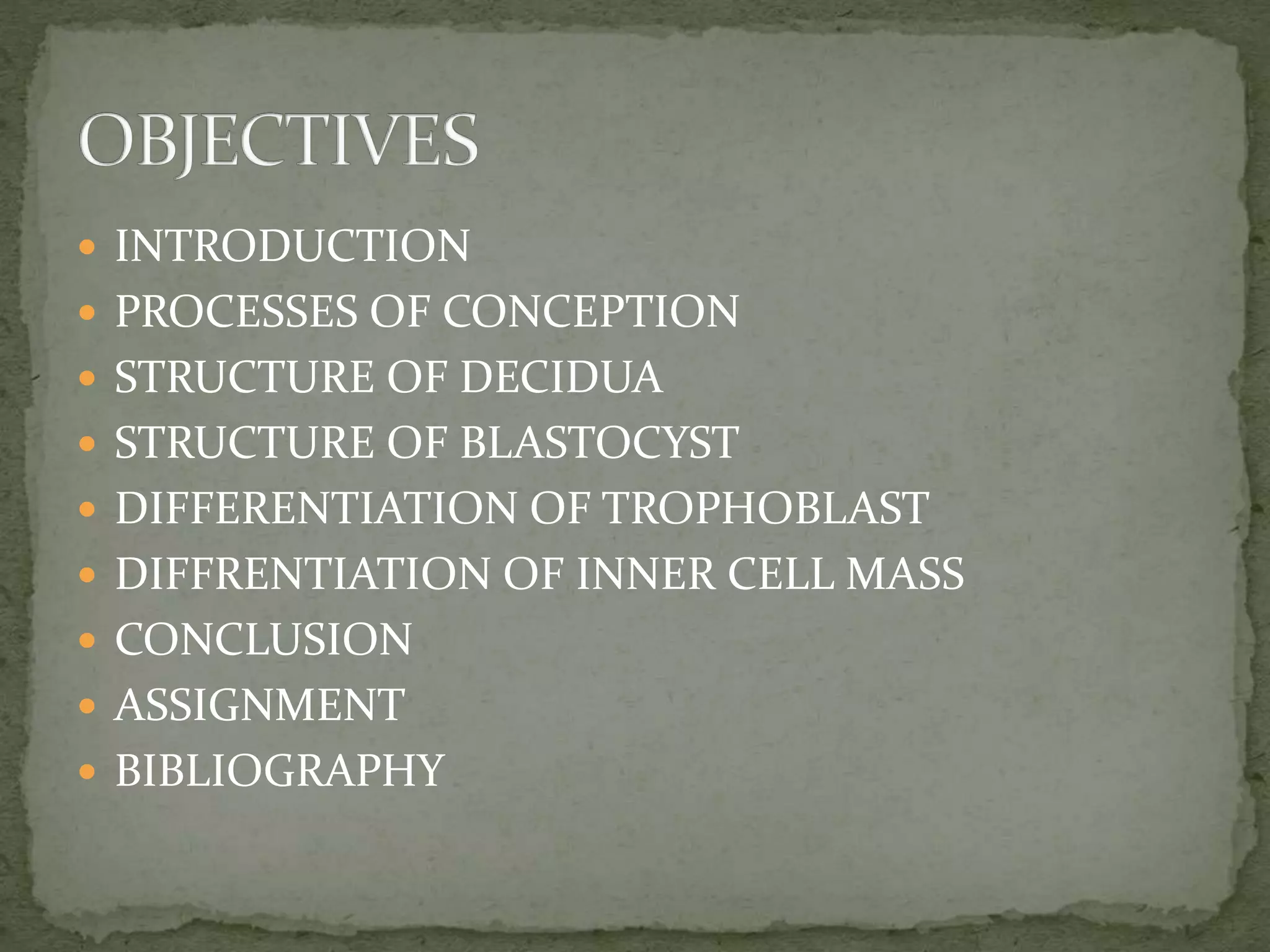
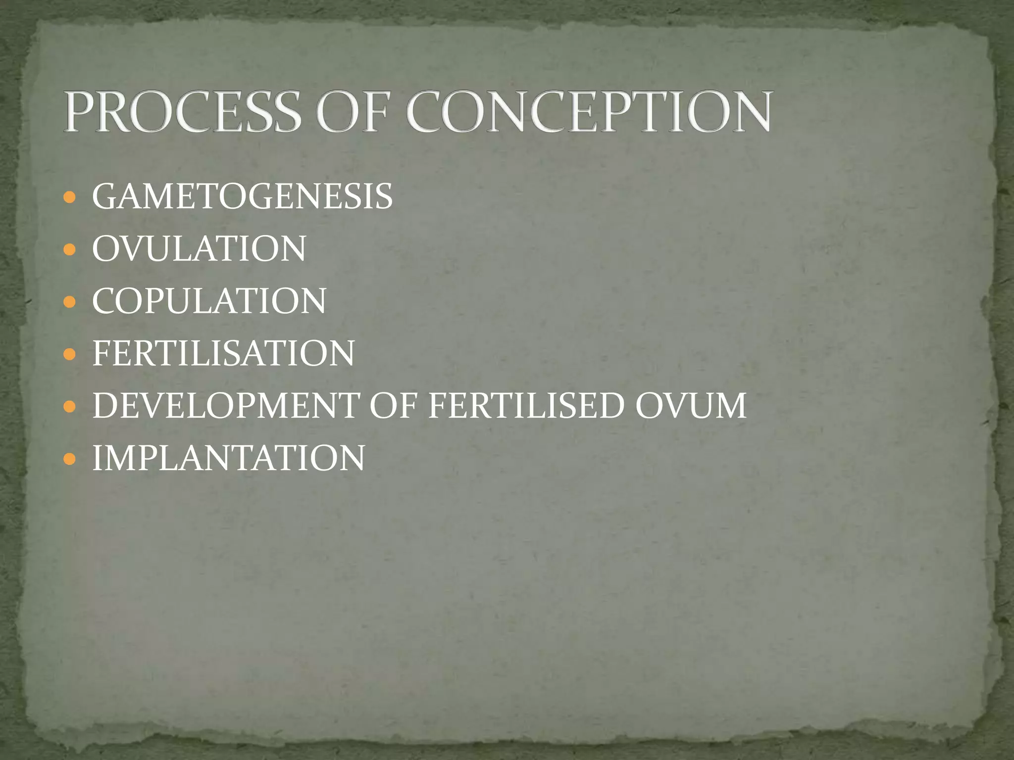
The document discusses the processes involved in conception, including gametogenesis, ovulation, copulation, fertilization, and implantation. It describes the formation of male and female gametes, ovulation and release of the ovum, fertilization occurring in the fallopian tubes, and cleavage and blastocyst formation. It then discusses implantation of the blastocyst in the uterine lining, formation of the decidua, and differentiation of the trophoblast and inner cell mass.
Presented by Srujani Swetasina Dash, this presentation discusses conception processes, structures involved, and concludes with assignment and bibliography.
Conception is defined as pregnancy, occurring when a fertilized ovum embeds in the uterus. Key processes include gametogenesis, ovulation, copulation, fertilization, and implantation.
Gametogenesis includes spermatogenesis (male gamete formation) and oogenesis (female gamete formation) within respective reproductive organs.
Ovulation involves releasing the ovum, while copulation is the sexual intercourse necessary for sperm to fertilize the ovum within specific timeframes.
Fertilization occurs at the ampulla of the Fallopian tube, where sperm penetrate the ovum, resulting in the formation of a zygote.
After fertilization, zygote undergoes cleavage formation, leading to stages such as blastomere and morula, which enter the uterine cavity.
Implantation involves the blastocyst attaching to the uterine lining, completed by the 10th to 11th day after fertilization.
Decidua comprises three layers, each with specific functions, crucial for the implantation and nutrition of early embryonic development.
Blastocyst consists of two parts: trophoblast and inner cell mass. Trophoblast differentiates to form chorionic structures essential for pregnancy.
Inner cell mass differentiates into bilaminar germ layers and later trilaminar layers, establishing foundational structures of the embryo.
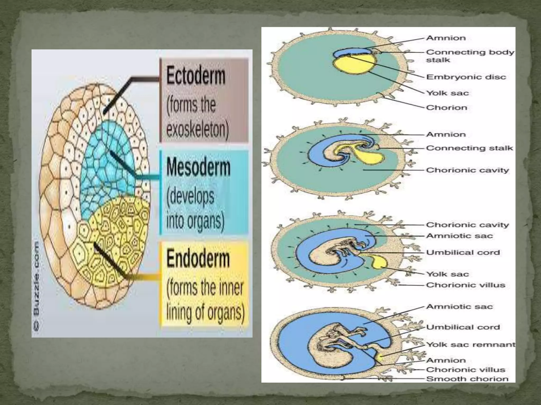
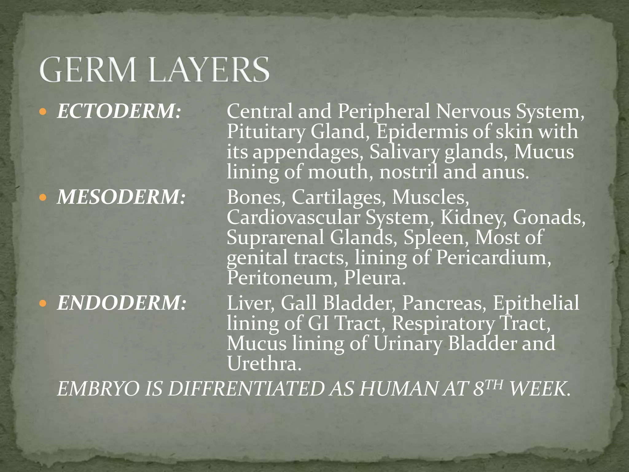
The ectoderm, mesoderm, and endoderm layers develop distinct organ systems and structures; embryo is recognized as human by the 8th week.
Conception is vital for life continuity; abnormalities in any process can lead to infertility.
Encourages preparation of notes on the processes involved in conception.
Lists textbooks and references for understanding obstetrics and midwifery related to conception.
An assessment activity designed to test knowledge on key terms and processes related to conception.
INTRODUCTION
PROCESSESOF CONCEPTION
STRUCTURE OF DECIDUA
STRUCTURE OF BLASTOCYST
DIFFERENTIATION OF TROPHOBLAST
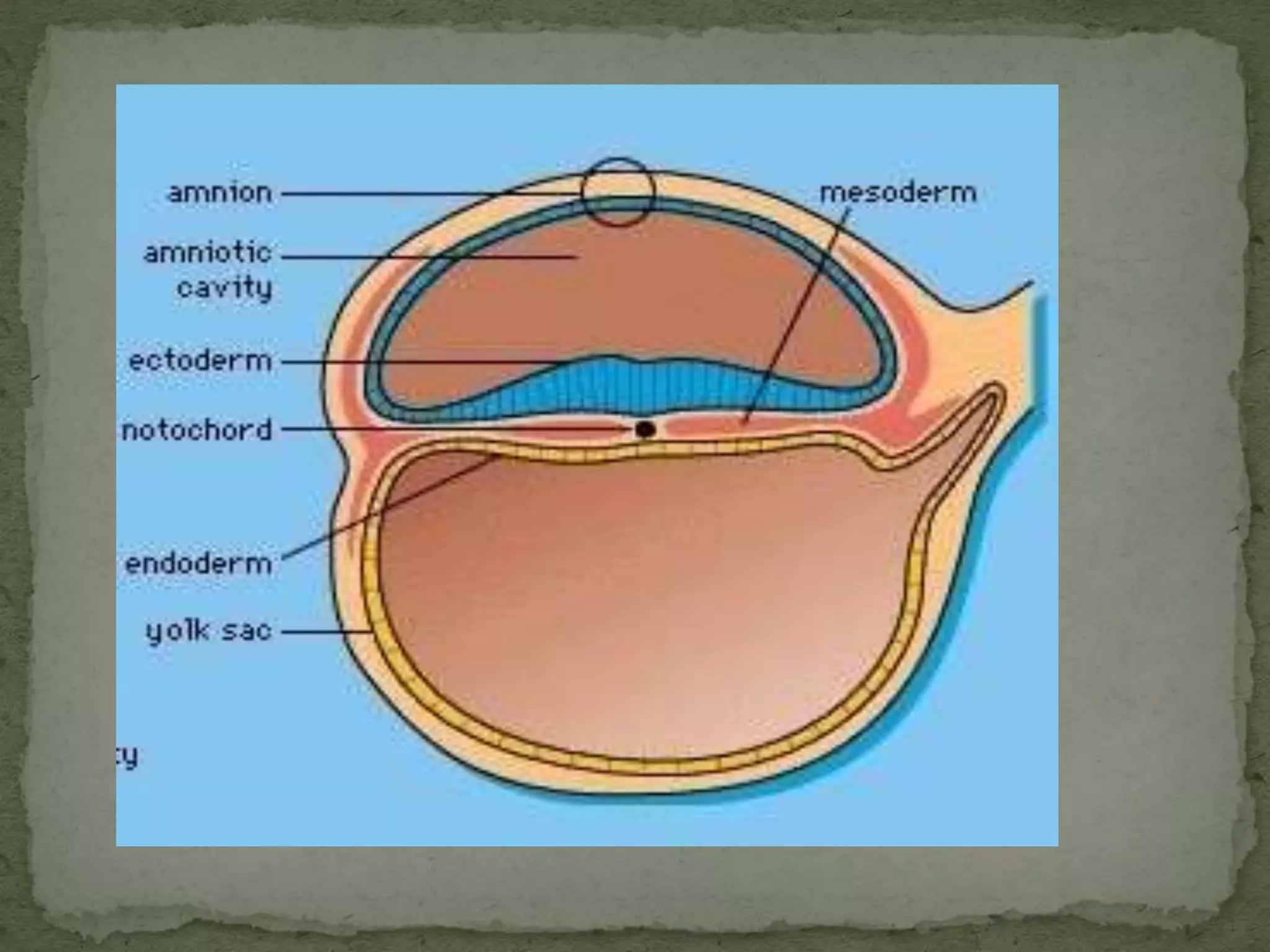
DIFFRENTIATION OF INNER CELL MASS
CONCLUSION
ASSIGNMENT
BIBLIOGRAPHY
3.
The term“conception” means to become pregnant.
Conception or pregnancy occurs when fertilized ovum
embeds in the uterus.
Numerous processes are directly or indirectly
responsible for conception.
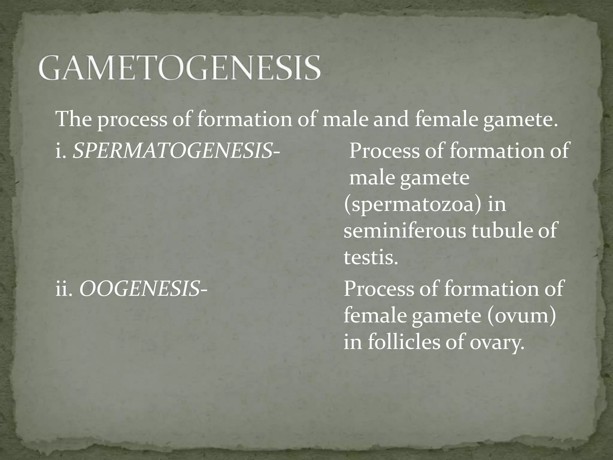
The process offormation of male and female gamete.
i. SPERMATOGENESIS- Process of formation of
male gamete
(spermatozoa) in
seminiferous tubule of
testis.
ii. OOGENESIS- Process of formation of
female gamete (ovum)
in follicles of ovary.
8.
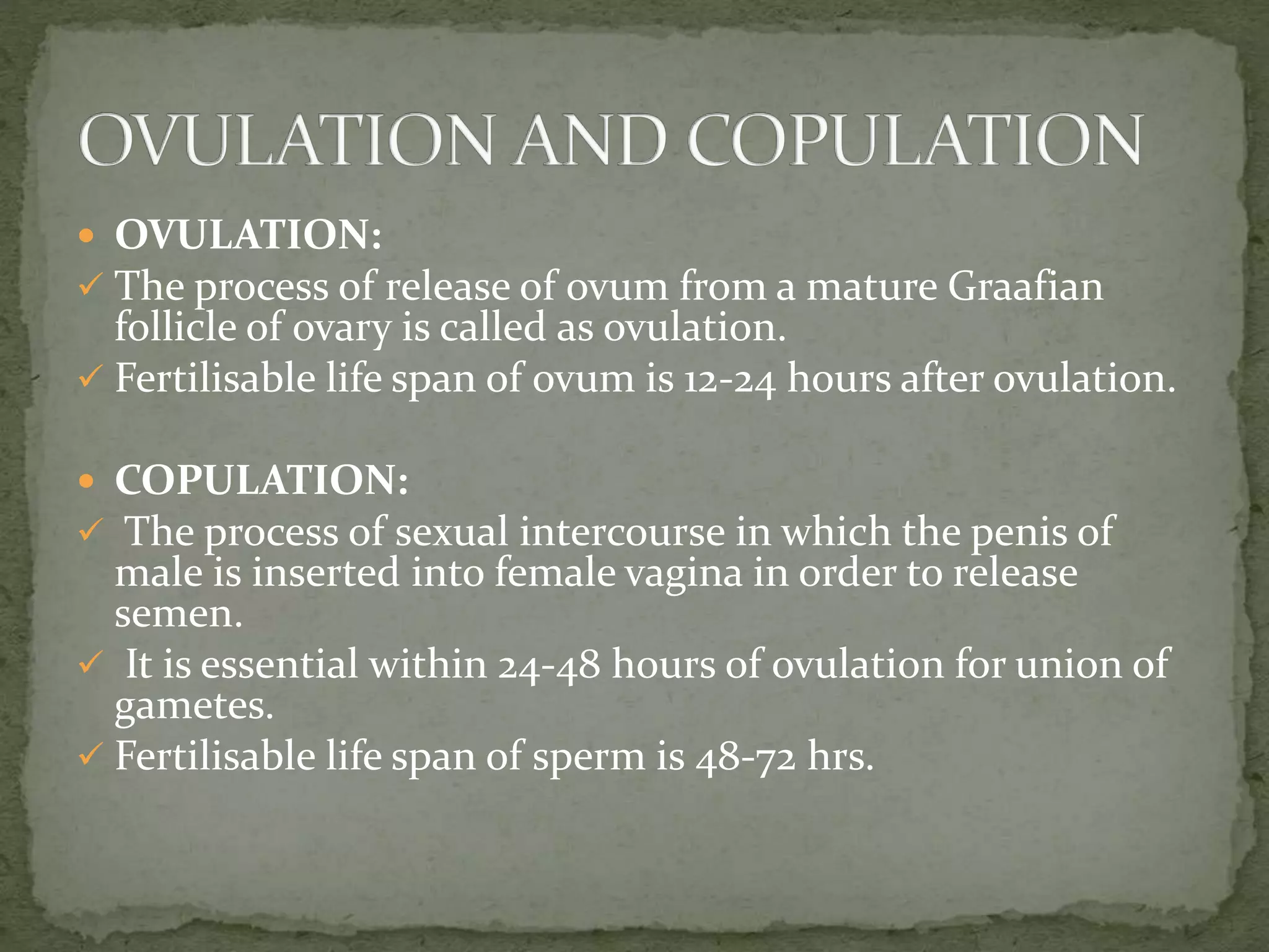
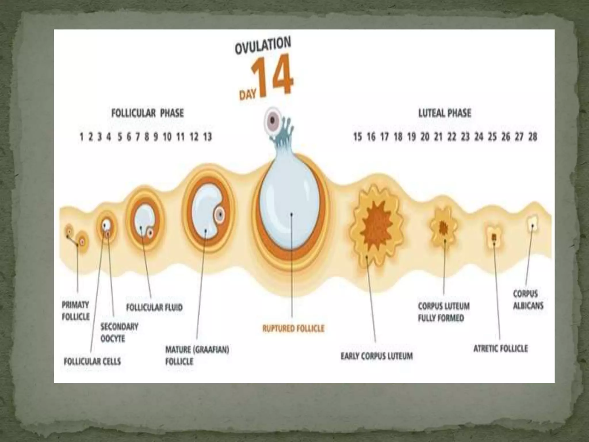
OVULATION:
Theprocess of release of ovum from a mature Graafian
follicle of ovary is called as ovulation.
Fertilisable life span of ovum is 12-24 hours after ovulation.
COPULATION:
The process of sexual intercourse in which the penis of
male is inserted into female vagina in order to release
semen.
It is essential within 24-48 hours of ovulation for union of
gametes.
Fertilisable life span of sperm is 48-72 hrs.
10.
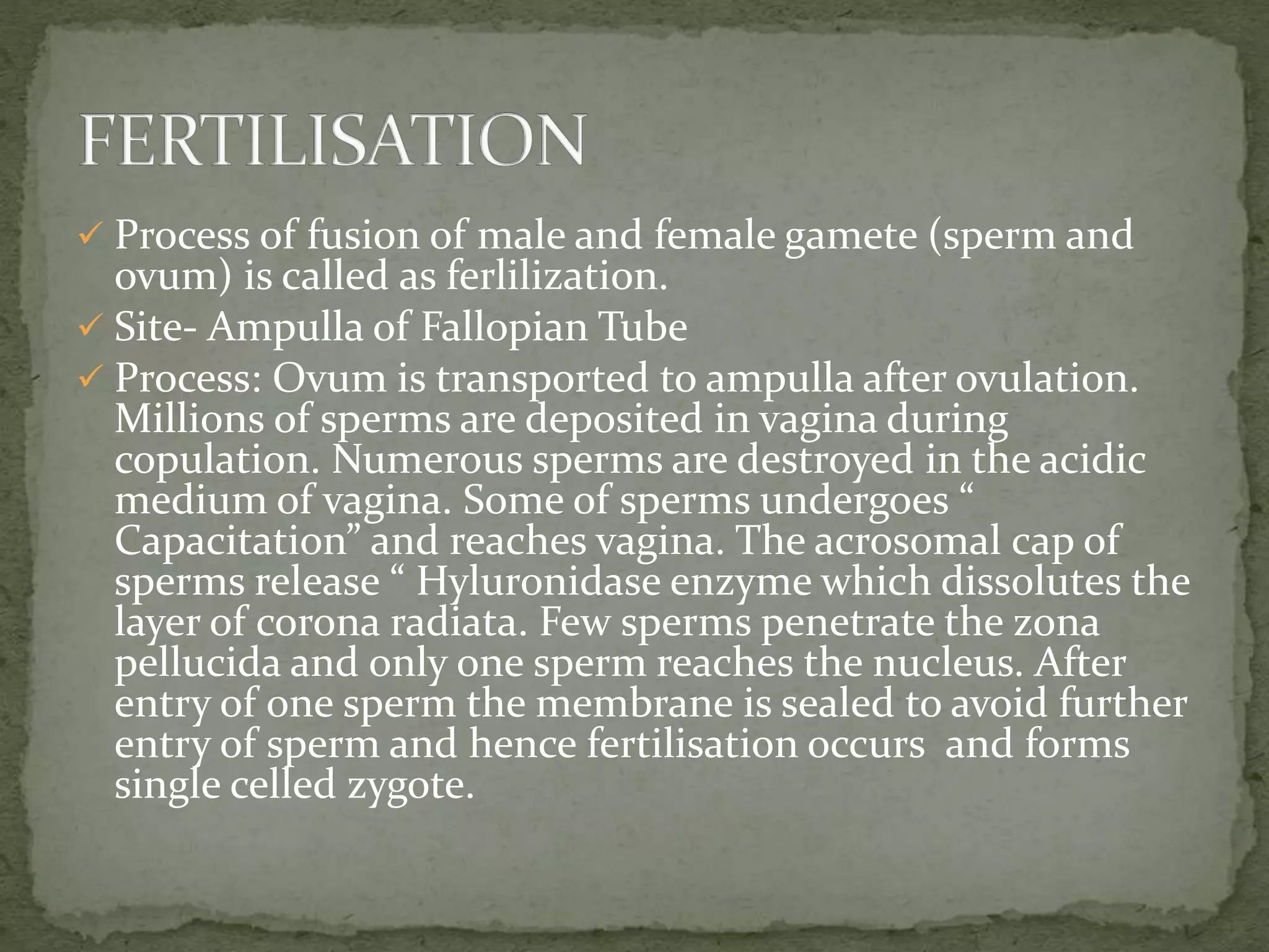
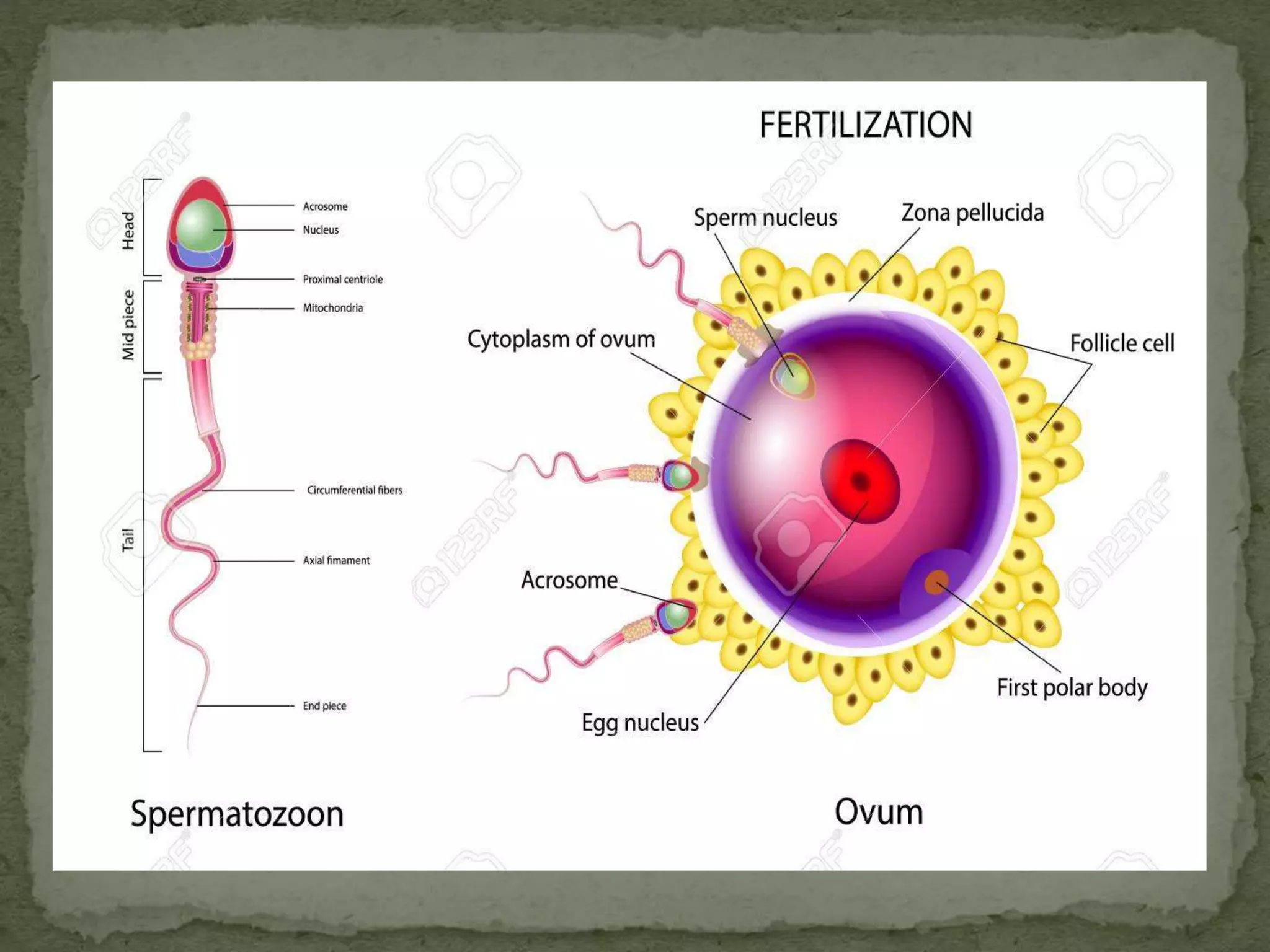
Process offusion of male and female gamete (sperm and
ovum) is called as ferlilization.
Site- Ampulla of Fallopian Tube
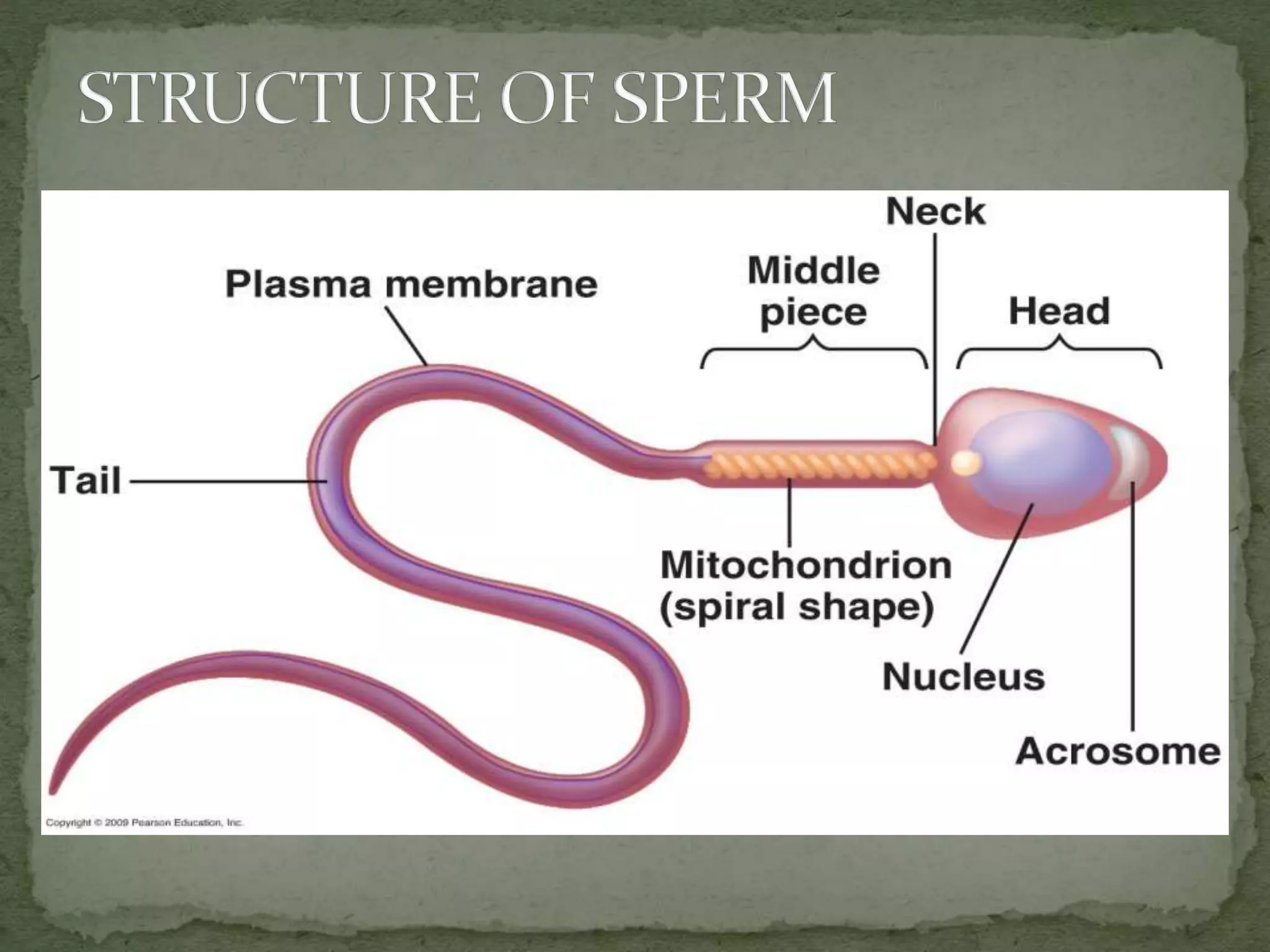
Process: Ovum is transported to ampulla after ovulation.
Millions of sperms are deposited in vagina during
copulation. Numerous sperms are destroyed in the acidic
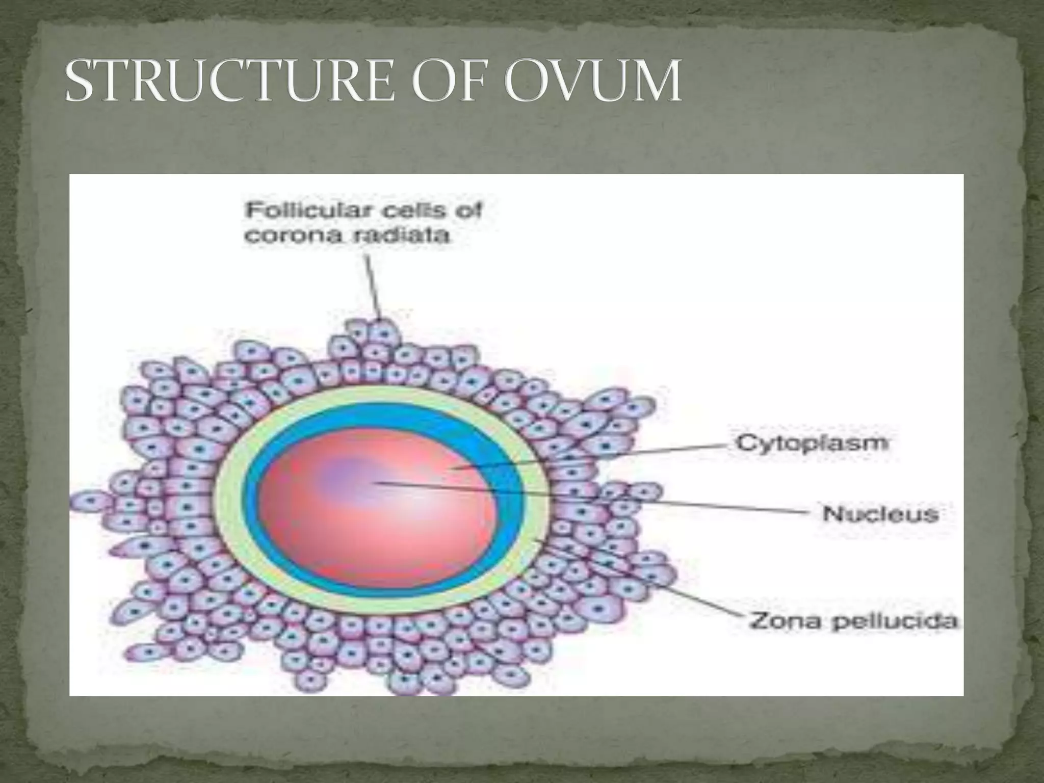
medium of vagina. Some of sperms undergoes “
Capacitation” and reaches vagina. The acrosomal cap of
sperms release “ Hyluronidase enzyme which dissolutes the
layer of corona radiata. Few sperms penetrate the zona
pellucida and only one sperm reaches the nucleus. After
entry of one sperm the membrane is sealed to avoid further
entry of sperm and hence fertilisation occurs and forms
single celled zygote.
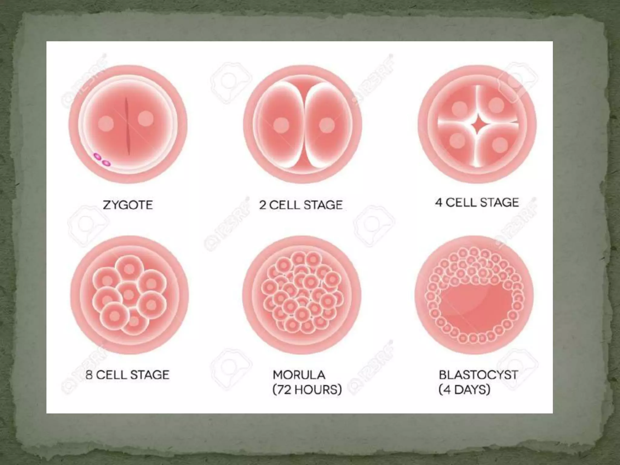
12.
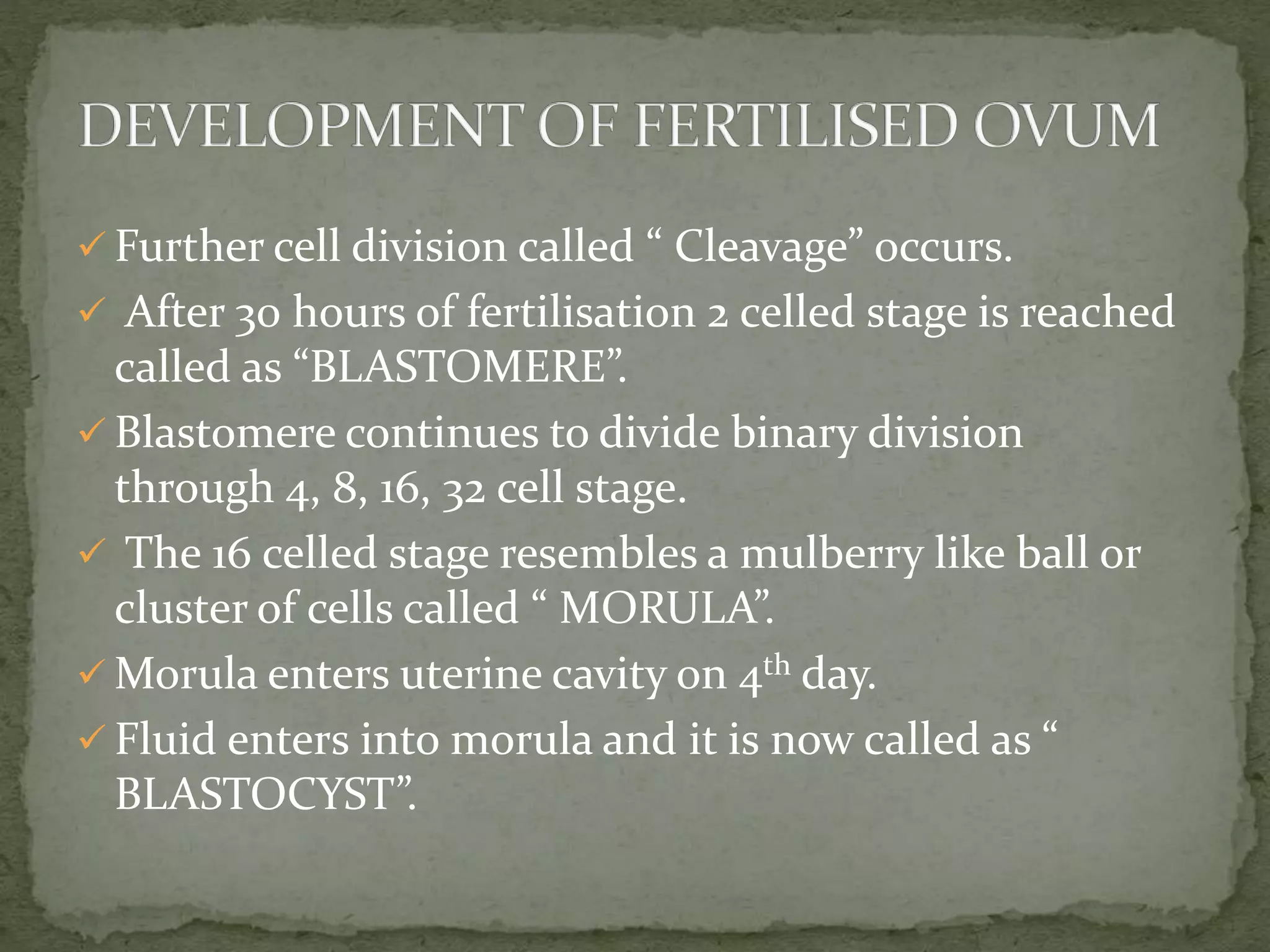
Further celldivision called “ Cleavage” occurs.
After 30 hours of fertilisation 2 celled stage is reached
called as “BLASTOMERE”.
Blastomere continues to divide binary division
through 4, 8, 16, 32 cell stage.
The 16 celled stage resembles a mulberry like ball or
cluster of cells called “ MORULA”.
Morula enters uterine cavity on 4th day.
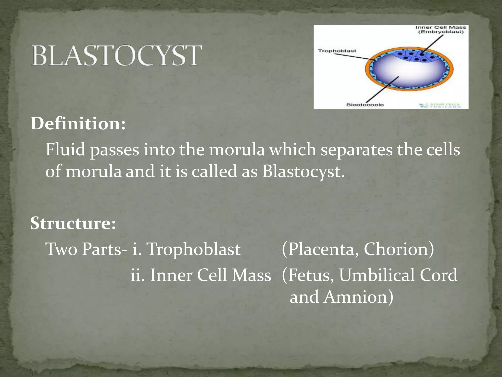
Fluid enters into morula and it is now called as “
BLASTOCYST”.
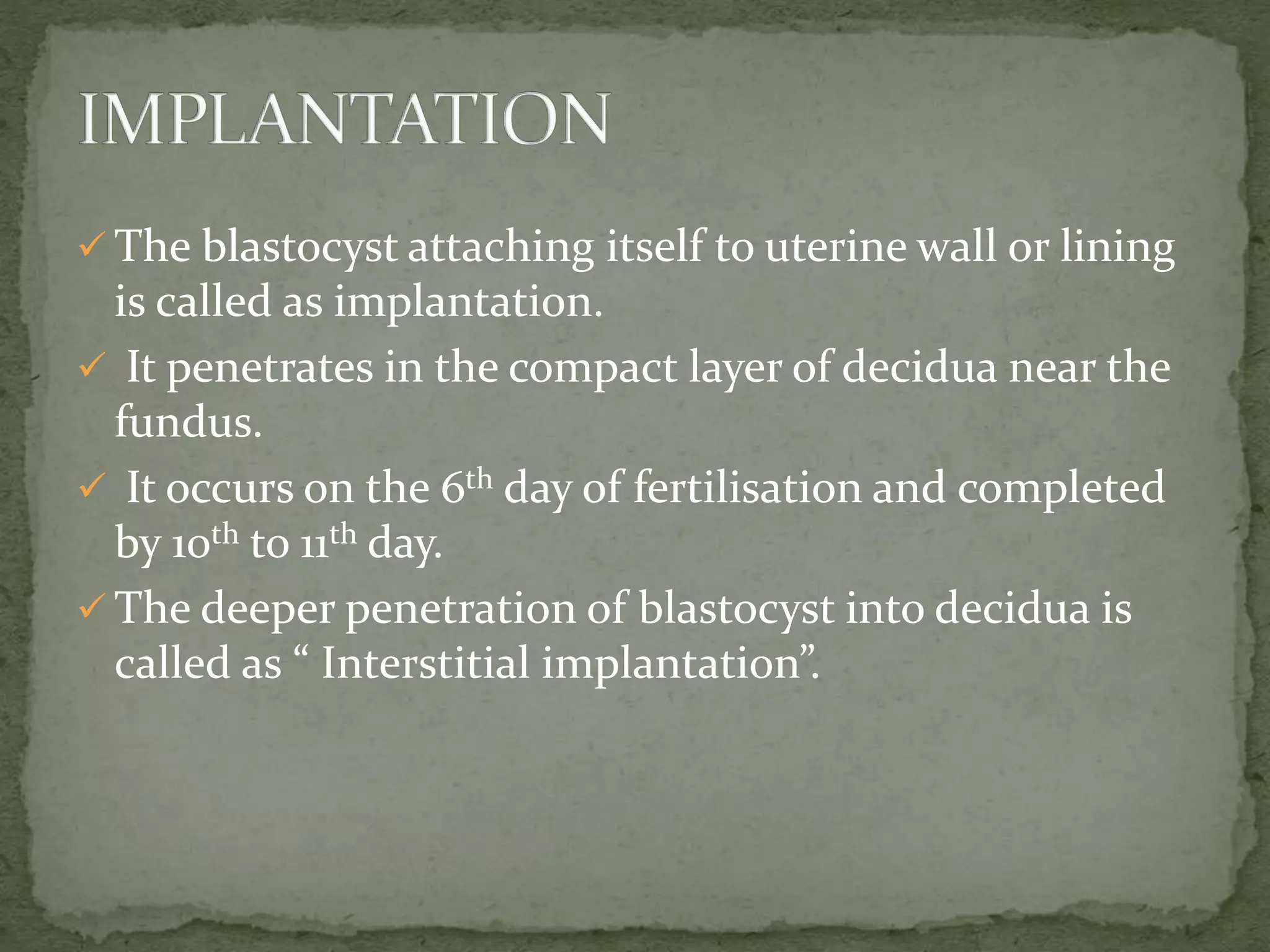
14.
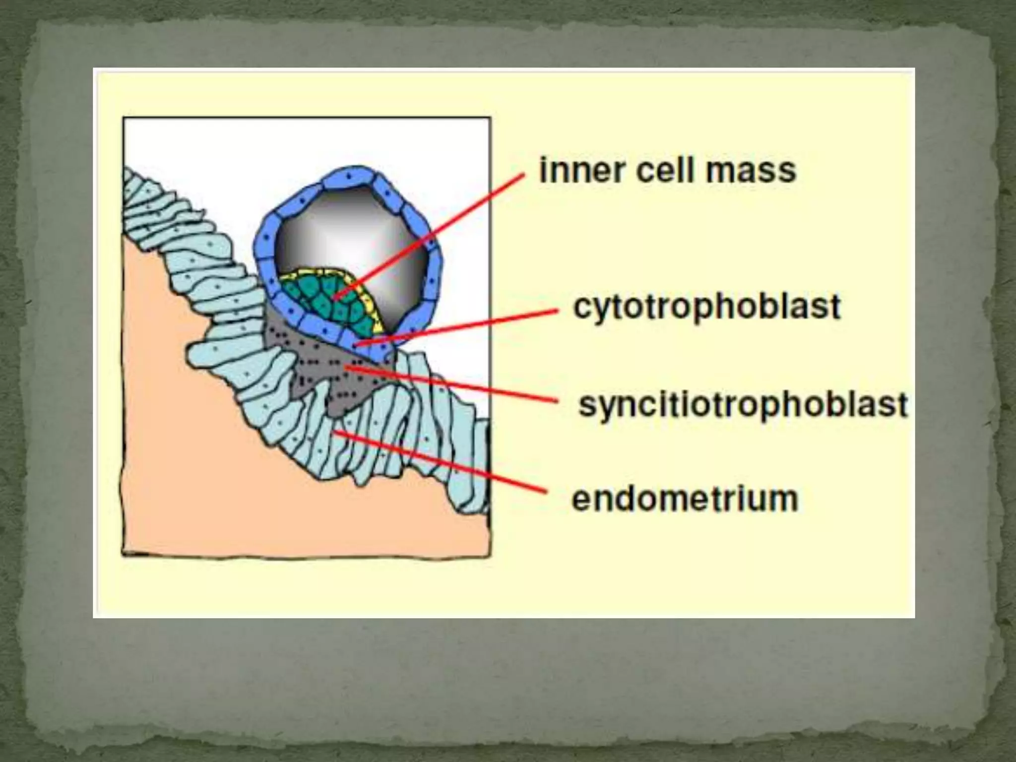
The blastocystattaching itself to uterine wall or lining
is called as implantation.
It penetrates in the compact layer of decidua near the
fundus.
It occurs on the 6th day of fertilisation and completed
by 10th to 11th day.
The deeper penetration of blastocyst into decidua is
called as “ Interstitial implantation”.
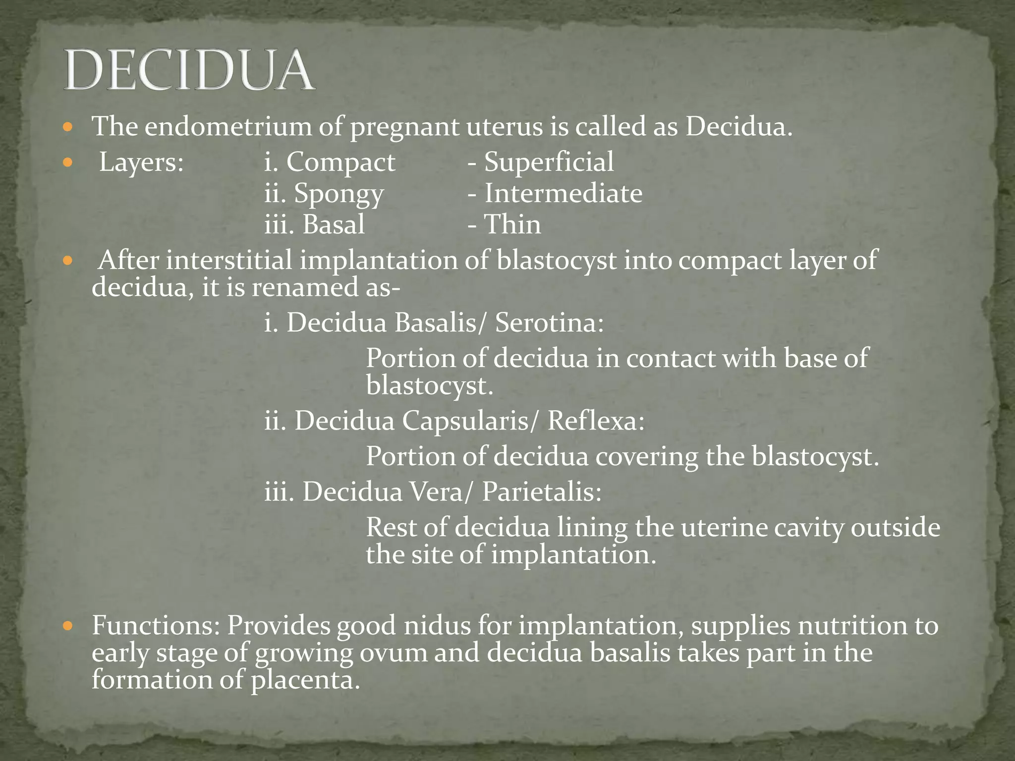
16.
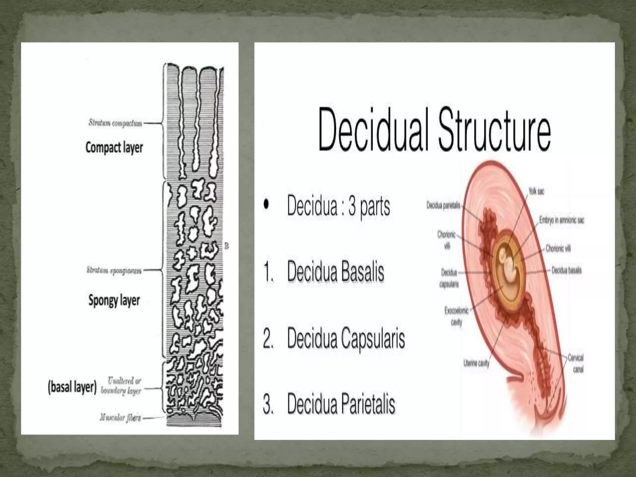
The endometriumof pregnant uterus is called as Decidua.
Layers: i. Compact - Superficial
ii. Spongy - Intermediate
iii. Basal - Thin
After interstitial implantation of blastocyst into compact layer of
decidua, it is renamed as-
i. Decidua Basalis/ Serotina:
Portion of decidua in contact with base of
blastocyst.
ii. Decidua Capsularis/ Reflexa:
Portion of decidua covering the blastocyst.
iii. Decidua Vera/ Parietalis:
Rest of decidua lining the uterine cavity outside
the site of implantation.
Functions: Provides good nidus for implantation, supplies nutrition to
early stage of growing ovum and decidua basalis takes part in the
formation of placenta.
18.
Definition:
Fluid passes intothe morula which separates the cells
of morula and it is called as Blastocyst.
Structure:
Two Parts- i. Trophoblast (Placenta, Chorion)
ii. Inner Cell Mass (Fetus, Umbilical Cord
and Amnion)
19.
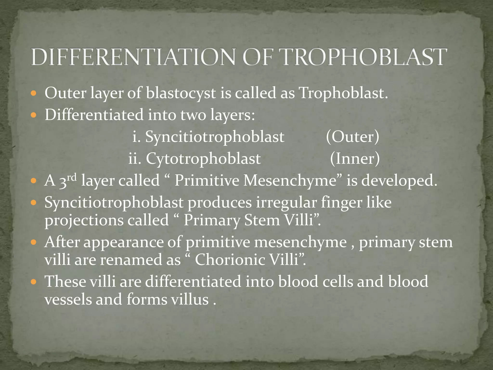
Outer layerof blastocyst is called as Trophoblast.
Differentiated into two layers:
i. Syncitiotrophoblast (Outer)
ii. Cytotrophoblast (Inner)
A 3rd layer called “ Primitive Mesenchyme” is developed.
Syncitiotrophoblast produces irregular finger like
projections called “ Primary Stem Villi”.
After appearance of primitive mesenchyme , primary stem
villi are renamed as “ Chorionic Villi”.
These villi are differentiated into blood cells and blood
vessels and forms villus .
21.
The cellssuspended in the blastocyst is termed as “ Inner
Cell Mass”.
The Inner Cell Mass is differentiated into bilaminar germ
layer- i.Ectoderm
ii.Endoderm.
The bilaminar germ disc is connected with trophoblast by
connecting stalk or body stalk.
A 3rd germ layer appears during 3rd week called mesoderm.
And now bilaminar germ layer becomes trilaminar germ
layer.
Two cavities appears one on each side of bilaminar germ
layer- i. Amniotic Cavity (Filled with amniotic fluid)
ii. Yolk Sac (Incorporated into gut)
Extra embronic coelom- A cavity external to developing
embryo. Also called as chorionic cavity.
24.
ECTODERM: Centraland Peripheral Nervous System,
Pituitary Gland, Epidermis of skin with
its appendages, Salivary glands, Mucus
lining of mouth, nostril and anus.
MESODERM: Bones, Cartilages, Muscles,
Cardiovascular System, Kidney, Gonads,
Suprarenal Glands, Spleen, Most of
genital tracts, lining of Pericardium,
Peritoneum, Pleura.
ENDODERM: Liver, Gall Bladder, Pancreas, Epithelial
lining of GI Tract, Respiratory Tract,
Mucus lining of Urinary Bladder and
Urethra.
EMBRYO IS DIFFRENTIATED AS HUMAN AT 8TH WEEK.
25.
Conception is essentialprocess for continuity of life.
Numerous processes together helps in the process of
conception. Failure or abnormality in any
physiological process can lead to non conception and
ultimately infertility.
Dutta D.C,“Textbook Of Obstetrics”, New Central
Book Agency(P)LTD, 6th edition, Pg.28-29
Jacob Annamma, “A Comprehensive Textbook of
Midwifery”, Jaypee Brothers Medical
Publishers(P)LTD, 2nd edition, Pg.75-78
Myles, “ Textbook for Midwives”, Churchill Livingstone
Publishers, 13th edition, Pg.143-147
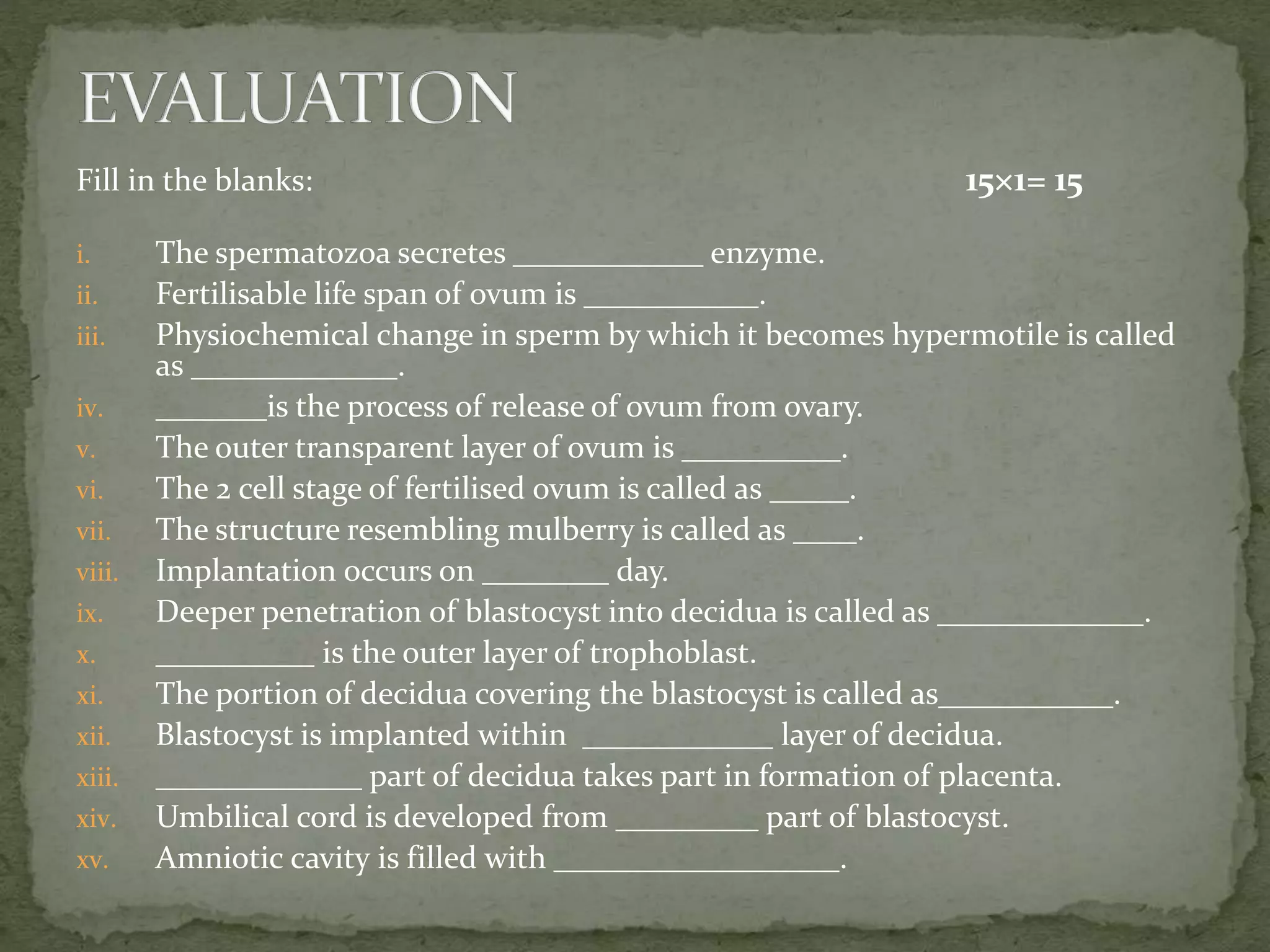
28.
Fill in theblanks: 15×1= 15
i. The spermatozoa secretes ____________ enzyme.
ii. Fertilisable life span of ovum is ___________.
iii. Physiochemical change in sperm by which it becomes hypermotile is called
as _____________.
iv. _______is the process of release of ovum from ovary.
v. The outer transparent layer of ovum is __________.
vi. The 2 cell stage of fertilised ovum is called as _____.
vii. The structure resembling mulberry is called as ____.
viii. Implantation occurs on ________ day.
ix. Deeper penetration of blastocyst into decidua is called as _____________.
x. __________ is the outer layer of trophoblast.
xi. The portion of decidua covering the blastocyst is called as___________.
xii. Blastocyst is implanted within ____________ layer of decidua.
xiii. _____________ part of decidua takes part in formation of placenta.
xiv. Umbilical cord is developed from _________ part of blastocyst.
xv. Amniotic cavity is filled with __________________.
29.
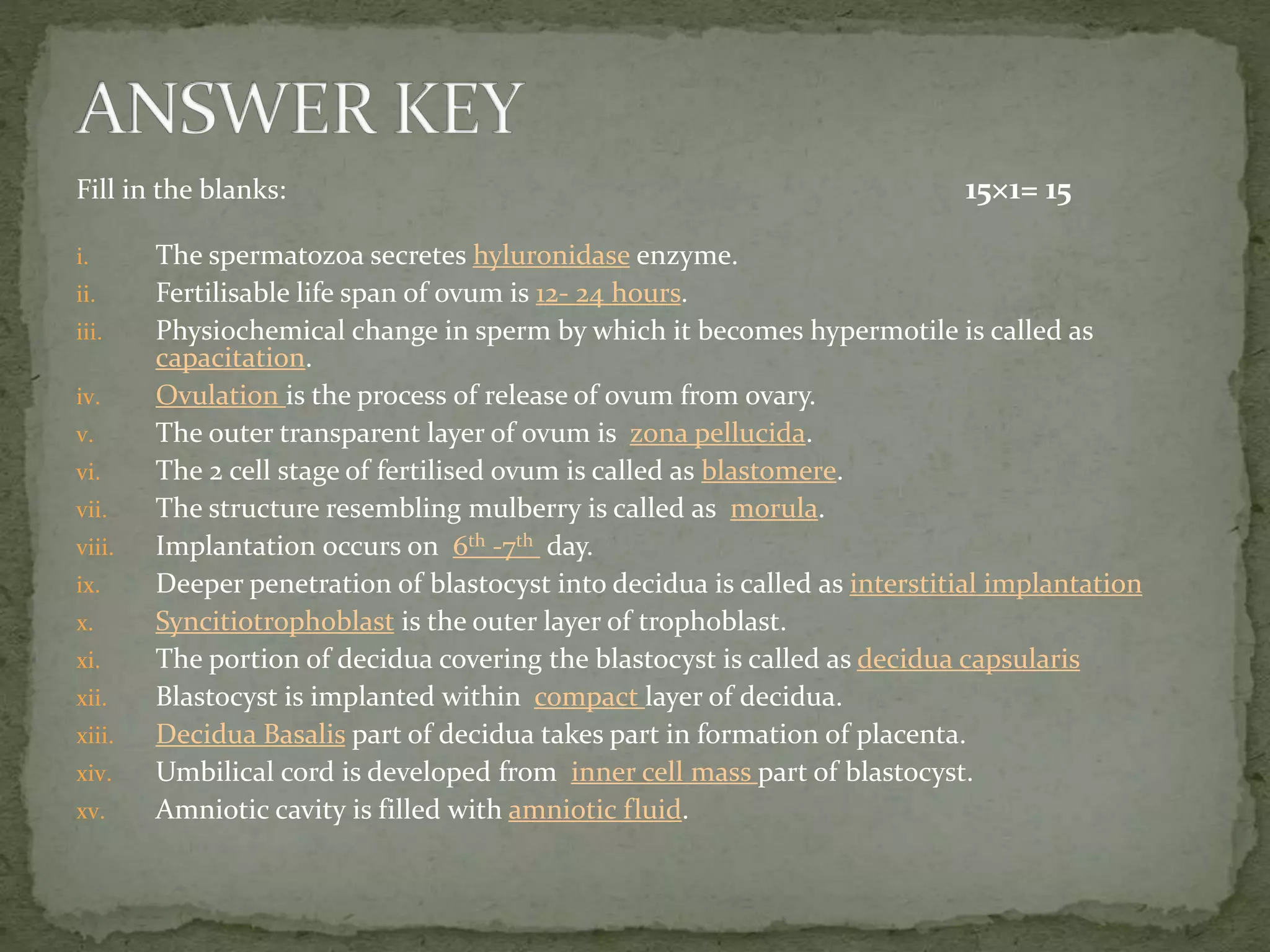
Fill in theblanks: 15×1= 15
i. The spermatozoa secretes hyluronidase enzyme.
ii. Fertilisable life span of ovum is 12- 24 hours.
iii. Physiochemical change in sperm by which it becomes hypermotile is called as
capacitation.
iv. Ovulation is the process of release of ovum from ovary.
v. The outer transparent layer of ovum is zona pellucida.
vi. The 2 cell stage of fertilised ovum is called as blastomere.
vii. The structure resembling mulberry is called as morula.
viii. Implantation occurs on 6th -7th day.
ix. Deeper penetration of blastocyst into decidua is called as interstitial implantation
x. Syncitiotrophoblast is the outer layer of trophoblast.
xi. The portion of decidua covering the blastocyst is called as decidua capsularis
xii. Blastocyst is implanted within compact layer of decidua.
xiii. Decidua Basalis part of decidua takes part in formation of placenta.
xiv. Umbilical cord is developed from inner cell mass part of blastocyst.
xv. Amniotic cavity is filled with amniotic fluid.
Editor's Notes
#11 Capacitation- Physiochemical change in sperm by which it becomes hypermotile and able to bind and fertilise ovum.