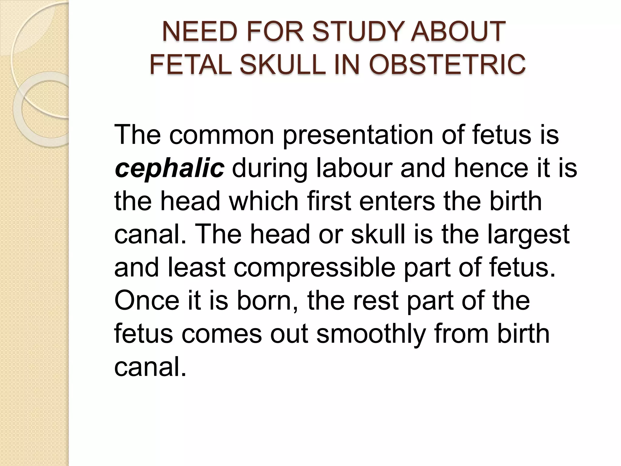
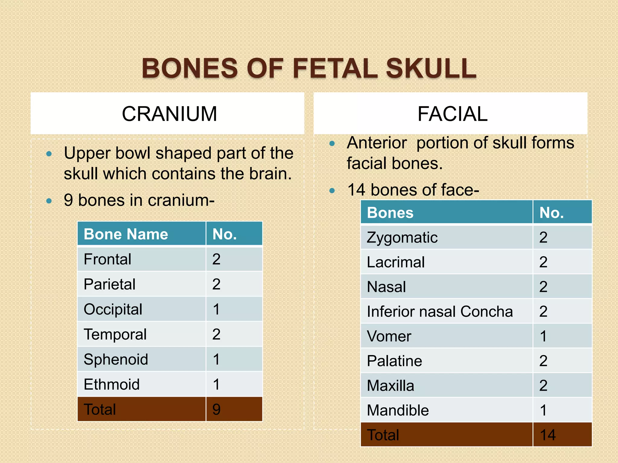
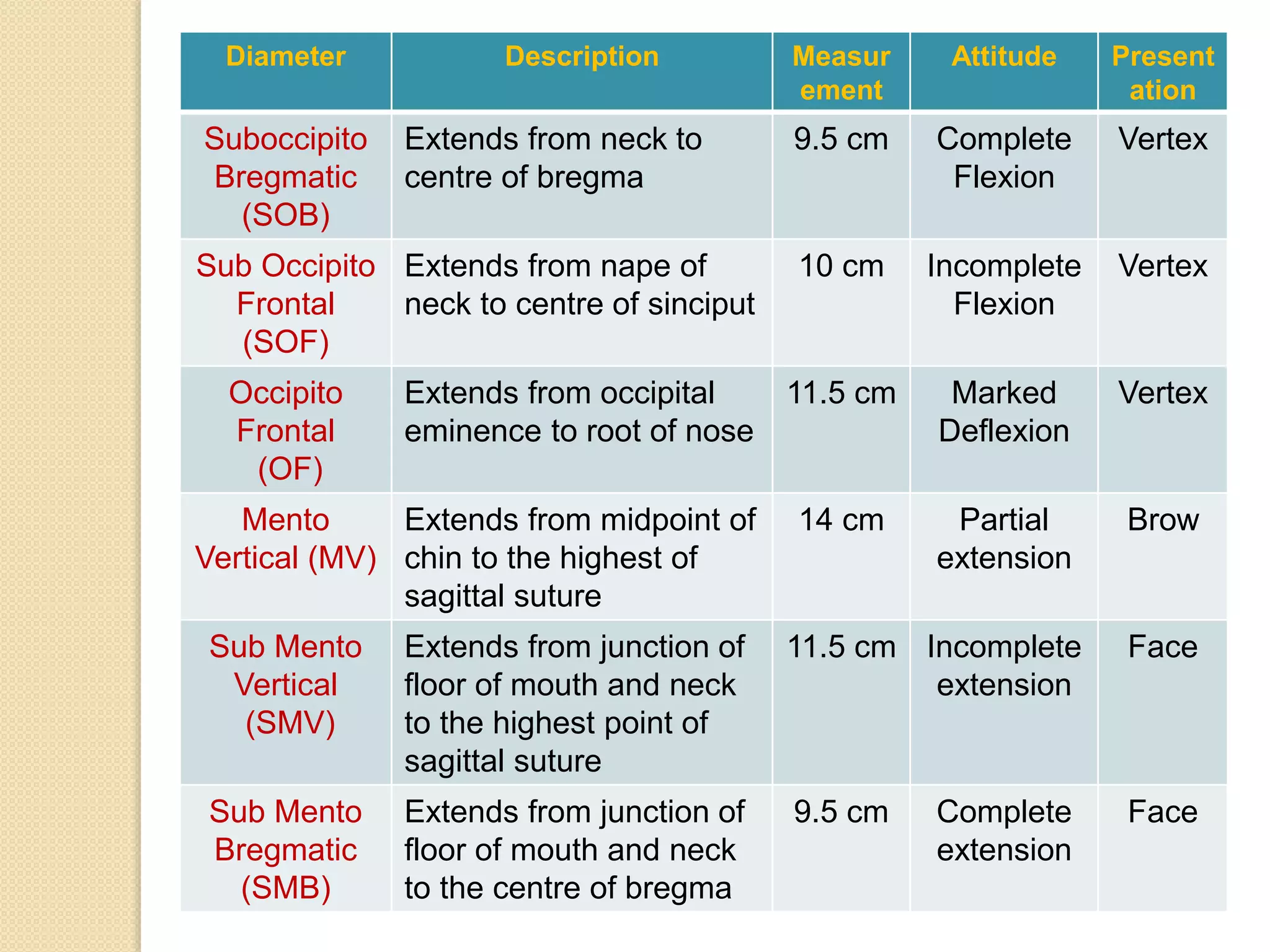
FETAL SKULL
PREPARED BY:
SRUJANI SWETASINA
DASH
NURSING TUTOR
G.N.M.INSTITUTE
A.N.M.M.C.H, GAYA
OBJECTIVES
 NEED FOR STUDY ABOUT FETAL SKULL
IN OBSTETRIC
 FUNCTIONS OF SKULL
 BONES OF FETAL SKULL
 SUTURES
 FONTANELLES
 AREAS OF SKULL
 DIAMETERS OF FETAL SKULL
 SUMMARY
 BIBLIOGRAPHY
NEED FOR STUDY ABOUT
FETAL SKULL IN OBSTETRIC
The common presentation of fetus is
cephalic during labour and hence it is
the head which first enters the birth
canal. The head or skull is the largest
and least compressible part of fetus.
Once it is born, the rest part of the
fetus comes out smoothly from birth
canal.
FUNCTIONS OF FETAL SKULL
 Cranium protects the brain.
 Bony eye sockets protects the eye
against injury
 Temporal bone protects the ear.
 Mandible helps in chewing of food.
 Skull or head is the most common
presenting part which comes out
through the birth canal.
BONES OF FETAL
SKULL
 Skull is divided into two parts-
a. CRANIUM
b. FACE
BONES OF FETAL SKULL
CRANIUM FACIAL
 Upper bowl shaped part of the
skull which contains the brain.
 9 bones in cranium-
 Anterior portion of skull forms
facial bones.
 14 bones of face-
Bone Name No.
Frontal 2
Parietal 2
Occipital 1
Temporal 2
Sphenoid 1
Ethmoid 1
Total 9
Bones No.
Zygomatic 2
Lacrimal 2
Nasal 2
Inferior nasal Concha 2
Vomer 1
Palatine 2
Maxilla 2
Mandible 1
Total 14
SUTURES
Immovable joint between skull bones
• Frontal Suture-
Frontal Bone + Frontal
Bone
• Coronal Suture-
Frontal Bone + Parietal
Bone
• Sagittal Suture
Parietal Bone + Parietal
Bone
• Lambdoid Suture
Parietal Bone + Occipital
Bone
• Squamous Suture
FONTANELLES
 Area where two or more sutures join.
 There are 6 fontanelles in skull but only two are of
obstetric importance.
ANTERIOR FONTANELLE POSTERIOR FONTANELLE
Also called as Bregma Also called as Lambda
Located anteriorly at junction of
sagittal, frontal and coronal
sutures.
Located posteriorly at sagittal
and lamdoidal sutures.
Diamond shape Traingle shape
2.5 cm long, 1.5 cm wide 1.2 cm long , 1.2 cm wide
Larger than lambda Smaller than bregma
Ossifies by 18 months after birth Ossifies by 6 weeks after birth
CLINICAL SIGNIFICANCE OF
SUTURES AND FONTANELLES
SUTURES
 Helps in moulding
neccessary during
vaginal delivery.
 Digital palpation of
sagittal suture
during p/v
examination gives
an idea of
engagement and
degree of internal
rotation.
FONTANELLES
 Facilitates
moulding
 Palpation denotes
degree of flexion
 Depressed
fontanelle indicates
dehydration
 Elevated fontanelle
indicates raised
ICP
 CSF can be drawn
AREAS OF SKULL
 Skull is divided into-
1.Vault
2.Base
3.Face
Areas/Landmarks of
Obstetrical Importance
AREA DESCRIPTION
VERTEX Quadrangular area bounded anteriorly by bregma
and coronal sutures and behind by the lambda and
lambdoid sutures and laterally by lines passing
through the pariatal eminences.
BROW Area bounded on one side by bregma and coronal
sutures and on other side by root of nose and supra
orbital ridges of either side.
FACE Area bounded on one side by root of the nose and
supraorbital ridges and on the other by the junction of
the floor of mouth and neck.
OCCIPUT Area of surrounded within occipital bone
SINCIPUT Area lying in front of bregma and corresponds to area
of brow
FETAL DIAMETERS
 It has two types of diameters-
a. Anteroposterior Diameters
b. Transverse Diameters
ANTERO POSTERIOR
DIAMETERS
Submento Vertical, 11.5
cm
Diameter Description Measur
ement
Attitude Present
ation
Suboccipito
Bregmatic
(SOB)
Extends from neck to
centre of bregma
9.5 cm Complete
Flexion
Vertex
Sub Occipito
Frontal
(SOF)
Extends from nape of
neck to centre of sinciput
10 cm Incomplete
Flexion
Vertex
Occipito
Frontal
(OF)
Extends from occipital
eminence to root of nose
11.5 cm Marked
Deflexion
Vertex
Mento
Vertical (MV)
Extends from midpoint of
chin to the highest of
sagittal suture
14 cm Partial
extension
Brow
Sub Mento
Vertical
(SMV)
Extends from junction of
floor of mouth and neck
to the highest point of
sagittal suture
11.5 cm Incomplete
extension
Face
Sub Mento
Bregmatic
(SMB)
Extends from junction of
floor of mouth and neck
to the centre of bregma
9.5 cm Complete
extension
Face
TRANSVERSE DIAMETERS
Diameter Description Measurement
Biparietal
Diameter
(BPD)
Extends between two parietal
eminences
9.5 cm
Super Sub
Parietal Diameter
(SSPD)
Extends from a point placed
below one parietal eminence to a
point placed above the opposite
side parietal eminence
8.5 cm
Bitemporal
Diameter
(BTD)
Extends from both antero inferior
ends of coronal suture
8 cm
Bi Mastoid
Diameter
(BMD)
Extends from both tip of mastoid
process
7.5 cm
SUMMARY
 Fetal skull is significant in obstetrical
standpoint because it is the first part and
hardest part which comes out during
delivery in normal cases and if it is
delivered smoothly then the rest of the
trunk follows smoothly as well.
 A midwife must be competent to
recognise areas, sutures, fontanelles of
fetal skull in order to understand the
attitude, degree of rotation, presentation
& presenting part, status of intracranial
pressure and able to detect deviations
from normal.
BIBLIOGRAPHY
 Dutta D.C, “Textbook Of Obstetrics”, Jaypee
Brothers , The Health Science Publishers, 8th
edition, Pg.96-98
 Jacob Annamma, “A Comprehensive
Textbook of Midwifery”, Jaypee Brothers
Medical Publishers(P)LTD, 2nd edition, Pg.43-
51
 Elizabeth Marie, “Midwifery for Nurses”, CBS
Publishers and Distributors Pvt Ltd, 2nd
edition, Pg.90-96
 Kaur Sandeep, “ Midwifery and
Gynaecological Nursing”, CBS Publishers
and Distributors Pvt Ltd, 1st edition, 2019,
Pg.39-40
Fetal skull.pptx

Fetal skull.pptx

  • 1.
    FETAL SKULL PREPARED BY: SRUJANISWETASINA DASH NURSING TUTOR G.N.M.INSTITUTE A.N.M.M.C.H, GAYA
  • 2.
    OBJECTIVES  NEED FORSTUDY ABOUT FETAL SKULL IN OBSTETRIC  FUNCTIONS OF SKULL  BONES OF FETAL SKULL  SUTURES  FONTANELLES  AREAS OF SKULL  DIAMETERS OF FETAL SKULL  SUMMARY  BIBLIOGRAPHY
  • 3.
    NEED FOR STUDYABOUT FETAL SKULL IN OBSTETRIC The common presentation of fetus is cephalic during labour and hence it is the head which first enters the birth canal. The head or skull is the largest and least compressible part of fetus. Once it is born, the rest part of the fetus comes out smoothly from birth canal.
  • 4.
    FUNCTIONS OF FETALSKULL  Cranium protects the brain.  Bony eye sockets protects the eye against injury  Temporal bone protects the ear.  Mandible helps in chewing of food.  Skull or head is the most common presenting part which comes out through the birth canal.
  • 5.
    BONES OF FETAL SKULL Skull is divided into two parts- a. CRANIUM b. FACE
  • 6.
    BONES OF FETALSKULL CRANIUM FACIAL  Upper bowl shaped part of the skull which contains the brain.  9 bones in cranium-  Anterior portion of skull forms facial bones.  14 bones of face- Bone Name No. Frontal 2 Parietal 2 Occipital 1 Temporal 2 Sphenoid 1 Ethmoid 1 Total 9 Bones No. Zygomatic 2 Lacrimal 2 Nasal 2 Inferior nasal Concha 2 Vomer 1 Palatine 2 Maxilla 2 Mandible 1 Total 14
  • 9.
    SUTURES Immovable joint betweenskull bones • Frontal Suture- Frontal Bone + Frontal Bone • Coronal Suture- Frontal Bone + Parietal Bone • Sagittal Suture Parietal Bone + Parietal Bone • Lambdoid Suture Parietal Bone + Occipital Bone • Squamous Suture
  • 10.
    FONTANELLES  Area wheretwo or more sutures join.  There are 6 fontanelles in skull but only two are of obstetric importance. ANTERIOR FONTANELLE POSTERIOR FONTANELLE Also called as Bregma Also called as Lambda Located anteriorly at junction of sagittal, frontal and coronal sutures. Located posteriorly at sagittal and lamdoidal sutures. Diamond shape Traingle shape 2.5 cm long, 1.5 cm wide 1.2 cm long , 1.2 cm wide Larger than lambda Smaller than bregma Ossifies by 18 months after birth Ossifies by 6 weeks after birth
  • 11.
    CLINICAL SIGNIFICANCE OF SUTURESAND FONTANELLES SUTURES  Helps in moulding neccessary during vaginal delivery.  Digital palpation of sagittal suture during p/v examination gives an idea of engagement and degree of internal rotation. FONTANELLES  Facilitates moulding  Palpation denotes degree of flexion  Depressed fontanelle indicates dehydration  Elevated fontanelle indicates raised ICP  CSF can be drawn
  • 12.
    AREAS OF SKULL Skull is divided into- 1.Vault 2.Base 3.Face
  • 13.
    Areas/Landmarks of Obstetrical Importance AREADESCRIPTION VERTEX Quadrangular area bounded anteriorly by bregma and coronal sutures and behind by the lambda and lambdoid sutures and laterally by lines passing through the pariatal eminences. BROW Area bounded on one side by bregma and coronal sutures and on other side by root of nose and supra orbital ridges of either side. FACE Area bounded on one side by root of the nose and supraorbital ridges and on the other by the junction of the floor of mouth and neck. OCCIPUT Area of surrounded within occipital bone SINCIPUT Area lying in front of bregma and corresponds to area of brow
  • 14.
    FETAL DIAMETERS  Ithas two types of diameters- a. Anteroposterior Diameters b. Transverse Diameters
  • 15.
  • 16.
    Diameter Description Measur ement AttitudePresent ation Suboccipito Bregmatic (SOB) Extends from neck to centre of bregma 9.5 cm Complete Flexion Vertex Sub Occipito Frontal (SOF) Extends from nape of neck to centre of sinciput 10 cm Incomplete Flexion Vertex Occipito Frontal (OF) Extends from occipital eminence to root of nose 11.5 cm Marked Deflexion Vertex Mento Vertical (MV) Extends from midpoint of chin to the highest of sagittal suture 14 cm Partial extension Brow Sub Mento Vertical (SMV) Extends from junction of floor of mouth and neck to the highest point of sagittal suture 11.5 cm Incomplete extension Face Sub Mento Bregmatic (SMB) Extends from junction of floor of mouth and neck to the centre of bregma 9.5 cm Complete extension Face
  • 17.
  • 18.
    Diameter Description Measurement Biparietal Diameter (BPD) Extendsbetween two parietal eminences 9.5 cm Super Sub Parietal Diameter (SSPD) Extends from a point placed below one parietal eminence to a point placed above the opposite side parietal eminence 8.5 cm Bitemporal Diameter (BTD) Extends from both antero inferior ends of coronal suture 8 cm Bi Mastoid Diameter (BMD) Extends from both tip of mastoid process 7.5 cm
  • 19.
    SUMMARY  Fetal skullis significant in obstetrical standpoint because it is the first part and hardest part which comes out during delivery in normal cases and if it is delivered smoothly then the rest of the trunk follows smoothly as well.  A midwife must be competent to recognise areas, sutures, fontanelles of fetal skull in order to understand the attitude, degree of rotation, presentation & presenting part, status of intracranial pressure and able to detect deviations from normal.
  • 20.
    BIBLIOGRAPHY  Dutta D.C,“Textbook Of Obstetrics”, Jaypee Brothers , The Health Science Publishers, 8th edition, Pg.96-98  Jacob Annamma, “A Comprehensive Textbook of Midwifery”, Jaypee Brothers Medical Publishers(P)LTD, 2nd edition, Pg.43- 51  Elizabeth Marie, “Midwifery for Nurses”, CBS Publishers and Distributors Pvt Ltd, 2nd edition, Pg.90-96  Kaur Sandeep, “ Midwifery and Gynaecological Nursing”, CBS Publishers and Distributors Pvt Ltd, 1st edition, 2019, Pg.39-40