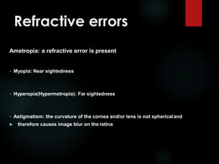
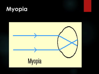
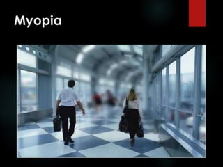
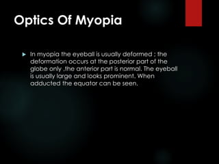
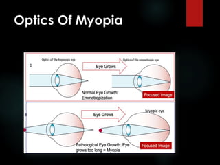
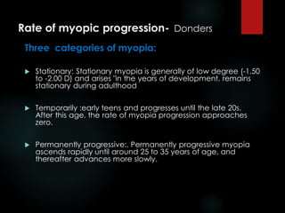
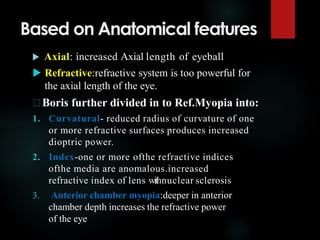
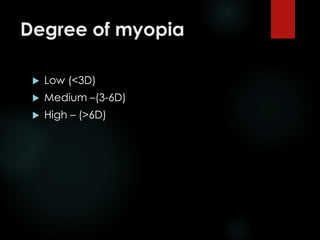
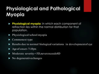
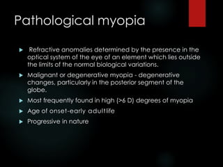
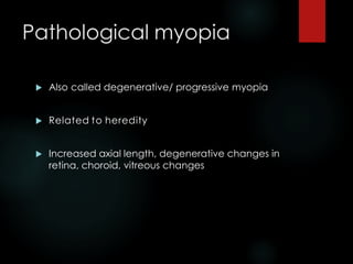
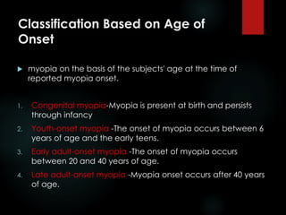
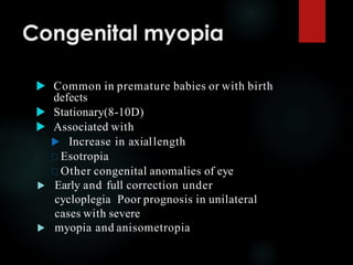
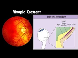
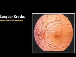
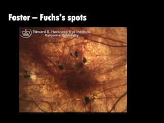
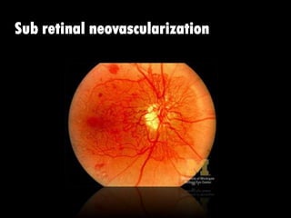
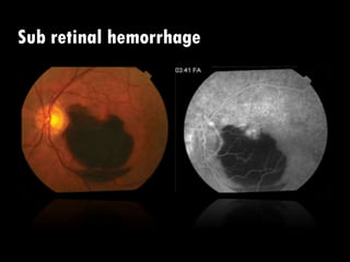
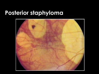
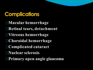
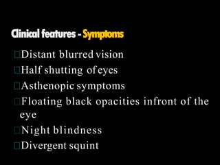
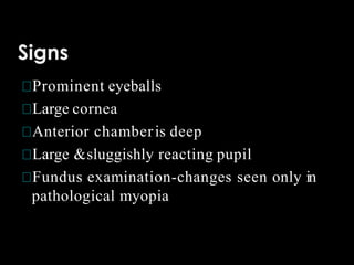
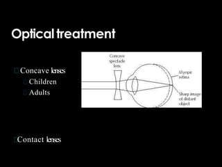
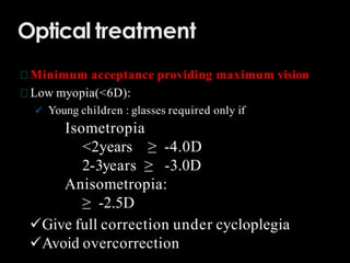
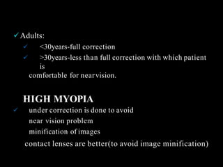
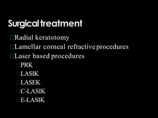
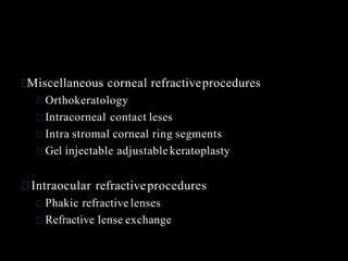
3. Refractive status of the eye
The ocular refractive status refers to the locus
within the eye conjugate with optical infinity
during minimal accommodation.* Under these
conditions:
In an emmetropic eye, incident parallel rays of
light are brought to a focus upon the retina.
In a hyperopic (or hypermetropic) eye, incident
parallel rays of light are brought to a focus behind
the retina.
In a myopic eye, incident parallel rays of light are
brought to a focus in front ofthe retina.
4. Refractive status of the eye
Ametropia, or the absence of emmetropia,
may be produced by variations in:
1. The relative location of the optical elements of
the eye with respect to the retina.
2. The relative refractive power of the optical
elements with respect to the location of the
retina
5. Refractive errors
Ametropia: a refractive error is present
• Myopia: Near sightedness
• Hyperopia(Hypermetropia): Far sightedness
• Astigmatism: the curvature of the cornea and/or lens is not sphericaland
therefore causes image blur on the retina
6. Refractive errors
• Anisometropia: a refractive power difference between
the 2 eyes (> 2D)
• Aniseikonia: a difference of image size between the 2
eyes as perceived by the patient
• Aphakia: (Phakos=lens), aphakia is no lens
• Pseudophakia: artificial lens in the eye
9. Myopia
Results from an eye having excessive
refractive power for its axial length.
This may be due either to the eye having
a relatively long axial length or to
increased dioptric power of one or more
of the refractive elements.
10. Myopia
Term myopia was derived by Galen from the
words myein ("to close") and ops ("eye").
Galen observed that nearsighted people partially
closed their eyes to see better.
11. Optics Of Myopia
In myopia the eyeball is usually deformed ; the
deformation occurs at the posterior part of the
globe only ,the anterior part is normal. The eyeball
is usually large and looks prominent. When
adducted the equator can be seen.
13. Classifications
Rate of myopic progression
Anatomical features of myopia
Degree of myopia
Physiological and pathological myopia
Hereditary and environmentally induced myopia
theory of myopic development
Age of myopia onset
14. Rate of myopic progression- Donders
Three categories of myopia:
Stationary: Stationary myopia is generally of low degree (-1.50
to -2.00 D) and arises "in the years of development. remains
stationary during adulthood
Temporarily :early teens and progresses until the late 20s.
After this age, the rate of myopia progression approaches
zero.
Permanently progressive:. Permanently progressive myopia
ascends rapidly until around 25 to 35 years of age, and
thereafter advances more slowly.
15. Based on Anatomical features
Axial: increased Axial length of eyeball
Refractive:refractive system is too powerful for
the axial length of the eye.
Boris further divided in to Ref.Myopia into:
1. Curvatural- reduced radius of curvature of one
or more refractive surfaces produces increased
dioptric power.
2. Index-one or more ofthe refractive indices
ofthe media are anomalous.increased
refractive index of lens withnuclear sclerosis
3. Anterior chamber myopia:deeper in anterior
chamber depth increases the refractive power
of the eye
17. Physiological and Pathological
Myopia
Physiological myopia: in which each component of
refraction lies within the normal distribution for that
population.
Physiological/school myopia
Commonest type
Results due to normal biological variations in developmentof eye
Age of onset- 7-10yrs
Moderate severity-<5D,neverexceeds8D
No degenerativechanges
18. Pathological myopia
Refractive anomalies determined by the presence in the
optical system of the eye of an element which lies outside
the limits of the normal biological variations.
Malignant or degenerative myopia - degenerative
changes, particularly in the posterior segment of the
globe.
Most frequently found in high (>6 D) degrees of myopia
Age of onset-early adultlife
Progressive in nature
19. Pathological myopia
Also called degenerative/ progressive myopia
Related to heredity
Increased axial length, degenerative changes in
retina, choroid, vitreous changes
20. Classification Based on Age of
Onset
myopia on the basis of the subjects' age at the time of
reported myopia onset.
1. Congenital myopia-Myopia is present at birth and persists
through infancy
2. Youth-onset myopia -The onset of myopia occurs between 6
years of age and the early teens.
3. Early adult-onset myopia -The onset of myopia occurs
between 20 and 40 years of age.
4. Late adult-onset myopia -Myopia onset occurs after 40 years
of age.
21. Congenital myopia
Common in premature babies or with birth
defects
Stationary(8-10D)
Associated with
Increase in axiallength
Esotropia
Other congenital anomalies of eye
Early and full correction under
cycloplegia Poor prognosis in unilateral
cases with severe
myopia and anisometropia
22. Other types myopia
Night myopia: increased myopia under low
iluminance conditions was first reported in 1789.
produced by an increased accommodative
response (typically on the order of 0.50 to 1.00 D)
under degraded stimulus conditions.
Pseudo myopia: results from a spasm of the ciliary
muscle
23. Pathological changes in
myopia
Optic disc appear large and pale
Degenerative changes in retina & choroid
Vitreous degeneration
Posterior staphyloma
34. Optical treatment
Minimum acceptance providing maximum vision
Low myopia(<6D):
Young children : glasses required only if
Isometropia
<2years ≥ -4.0D
2-3years ≥ -3.0D
Anisometropia:
≥ -2.5D
Give full correction under cycloplegia
Avoid overcorrection
35. Adults:
<30years-full correction
>30years-less than full correction with which patient
is
comfortable for nearvision.
HIGH MYOPIA
under correction is done to avoid
near vision problem
minification of images
contact lenses are better(to avoid image minification)