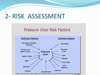
By
DR Kabiru Salisu NOHD
 INTRODUCTION
 MANAGEMENT
- Diagnosis
- Treatment
 Complication
 CONCLUSION
 REFERANCES
INTRODUCTION
 Definition
- This is a representative of
traumatic ulcer due to
direct pressure on bony
tissues or shearing forces
resulting in microvascular
compromise leading to
tissue necrosis and
ulceration
- Decubitus Ulcer;- Latin
decumbere means "to lie
down
History
- Thompson Rowling 1861
- Devis 1938
Epidemiology
Pressure sores are common conditions among patients
hospitalized in acute- and chronic-care facilities
- Hospitalised 3-10%
- Studies reported prevalence rates as high as 25-33% In
SCI
- 57.1% FMC Gombe and NOHI
Multidisplinary
 Plastic surgeon
 Neurosurgeon
 Orthopedic surgeon
 Nurses
 Social workers
 Physician
 Dietitian
 Physiotherapy
A- Diagnosis
Aim at:
- Determining the vulnerable individuals
- Risk assessment
- Skin assessment
- Ulcer assessment
1- People vulnerable to pressure ulcers
- Post surgery
- Critical care
- Orthopaedic patient
-Spinal injured
- Diabetes
- Peripheral vascular disease
- Previous history of pressure ulcers
-Extremes of age.
2- RISK ASSESSMENT
RISK SCORING
Score ranges between 4-20, the higher the score the
lower the risk
<14 – Greatest risk
14-18 – Moderate risk
18-20 – Minimal risk
Inspect the vulnerable areas
Characterised the ulcer if present
Look for:
– persistent erythema
– non-blanching hyperaemia
– blisters
– localised heat
– localised oedema
– localised induration
– purplish/bluish localised
areas
–localised coldness
- site, Size, shape,
surrounding, edge , base &
color
- Discharge
- Bleeding
- Necrotic tissue
- Odour
4- classify the ulcer
Investigation
- FBC
- Wound swab M/C/S
- Serum protein (Albumin / transferin)
- E/U
- X-Ray
- Biopsy
A- NON OPERATIVE
PREVENTION
1- Repositioning 2- Protect bony areas
- frequency-
2hrly on bed
15min wheelchair
- Assisted/ by self
- Devices eg specialise
wheelchair or
mattress eg trapeze
bar
 Special cushions
 foam mattress pads,
air-filled mattresses
 water-filled
mattresses/ gloves
 Bed sheet should be
smooth
3- Skin care
4- Improve Nutrition
- Bathing
- Skin protecting agent
eg talcum powder
- frequent skin
inspection
- Managing
incontinence/ UTI
- Good diet
- Dietary suppliment
vit. C, A, and Zinc
- Feeding Assistance
 5 - Early mobilization
 6- Quit smoking
 7- Control spasticity
 8- Adequate pain control
 9- psychological counseling
10- pressure measurement
Pressure mat / map
Pressure mapping system
Superficial sores (stage 1and 2)
I. Take all preventive measures
2. wound dressing using aseptic technique sing N/S,
hydrocolloid dressing , antibiotic gels/gauze or
adhesive dressing
3. Minimal wound debridement
4. Determine presence of infection and treat
5. Avoid urine or faecal contamination of wound
6. Keep record of wound size , shape and other changes
 7. Tetanus prophylaxis
 8. Antibiotic/ suppliments
Operative treatment (stage
III/IV)
A- preoperative considerations
- Determine whether the underlying cause can be eliminated post
operatively
- Patient / care giver EDUCATION about the treatment
- Nutritional Consideration ( SA – 3.5g/100ml & transferin 220g%)
- Sterilize the urinary tract
- Treat spasticity
- Radical wound debridement
- Determine presence of osteomylitis
Intraoperative
 Excision of the ulcer, surrounding scar, underlying
bursa, and soft-tissue calcification,
• Radical removal of underlying bone and any heterotopic
ossification
 padding of bone stumps and filling dead space
with fascia or muscle flaps
 Resurfacing with large regional pedicle flap
 Grafting the donor site of the flap with thick split skin
Example of flap to be raise include;
- Tensor faciae latae flap
- Transverse lumbosecral flap
- Sliding gluteal flap
- Hamstring V-Y advancement flap
- Rhomboid double Z plasty
- Gluteal maximus island flap
Post operative measures
 Prevent pressure or
shearing force
 Drain
 Sitting begin at 4-6wks
- Initially 10min once or
twice daily
- Sitting period increase
gradually up to 2hrs
 Improve nutrition
complications
 Osteomylitis
 Pyoarthrosis
 Anaemia
 Urethral fistula
 Recurrence
 Autonomic dysreflexia
 Malignant transformation
 Depressive illness
CONCLUSION
 Pressure ulcer management is challenging both to the
patient and the managing team. Is associated with
high morbidity, mortality and economic burden.
ALWAYS REMEMBER THAT IT IS EASIER
PREVENTED THAN TREATED.
References
 Al-fallouji M. A: post graduate surgery, 2nd edition, Read publishing Ltd. 1998
 Onche I.I, Yiltok S.J, Obiano S. K; pressure ulcer in spinal cord injury patient
in gombe Nigeria, nigerian journal of orthopaedics and trauma Vol-3 ,2004
 Idowu O.k, yinusa W; Risk factors of pressure ulcer in resource constrained
spinal injury service, nature.com, j an. 2011
 Douglas A. H ; principles of pressure management, national institute on
disability and rehabilitation research, sept. 1999
 Jeffrey E.J. etal; Pressure sore, baylor university medical centre, vol 9,2003
 Bed sore by Mayo foundation for medical education and research, march 2011
 Bradon J. W; surgical treatment of pressure ulcer, medscape 2011
 Prevention and treatment of pressure ulcer, clinical guideline 29, royal collage
of nursing 2006
 John L. Ziller; pressure ulcer, JAMA patient page , the journal of American
medical associationVol296, Aug. 2006
Pressure sore

Pressure sore

  • 1.
  • 2.
     INTRODUCTION  MANAGEMENT -Diagnosis - Treatment  Complication  CONCLUSION  REFERANCES
  • 3.
    INTRODUCTION  Definition - Thisis a representative of traumatic ulcer due to direct pressure on bony tissues or shearing forces resulting in microvascular compromise leading to tissue necrosis and ulceration - Decubitus Ulcer;- Latin decumbere means "to lie down
  • 4.
    History - Thompson Rowling1861 - Devis 1938
  • 5.
    Epidemiology Pressure sores arecommon conditions among patients hospitalized in acute- and chronic-care facilities - Hospitalised 3-10% - Studies reported prevalence rates as high as 25-33% In SCI - 57.1% FMC Gombe and NOHI
  • 7.
    Multidisplinary  Plastic surgeon Neurosurgeon  Orthopedic surgeon  Nurses  Social workers  Physician  Dietitian  Physiotherapy
  • 8.
    A- Diagnosis Aim at: -Determining the vulnerable individuals - Risk assessment - Skin assessment - Ulcer assessment
  • 9.
    1- People vulnerableto pressure ulcers - Post surgery - Critical care - Orthopaedic patient -Spinal injured - Diabetes - Peripheral vascular disease - Previous history of pressure ulcers -Extremes of age.
  • 10.
  • 11.
  • 12.
    Score ranges between4-20, the higher the score the lower the risk <14 – Greatest risk 14-18 – Moderate risk 18-20 – Minimal risk
  • 13.
  • 14.
    Characterised the ulcerif present Look for: – persistent erythema – non-blanching hyperaemia – blisters – localised heat – localised oedema – localised induration – purplish/bluish localised areas –localised coldness - site, Size, shape, surrounding, edge , base & color - Discharge - Bleeding - Necrotic tissue - Odour
  • 15.
  • 16.
    Investigation - FBC - Woundswab M/C/S - Serum protein (Albumin / transferin) - E/U - X-Ray - Biopsy
  • 17.
  • 18.
    PREVENTION 1- Repositioning 2-Protect bony areas - frequency- 2hrly on bed 15min wheelchair - Assisted/ by self - Devices eg specialise wheelchair or mattress eg trapeze bar  Special cushions  foam mattress pads, air-filled mattresses  water-filled mattresses/ gloves  Bed sheet should be smooth
  • 19.
    3- Skin care 4-Improve Nutrition - Bathing - Skin protecting agent eg talcum powder - frequent skin inspection - Managing incontinence/ UTI - Good diet - Dietary suppliment vit. C, A, and Zinc - Feeding Assistance
  • 20.
     5 -Early mobilization  6- Quit smoking  7- Control spasticity  8- Adequate pain control  9- psychological counseling
  • 21.
  • 22.
  • 23.
  • 24.
    Superficial sores (stage1and 2) I. Take all preventive measures 2. wound dressing using aseptic technique sing N/S, hydrocolloid dressing , antibiotic gels/gauze or adhesive dressing 3. Minimal wound debridement 4. Determine presence of infection and treat 5. Avoid urine or faecal contamination of wound 6. Keep record of wound size , shape and other changes
  • 25.
     7. Tetanusprophylaxis  8. Antibiotic/ suppliments
  • 26.
    Operative treatment (stage III/IV) A-preoperative considerations - Determine whether the underlying cause can be eliminated post operatively - Patient / care giver EDUCATION about the treatment - Nutritional Consideration ( SA – 3.5g/100ml & transferin 220g%) - Sterilize the urinary tract - Treat spasticity - Radical wound debridement - Determine presence of osteomylitis
  • 27.
    Intraoperative  Excision ofthe ulcer, surrounding scar, underlying bursa, and soft-tissue calcification, • Radical removal of underlying bone and any heterotopic ossification  padding of bone stumps and filling dead space with fascia or muscle flaps  Resurfacing with large regional pedicle flap  Grafting the donor site of the flap with thick split skin
  • 28.
    Example of flapto be raise include; - Tensor faciae latae flap - Transverse lumbosecral flap - Sliding gluteal flap - Hamstring V-Y advancement flap - Rhomboid double Z plasty - Gluteal maximus island flap
  • 29.
    Post operative measures Prevent pressure or shearing force  Drain  Sitting begin at 4-6wks - Initially 10min once or twice daily - Sitting period increase gradually up to 2hrs  Improve nutrition
  • 30.
    complications  Osteomylitis  Pyoarthrosis Anaemia  Urethral fistula  Recurrence  Autonomic dysreflexia  Malignant transformation  Depressive illness
  • 31.
    CONCLUSION  Pressure ulcermanagement is challenging both to the patient and the managing team. Is associated with high morbidity, mortality and economic burden. ALWAYS REMEMBER THAT IT IS EASIER PREVENTED THAN TREATED.
  • 32.
    References  Al-fallouji M.A: post graduate surgery, 2nd edition, Read publishing Ltd. 1998  Onche I.I, Yiltok S.J, Obiano S. K; pressure ulcer in spinal cord injury patient in gombe Nigeria, nigerian journal of orthopaedics and trauma Vol-3 ,2004  Idowu O.k, yinusa W; Risk factors of pressure ulcer in resource constrained spinal injury service, nature.com, j an. 2011  Douglas A. H ; principles of pressure management, national institute on disability and rehabilitation research, sept. 1999  Jeffrey E.J. etal; Pressure sore, baylor university medical centre, vol 9,2003  Bed sore by Mayo foundation for medical education and research, march 2011  Bradon J. W; surgical treatment of pressure ulcer, medscape 2011  Prevention and treatment of pressure ulcer, clinical guideline 29, royal collage of nursing 2006  John L. Ziller; pressure ulcer, JAMA patient page , the journal of American medical associationVol296, Aug. 2006

Editor's Notes

  • #4 (while lying supine or sitting dawn) or (while sliding down a bed or a chair).
  • #5 Pressure sores are an ancient problem, observed at autopsy of Egyptian mummies in 1861 by Thompson Rowling, In 1938, Davis was the first to suggest replacing the unstable scar of a healed pressure sore with a flap of tissue.[1] In 1947, Kostrubala and Greeley recommended excising the bony prominence and adding padding for the exposed bone with local fascia or muscle-fascia flaps
  • #6 Studies have suggested that, at any given time, 3-10% of hospitalized persons have pressure sores
  • #12 Scores range from 5 (greatest risk) to 20 (least risk). In a series of 250 geriatric patients, 24% developed pressure ulcers at some time during their hospital stay. Patientswith a score of 11 or less had a 48% incidence of pressure ulcer; those with a score of 12 to 14 had a 32% incidence; and when the score was 18 or greater, only 5% of patients developed pressure ulcers. Gosnell107 added nutritional status of the patient as a variable in pressure sore risk calculation. His method is often referred to as the “modified Norton scale.”
  • #13 Score < 11 assocd with about 50% risk Score > 18 assocd with < 5% risk Braden, Gosnell, Knoll, Norton, Waterlow, and Douglas.
  • #15 localised coolness showed tissue death occurs
  • #19 Repositioning devices. People with enough upper body strength may be able to reposition themselves with the assistance of a device such as a trapeze bar. Using bed linens to help lift and reposition a person can reduce friction and shearing. Special mattresses and support surfaces. Special cushions, foam mattress pads, air-filled mattresses and water-filled mattresses can help a person lie in an appropriate position, relieve pressure and protect vulnerable areas from damage. Your doctor or other care team member can recommend an appropriate mattress or surface.
  • #23 One clear advantage of pressuring mapping devices is that they provide a visual image of the pressure profile (map) of a person’s interface pressure distribution. The left diagram shows a pressure mat which has more than 200 individual measurement cells. The right diagram shows the read out on the computer screen of the pressure map from the individual cells. The dark green areas are the maximum pressure areas which normally fall under the ischial tuberosities (the sitting bones). Cushion selection attempts to minimize these high pressure areas.
  • #24 One clear advantage of pressuring mapping devices is that they provide a visual image of the pressure profile (map) of a person’s interface pressure distribution. The left diagram shows a pressure mat which has more than 200 individual measurement cells. The right diagram shows the read out on the computer screen of the pressure map from the individual cells. The dark green areas are the maximum pressure areas which normally fall under the ischial tuberosities (the sitting bones). Cushion selection attempts to minimize these high pressure areas.
  • #25 The most common organisms found in pressure sores include Staphylococcus aureus, Proteus mirabilis, Pseudomonas aeruginosa, Bacteroides fragilis, and Bacteroides asaccharolyticus.
  • #27 Han and colleagues166 managed their patients with a delayed two-stage operative plan. The first operation consisted of wound debridement and Jamshidi core needle bone biopsy. The second stage consisted of definitive musculocutaneous flap closure if the biopsy results were negative for osteomyelitis. When biopsy results were positive, flap closure was delayed for 6 weeks and intravenous antibiotics were administered in the interim in hopes of eliminating the osteomyelitis.