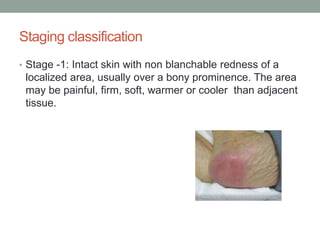
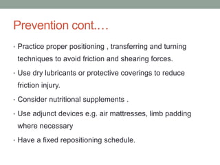
PRESSURE SORES
Chosen Care Group Ltd.
OBJECTIVES
• Definition
• Epidemiology
• Pathogenesis
• Risk Factors
• Stage and Risk Assessment
• Prevention
• Management
Definition
• A Pressure sore is a localized Injury to the skin or the
underlying tissue as a result of unrelieved pressure.
• The other names are Decubitus ulcer or bed sore.
EPIDEMIOLOGY
• Between 1 to 3 million UK affected
• 11-18 % nursing home residents
• 9-60% hospital
• 3-18% home
• Health care expenditure 1.4 to 2.1 Billion pounds per year.
PATHOGENESIS
• Prolonged pressure
• Friction
• Shearing forces
• Moisture
COMMON SITES
• Commonly occur at bony prominences, example: heels
• 95% occur in the caudal aspect of the body; 65% in the
pelvic area; 30% on the lower limbs.
Common sites
Intrinsic risk factors
• Limited mobility
• Spinal cord Injury
• Pain
• Alzheimer's disease
• Fractures
• Post Surgical
• Coma or Sedation
• Parkinson Disease
INTRINSIC RISK FACTORS
• Poor Nutrition
• Anorexia
• Poor Dentition
• Poverty or lack of access to food
• Dietary Restriction
Intrinsic risk factors
• Dementia
• Diabetes
• Depression
• Renal disease
• Cancer
Extrinsic risk factors
• Pressure from external Surface e.g. bed, chair
• Friction from being unable to move well
• Shear forces from involuntary movement
• Moisture – Bowel or bladder Incontinence, Perspiration,
Wound Drainage
Staging classification
• Stage -1: Intact skin with non blanchable redness of a
localized area, usually over a bony prominence. The area
may be painful, firm, soft, warmer or cooler than adjacent
tissue.
Staging classification
• Stage 2 – Partial thickness skin loss, presenting as
shallow open ulcer with a red pink wound bed without
slough(pus).May also present as an intact or open serum
filled blister.
Staging classification
• Stage 3 – Full thickness skin loss. Fat may be visible but
bone , tendon or muscle tissue are not ,slough may be
present.
Staging classification
• Stage 4 – Full thickness tissue loss with exposed bone,
tendon or muscle. Slough or eschar may be present.
prevention
Aims:
• Reduce pressure and shearing effects
• Reduce Moisture
• General skin care
• Nutrition
• Co Morbidities
• Involve Patient, family, care givers
prevention
• Daily skin inspection
• Bathing and skin cleaning frequency
• Moisturize skin; avoid hot water or harsh solutions
• Assess and treat incontinence: use tropical barriers or absorbent padding when
needed
• Proper repositioning frequently
• Avoid manipulating bony prominenses
Prevention cont.…
• Practice proper positioning , transferring and turning
techniques to avoid friction and shearing forces.
• Use dry lubricants or protective coverings to reduce
friction injury.
• Consider nutritional supplements .
• Use adjunct devices e.g. air mattresses, limb padding
where necessary
• Have a fixed repositioning schedule.
management
• Based on staging and Investigation
• Wound swabs and cultures shows mixed growth
• Clean Barrier
• Antibiotic where appropriate
• Debride necrotic tissue
complications
• Sepsis, Cellulitis, endocarditis, meningitis
• Fistula formation
• Septic arthritis
• Sinus tracts
conclusion
• Risk
• Prevention
• Identify early
• Manage
The end
ANY QUESTIONS?
Thank you

Pressure Sore

  • 1.
  • 2.
    OBJECTIVES • Definition • Epidemiology •Pathogenesis • Risk Factors • Stage and Risk Assessment • Prevention • Management
  • 3.
    Definition • A Pressuresore is a localized Injury to the skin or the underlying tissue as a result of unrelieved pressure. • The other names are Decubitus ulcer or bed sore.
  • 4.
    EPIDEMIOLOGY • Between 1to 3 million UK affected • 11-18 % nursing home residents • 9-60% hospital • 3-18% home • Health care expenditure 1.4 to 2.1 Billion pounds per year.
  • 5.
    PATHOGENESIS • Prolonged pressure •Friction • Shearing forces • Moisture
  • 6.
    COMMON SITES • Commonlyoccur at bony prominences, example: heels • 95% occur in the caudal aspect of the body; 65% in the pelvic area; 30% on the lower limbs.
  • 7.
  • 8.
    Intrinsic risk factors •Limited mobility • Spinal cord Injury • Pain • Alzheimer's disease • Fractures • Post Surgical • Coma or Sedation • Parkinson Disease
  • 9.
    INTRINSIC RISK FACTORS •Poor Nutrition • Anorexia • Poor Dentition • Poverty or lack of access to food • Dietary Restriction
  • 10.
    Intrinsic risk factors •Dementia • Diabetes • Depression • Renal disease • Cancer
  • 11.
    Extrinsic risk factors •Pressure from external Surface e.g. bed, chair • Friction from being unable to move well • Shear forces from involuntary movement • Moisture – Bowel or bladder Incontinence, Perspiration, Wound Drainage
  • 12.
    Staging classification • Stage-1: Intact skin with non blanchable redness of a localized area, usually over a bony prominence. The area may be painful, firm, soft, warmer or cooler than adjacent tissue.
  • 13.
    Staging classification • Stage2 – Partial thickness skin loss, presenting as shallow open ulcer with a red pink wound bed without slough(pus).May also present as an intact or open serum filled blister.
  • 14.
    Staging classification • Stage3 – Full thickness skin loss. Fat may be visible but bone , tendon or muscle tissue are not ,slough may be present.
  • 15.
    Staging classification • Stage4 – Full thickness tissue loss with exposed bone, tendon or muscle. Slough or eschar may be present.
  • 16.
    prevention Aims: • Reduce pressureand shearing effects • Reduce Moisture • General skin care • Nutrition • Co Morbidities • Involve Patient, family, care givers
  • 17.
    prevention • Daily skininspection • Bathing and skin cleaning frequency • Moisturize skin; avoid hot water or harsh solutions • Assess and treat incontinence: use tropical barriers or absorbent padding when needed • Proper repositioning frequently • Avoid manipulating bony prominenses
  • 18.
    Prevention cont.… • Practiceproper positioning , transferring and turning techniques to avoid friction and shearing forces. • Use dry lubricants or protective coverings to reduce friction injury. • Consider nutritional supplements . • Use adjunct devices e.g. air mattresses, limb padding where necessary • Have a fixed repositioning schedule.
  • 22.
    management • Based onstaging and Investigation • Wound swabs and cultures shows mixed growth • Clean Barrier • Antibiotic where appropriate • Debride necrotic tissue
  • 23.
    complications • Sepsis, Cellulitis,endocarditis, meningitis • Fistula formation • Septic arthritis • Sinus tracts
  • 24.
    conclusion • Risk • Prevention •Identify early • Manage
  • 25.
  • 26.