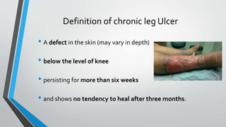
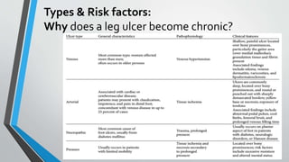
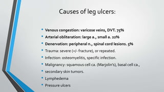
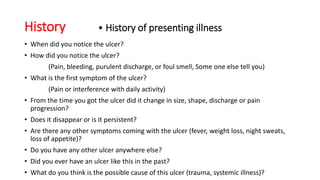
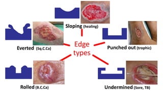
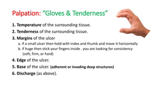
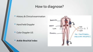
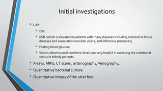
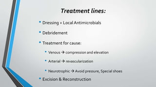
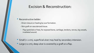
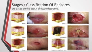
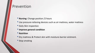
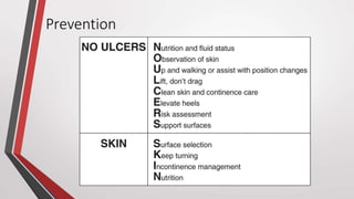
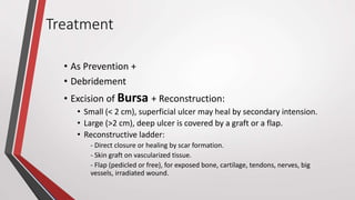
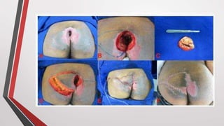
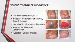
Chronic Leg Ulcers
& Pressure Sore
Professor Doctor
Mohamed Ahmed El Rouby
Ain Shams University
Remember!
• Structure of the skin
• Functions of the skin
• Inflammation
• Types of necrosis
Chronic Leg Ulcers
Definition of chronic leg Ulcer
• A defect in the skin (may vary in depth)
• below the level of knee
• persisting for more than six weeks
• and shows no tendency to heal after three months.
Types & Risk factors:
Why does a leg ulcer become chronic?
Causes of leg ulcers:
• Venous congestion: varicose veins, DVT. 75%
• Arterial obliteration: large a., small a. 22%
• Denervation: peripheral n., spinal cord lesions. 5%
• Trauma: severe (+/- fracture), or repeated.
• Infection: osteomyelitis, specific infection.
• Malignancy: squamous cell ca. (Marjolin’s), basal cell ca.,
• secondary skin tumors.
• Lymphedema
• Pressure ulcers
Management
1. Diagnosis (history, examination, investigations, DD)
2. Treatment (general, specific)
3. Treatment of complications
Clinical Examination of
Ulcer
Leg ulcer / pressure sore
History
Before History and Examination:
1- Introduce yourself
• ‫عليكم‬ ‫السالم‬
• ‫دكتور‬ ‫أنا‬(‫اسمك‬)
• ‫سنة‬---‫الطب‬ ‫كلية‬ ‫في‬
2- Tell the patient you will take the history of his/her ulcer
• ‫عندك‬ ‫اللي‬ ‫القرحة‬ ‫على‬ ‫سأكشف‬ ‫هللا‬ ‫شاء‬ ‫ان‬
3- Take verbal consent.
History
• Personal Data
• Name
• Age
• Nationality
• Marital status
• Occupation
• Special habits
• Complaint
History • History of presenting illness
• When did you notice the ulcer?
• How did you notice the ulcer?
(Pain, bleeding, purulent discharge, or foul smell, Some one else tell you)
• What is the first symptom of the ulcer?
(Pain or interference with daily activity)
• From the time you got the ulcer did it change in size, shape, discharge or pain
progression?
• Does it disappear or is it persistent?
• Are there any other symptoms coming with the ulcer (fever, weight loss, night sweats,
loss of appetite)?
• Do you have any other ulcer anywhere else?
• Did you ever have an ulcer like this in the past?
• What do you think is the possible cause of this ulcer (trauma, systemic illness)?
History
• Past History
• Medical: were hospitalized for a long time?
• Risk factors (DM, HTN, Hyperlipidemia, Heart Disease, Vascular Disease)
• Medication
• Allergy
• Surgical
• Family History
Summarize History
• Name
• Age
• Chief complaint
• Duration
• Important negatives
Examination
Before Examination:
1. Washing your hands.
2. Introducing yourself & confirm the patients ID
3. Explaining your procedure.
4. Taking verbal consent.
5. Privacy.
6. Positioning.
7. Exposing the ulcer (both sides e.g. both legs).
Clinical Examination:
•Inspection
•Palpation
•Missing things (clinical expertise)
Terms
Surrounding
Skin
Margin
Base
Floor
Border
Edge
Inspection
• Ss:
1. Site, Single or multiple
2. Size: 2dimentional & depth (3D),
3. Shape of the margin (regular or irregular)
4. Surrounding skin
• Margins: color changes , necrosis , pigmentation
• Edge: sloping, punched out, undermined, rolled, everted.
• Floor/Base: color, granulation tissue (important) , dead tissue, blood
, bone , tendon.
• Discharge (color, amount, and smell): serous, sanguineous,
serosanguineous, or purulent.
Edge
types
Sloping (healing(
Punched out (trophic)
Undermined (Sore, TB)Rolled (B.C.Ca)
Everted (Sq.C.Ca)
Palpation: “Gloves & Tenderness”
1. Temperature of the surrounding tissue.
2. Tenderness of the surrounding tissue.
3. Margins of the ulcer
a. if a small ulcer then hold with index and thumb and move it horizontally
b. if huge then stick your fingers inside . you are looking for consistency
(soft, firm, or hard)
4. Edge of the ulcer.
5. Base of the ulcer. (adherent or invading deep structures)
6. Discharge (as above).
Missing things ?!
Clinical Expertise
• Local lymph nodes
• Blood supply: pulses and veins.
• Innervation by a cotton or tongue depressor & compare to other side
• Assess the Range of motion of the surrounding 2 joints passively &
actively
• General examination.
• Thank the patient.
How to diagnose?
• History & Clinical examination
• Hand held Doppler
• Color Doppler US
• Ankle-Brachial index
Initial investigations
• Lab:
• CBC
• ESR (which is elevated in patients with many diseases including connective tissue
diseases and associated vasculitic ulcers, and infectious processes),
• Fasting blood glucose.
• Serum albumin and transferrin levels are very helpful in assessing the nutritional
status in elderly patients.
• X-rays, MRIs, CT scans , arteriography,Venography..
• Quantitative bacterial culture
• Quantitative biopsy of the ulcer bed
Treatment lines:
• Dressing + Local Antimicrobials
• Debridement
• Treatment for cause:
• Venous  compression and elevation
• Arterial  revascularization
• Neurotrophic  Avoid pressure, Special shoes
• Excision & Reconstruction
Excision & Reconstruction:
• Reconstructive ladder:
- Direct closure or healing by scar formation.
- Skin graft on vascularized tissue.
- Flap (pedicled or free), for exposed bone, cartilage, tendons, nerves, big vessels,
irradiated wound.
• Small (< 2 cm), superficial ulcer may heal by secondary intension.
• Large (>2 cm), deep ulcer is covered by a graft or a flap.
Pressure Sore
DEFINITION: Pressure Ulcer or Pressure Sore or
Decubitus Ulcer or Bedsore
• is a localized injury to the skin and other underlying tissue, usually over a
body prominence, as a result of prolonged unrelieved pressure.
• RF:
1. Friction 2. Shear
3. Impaired Sensory Perception 4. Impaired Physical Mobility
5. Altered Level Of Consciousness 6. Fecal And Urinary Incontinence
7. Malnutrition 8. Dehydration
9. Excessive Body Heat 10.Advanced Age
11.Chronic Medical Conditions- Diabetes, Cardiovascular Diseases.
Common Sites
• Hip and buttock 70%
• Ischial tuberosity, trochanteric and
sacral
• Lower extremities 15-25%
• Malleolar, heel, patellar and pretibial
• Others
• Nose, chin, forehead, occiput, chest,
back, elbow
Stages / Classification Of Bedsores
are based on the depth of tissue destroyed.
Complications
• Cellulitis
• Bone and joint infections
• Sepsis
• Cancer
Prevention
• Nursing: Change position /2 hours
• Use pressure-relieving devices such as air mattress, water mattress
• Daily Skin inspection
• Improve general condition
• Nutrition
• Dry mattress & Protect skin with moisture-barrier ointment.
• Stop smoking
Prevention
Treatment
• As Prevention +
• Debridement
• Excision of Bursa + Reconstruction:
• Small (< 2 cm), superficial ulcer may heal by secondary intension.
• Large (>2 cm), deep ulcer is covered by a graft or a flap.
• Reconstructive ladder:
- Direct closure or healing by scar formation.
- Skin graft on vascularized tissue.
- Flap (pedicled or free), for exposed bone, cartilage, tendons, nerves, big
vessels, irradiated wound.
Recent treatment modalities:
• Mechanical (Expander, VAC).
• Biological (Cultured keratinocytes,
Growth factors).
• Low Intensity Ultrasonic Stimulation
• Intermittent Pneumatic
Compression
• Hyperbaric Oxygen Therapy
THANKYOU
FORYOUR ATTENTION

Chronic leg ulcers & pressure sore

  • 1.
    Chronic Leg Ulcers &Pressure Sore Professor Doctor Mohamed Ahmed El Rouby Ain Shams University
  • 2.
    Remember! • Structure ofthe skin • Functions of the skin • Inflammation • Types of necrosis
  • 3.
  • 4.
    Definition of chronicleg Ulcer • A defect in the skin (may vary in depth) • below the level of knee • persisting for more than six weeks • and shows no tendency to heal after three months.
  • 5.
    Types & Riskfactors: Why does a leg ulcer become chronic?
  • 6.
    Causes of legulcers: • Venous congestion: varicose veins, DVT. 75% • Arterial obliteration: large a., small a. 22% • Denervation: peripheral n., spinal cord lesions. 5% • Trauma: severe (+/- fracture), or repeated. • Infection: osteomyelitis, specific infection. • Malignancy: squamous cell ca. (Marjolin’s), basal cell ca., • secondary skin tumors. • Lymphedema • Pressure ulcers
  • 7.
    Management 1. Diagnosis (history,examination, investigations, DD) 2. Treatment (general, specific) 3. Treatment of complications
  • 8.
    Clinical Examination of Ulcer Legulcer / pressure sore
  • 9.
  • 10.
    Before History andExamination: 1- Introduce yourself • ‫عليكم‬ ‫السالم‬ • ‫دكتور‬ ‫أنا‬(‫اسمك‬) • ‫سنة‬---‫الطب‬ ‫كلية‬ ‫في‬ 2- Tell the patient you will take the history of his/her ulcer • ‫عندك‬ ‫اللي‬ ‫القرحة‬ ‫على‬ ‫سأكشف‬ ‫هللا‬ ‫شاء‬ ‫ان‬ 3- Take verbal consent.
  • 11.
    History • Personal Data •Name • Age • Nationality • Marital status • Occupation • Special habits • Complaint
  • 12.
    History • Historyof presenting illness • When did you notice the ulcer? • How did you notice the ulcer? (Pain, bleeding, purulent discharge, or foul smell, Some one else tell you) • What is the first symptom of the ulcer? (Pain or interference with daily activity) • From the time you got the ulcer did it change in size, shape, discharge or pain progression? • Does it disappear or is it persistent? • Are there any other symptoms coming with the ulcer (fever, weight loss, night sweats, loss of appetite)? • Do you have any other ulcer anywhere else? • Did you ever have an ulcer like this in the past? • What do you think is the possible cause of this ulcer (trauma, systemic illness)?
  • 13.
    History • Past History •Medical: were hospitalized for a long time? • Risk factors (DM, HTN, Hyperlipidemia, Heart Disease, Vascular Disease) • Medication • Allergy • Surgical • Family History
  • 14.
    Summarize History • Name •Age • Chief complaint • Duration • Important negatives
  • 15.
  • 16.
    Before Examination: 1. Washingyour hands. 2. Introducing yourself & confirm the patients ID 3. Explaining your procedure. 4. Taking verbal consent. 5. Privacy. 6. Positioning. 7. Exposing the ulcer (both sides e.g. both legs).
  • 17.
  • 18.
  • 19.
  • 20.
    Inspection • Ss: 1. Site,Single or multiple 2. Size: 2dimentional & depth (3D), 3. Shape of the margin (regular or irregular) 4. Surrounding skin • Margins: color changes , necrosis , pigmentation • Edge: sloping, punched out, undermined, rolled, everted. • Floor/Base: color, granulation tissue (important) , dead tissue, blood , bone , tendon. • Discharge (color, amount, and smell): serous, sanguineous, serosanguineous, or purulent.
  • 21.
    Edge types Sloping (healing( Punched out(trophic) Undermined (Sore, TB)Rolled (B.C.Ca) Everted (Sq.C.Ca)
  • 22.
    Palpation: “Gloves &Tenderness” 1. Temperature of the surrounding tissue. 2. Tenderness of the surrounding tissue. 3. Margins of the ulcer a. if a small ulcer then hold with index and thumb and move it horizontally b. if huge then stick your fingers inside . you are looking for consistency (soft, firm, or hard) 4. Edge of the ulcer. 5. Base of the ulcer. (adherent or invading deep structures) 6. Discharge (as above).
  • 23.
  • 24.
    Clinical Expertise • Locallymph nodes • Blood supply: pulses and veins. • Innervation by a cotton or tongue depressor & compare to other side • Assess the Range of motion of the surrounding 2 joints passively & actively • General examination. • Thank the patient.
  • 25.
    How to diagnose? •History & Clinical examination • Hand held Doppler • Color Doppler US • Ankle-Brachial index
  • 26.
    Initial investigations • Lab: •CBC • ESR (which is elevated in patients with many diseases including connective tissue diseases and associated vasculitic ulcers, and infectious processes), • Fasting blood glucose. • Serum albumin and transferrin levels are very helpful in assessing the nutritional status in elderly patients. • X-rays, MRIs, CT scans , arteriography,Venography.. • Quantitative bacterial culture • Quantitative biopsy of the ulcer bed
  • 27.
    Treatment lines: • Dressing+ Local Antimicrobials • Debridement • Treatment for cause: • Venous  compression and elevation • Arterial  revascularization • Neurotrophic  Avoid pressure, Special shoes • Excision & Reconstruction
  • 28.
    Excision & Reconstruction: •Reconstructive ladder: - Direct closure or healing by scar formation. - Skin graft on vascularized tissue. - Flap (pedicled or free), for exposed bone, cartilage, tendons, nerves, big vessels, irradiated wound. • Small (< 2 cm), superficial ulcer may heal by secondary intension. • Large (>2 cm), deep ulcer is covered by a graft or a flap.
  • 29.
  • 30.
    DEFINITION: Pressure Ulceror Pressure Sore or Decubitus Ulcer or Bedsore • is a localized injury to the skin and other underlying tissue, usually over a body prominence, as a result of prolonged unrelieved pressure. • RF: 1. Friction 2. Shear 3. Impaired Sensory Perception 4. Impaired Physical Mobility 5. Altered Level Of Consciousness 6. Fecal And Urinary Incontinence 7. Malnutrition 8. Dehydration 9. Excessive Body Heat 10.Advanced Age 11.Chronic Medical Conditions- Diabetes, Cardiovascular Diseases.
  • 33.
    Common Sites • Hipand buttock 70% • Ischial tuberosity, trochanteric and sacral • Lower extremities 15-25% • Malleolar, heel, patellar and pretibial • Others • Nose, chin, forehead, occiput, chest, back, elbow
  • 34.
    Stages / ClassificationOf Bedsores are based on the depth of tissue destroyed.
  • 35.
    Complications • Cellulitis • Boneand joint infections • Sepsis • Cancer
  • 36.
    Prevention • Nursing: Changeposition /2 hours • Use pressure-relieving devices such as air mattress, water mattress • Daily Skin inspection • Improve general condition • Nutrition • Dry mattress & Protect skin with moisture-barrier ointment. • Stop smoking
  • 37.
  • 38.
    Treatment • As Prevention+ • Debridement • Excision of Bursa + Reconstruction: • Small (< 2 cm), superficial ulcer may heal by secondary intension. • Large (>2 cm), deep ulcer is covered by a graft or a flap. • Reconstructive ladder: - Direct closure or healing by scar formation. - Skin graft on vascularized tissue. - Flap (pedicled or free), for exposed bone, cartilage, tendons, nerves, big vessels, irradiated wound.
  • 40.
    Recent treatment modalities: •Mechanical (Expander, VAC). • Biological (Cultured keratinocytes, Growth factors). • Low Intensity Ultrasonic Stimulation • Intermittent Pneumatic Compression • Hyperbaric Oxygen Therapy
  • 41.