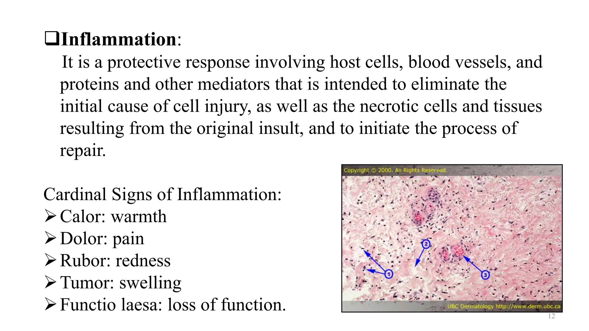
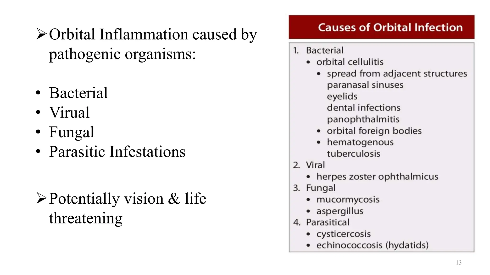
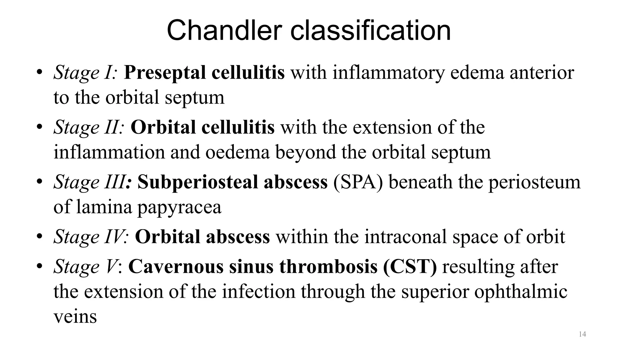
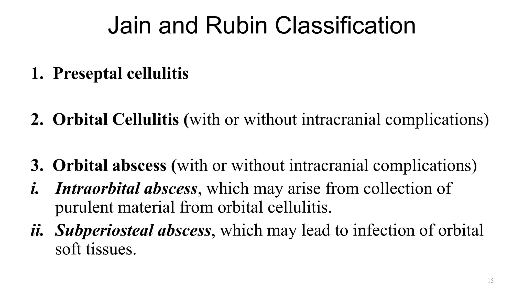
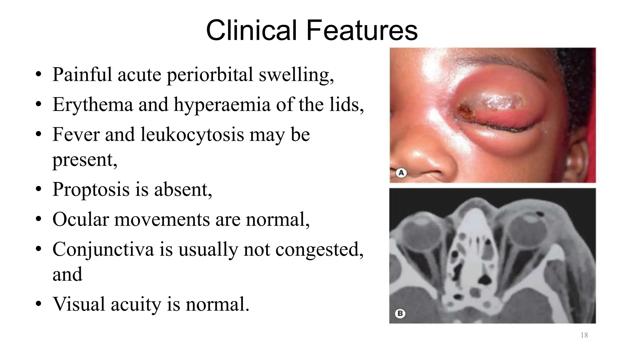
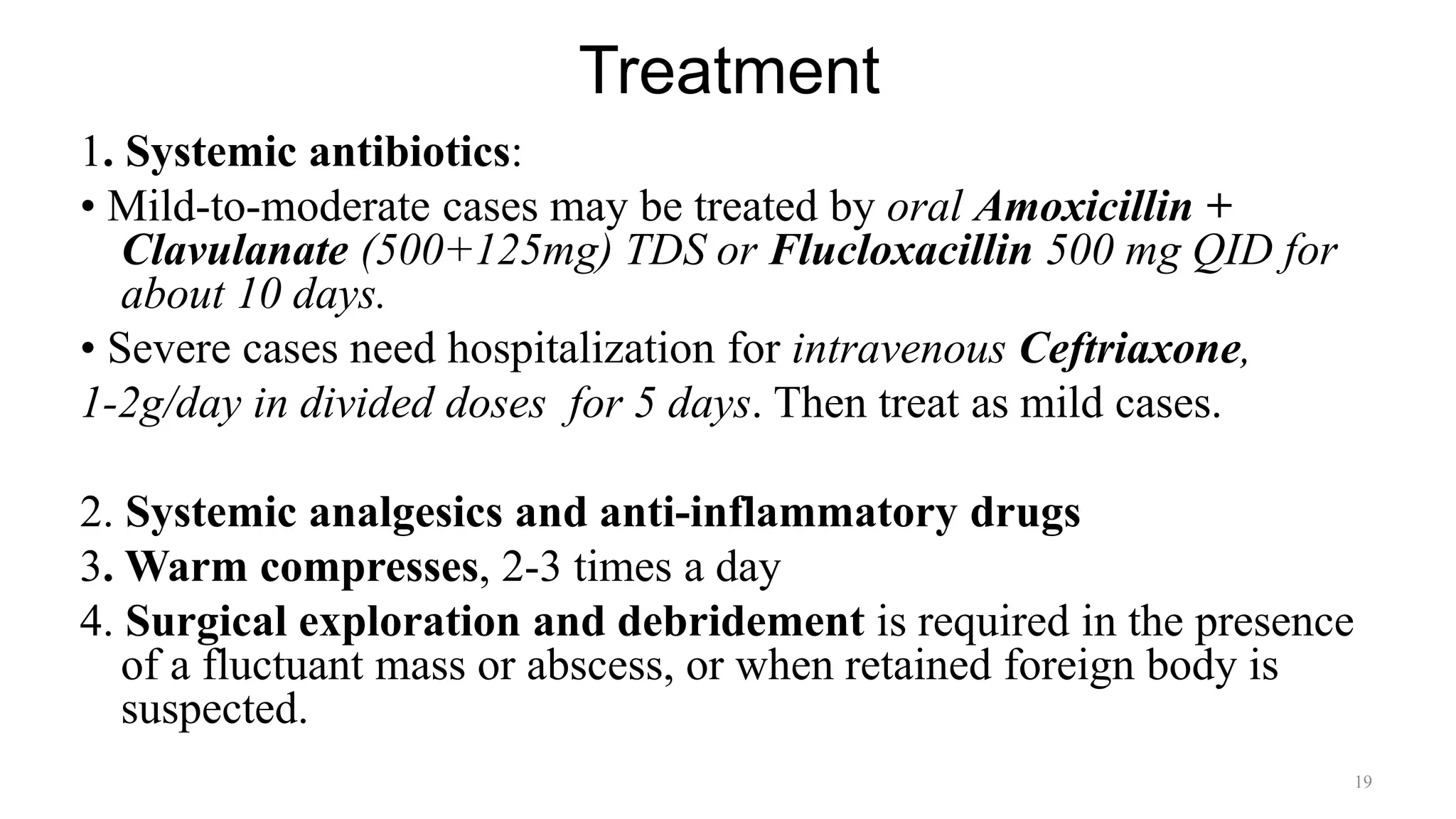
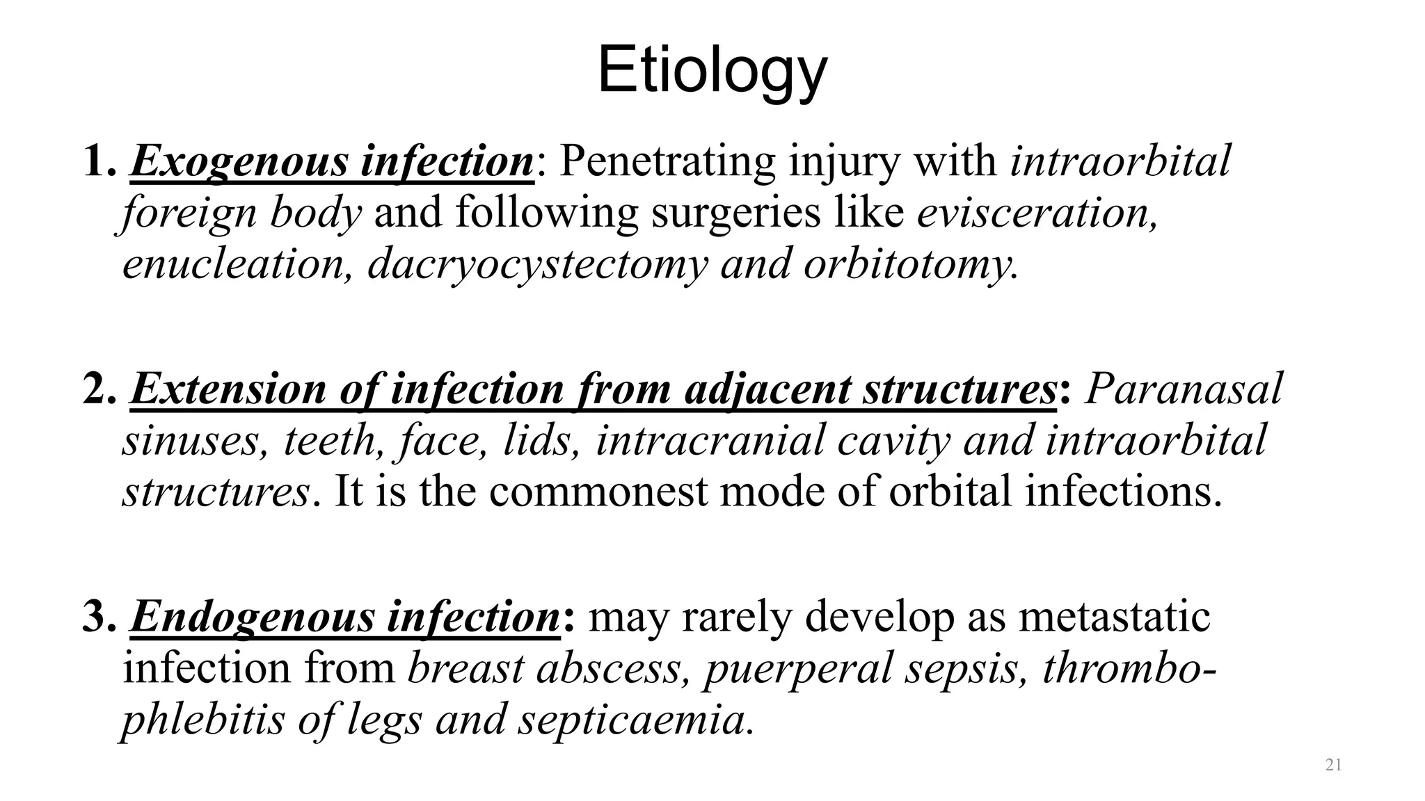
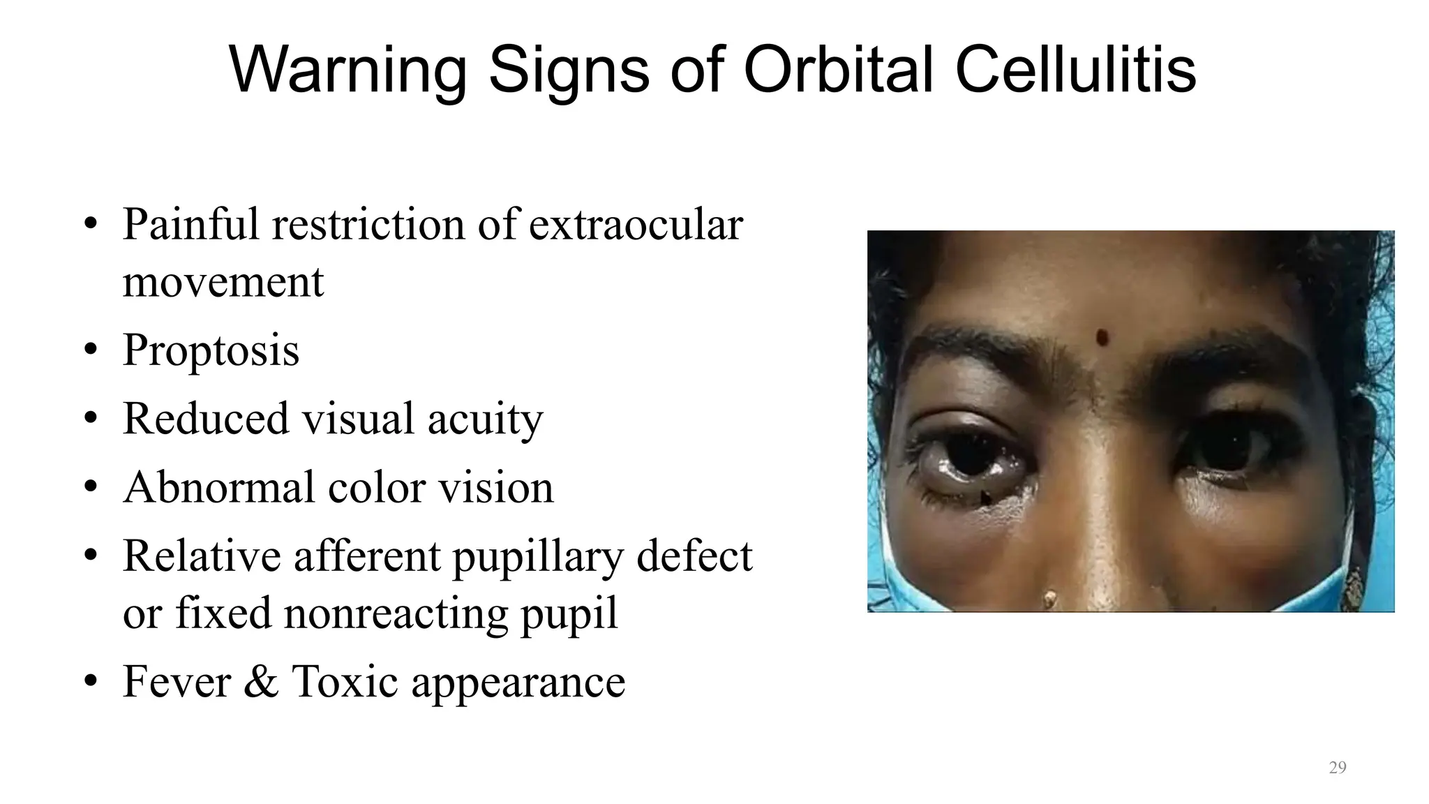
Infections of Orbit:
Preseptal & Orbital Cellulitis
Dr. PRABHAT DEVKOTA
MBBS(TU), MD(NAMS)
1
Content
• Applied Anatomy
• Preseptal Cellulitis
• Bacterial Orbital Cellulitis
• Necrotizing Orbital Cellulitis
• Orbital Tuberculosis
• Fungal Orbital Cellulitis
• Orbital Parasitic Infections
2
Eyelid Anatomy
• Skin and Subcutaneous tissue
• Muscles of Protraction
• Orbital Septum
• Orbital Fat
• Muscles of Retraction
• Tarsus
• Conjunctiva
3
4
Orbital Septum
• Thin, fibrous, multilayered
membrane.
• Sup. orbital septum begins at the
arcus marginalis along the orbital
rim into the anterior surface of the
levator aponeurosis inserted about
3-5 mm above the tarsal plate.
• Inf. orbital septum fuses with the
capsulopalpebral fascia several
millimeters below the tarsus.
5
Orbit Anatomy
Roof:
-Frontal bone
-Lesser wing of the
sphenoid bone
Medial Wall:
-Frontal process of the
maxillary bone
-Lacrimal bone
-Ethmoid bone
-Body of the sphenoid
bone
Floor:
-Maxillary bone
-Zygomatic bone
-Palatine bone
Lateral Wall:
-Zygomatic bone
-Greater wing of the
sphenoid bone
6
Medial wall of Orbit
Spread of infection occurs easily from the
ethmoid sinuses to the medial Orbit:-
• Thin Lamina Papyracea
• Multiple foramina for passage of blood
vessels and nerves.
• Valveless veins between the orbit and
adjacent structures allowing for bidirectional
passage of pathogens.
• Congenital bony defects (Zuckerkandl
dehiscences) & acquired bony defects may be
present.
7
8
Orbital Spaces
The four recti muscles arise from the
annulus of Zinn at the orbital apex and
course anteriorly to insert onto the sclera.
The intermuscular septum between the recti
form a cone that divides the orbit into:
The Extraconal Space lies between the
bony orbital walls and the annulus of Zinn.
The Intraconal Space lies within the
annulus, extending form the retrobulbar
surface to the orbital apex.
9
The Subperiosteal Space:
It is a potential space between the periorbita and the bony
orbital walls. The periorbita is loosely attached to the orbital
bones except at the suture lines & orbital margin.
The Sub-Tenon's Space:
It is a potential space between the globe and the Tenon's
capsule. It is a fibroelastic membrane that extends from the
dura of the optic nerve to the globe to fuse with it just behind
the corneo-scleral limbus.
10
The Paranasal Sinuses
• Frontal sinuses: Begin pneumatization at
5-6 years of age.
• Maxillary sinuses: Begin pneumatization
in utero; the first sinuses to fully
pneumatize.
• Ethmoid sinuses: Composed of three to
four air cells by birth, continue to expand,
and pneumatize till adulthood.
• Sphenoid sinuses: Start pneumatization at
3 months of age, and reach full size by 12
years
11
Inflammation:
It is a protective response involving host cells, blood vessels, and
proteins and other mediators that is intended to eliminate the
initial cause of cell injury, as well as the necrotic cells and tissues
resulting from the original insult, and to initiate the process of
repair.
Cardinal Signs of Inflammation:
Calor: warmth
Dolor: pain
Rubor: redness
Tumor: swelling
Functio laesa: loss of function. 12
Orbital Inflammation caused by
pathogenic organisms:
• Bacterial
• Virual
• Fungal
• Parasitic Infestations
Potentially vision & life
threatening
13
Chandler classification
• Stage I: Preseptal cellulitis with inflammatory edema anterior
to the orbital septum
• Stage II: Orbital cellulitis with the extension of the
inflammation and oedema beyond the orbital septum
• Stage III: Subperiosteal abscess (SPA) beneath the periosteum
of lamina papyracea
• Stage IV: Orbital abscess within the intraconal space of orbit
• Stage V: Cavernous sinus thrombosis (CST) resulting after
the extension of the infection through the superior ophthalmic
veins
14
Jain and Rubin Classification
1. Preseptal cellulitis
2. Orbital Cellulitis (with or without intracranial complications)
3. Orbital abscess (with or without intracranial complications)
i. Intraorbital abscess, which may arise from collection of
purulent material from orbital cellulitis.
ii. Subperiosteal abscess, which may lead to infection of orbital
soft tissues.
15
Preseptal Cellulitis
• Cellulitis: Latin; cellula(cell)+ itis(inflammation), misnomer
Cellulitis is a diffuse acute inflammatory infectious process that
involves the dermal and subcutaneous layers of the skin.
• Preseptal cellulitis refers to infection of the subcutaneous tissues
anterior to the orbital septum.
• The facial veins are valveless and preseptal cellulitis may spread
posteriorly to produce orbital cellulitis.
• Etiology:
-Staphylococcus aureus
-Streptococcus pyogenes and
-Haemophilus influenzae.
16
Modes of Infection
The organisms may invade the preseptal tissue by any of the
following modes:-
1. Exogenous infection: may result following skin lacerations,
insect bites and eyelid surgery.
2. Extension from adjacent infections: such as from an acute
hordeolum , dacryocystitis, conjunctivitis, sinusitis.
3. Endogenous infection: may occur by haematogenous spread
from remote infection of the middle ear or upper respiratory
tract infection(URTI).
17
Clinical Features
• Painful acute periorbital swelling,
• Erythema and hyperaemia of the lids,
• Fever and leukocytosis may be
present,
• Proptosis is absent,
• Ocular movements are normal,
• Conjunctiva is usually not congested,
and
• Visual acuity is normal.
18
Treatment
1. Systemic antibiotics:
• Mild-to-moderate cases may be treated by oral Amoxicillin +
Clavulanate (500+125mg) TDS or Flucloxacillin 500 mg QID for
about 10 days.
• Severe cases need hospitalization for intravenous Ceftriaxone,
1-2g/day in divided doses for 5 days. Then treat as mild cases.
2. Systemic analgesics and anti-inflammatory drugs
3. Warm compresses, 2-3 times a day
4. Surgical exploration and debridement is required in the presence
of a fluctuant mass or abscess, or when retained foreign body is
suspected.
19
Bacterial Orbital Cellulitis
• Orbital cellulitis refers to serious infection
of the soft tissues of the orbit behind the
orbital septum.
• medical emergency: sight/life threatening
• most common cause of unilateral proptosis
in children
• may progress to a subperiosteal or, orbital
abscess.
• team effort required between
Ophthalmologist + Physician + ENT
surgeon+ Neurosurgeon
20
Etiology
1. Exogenous infection: Penetrating injury with intraorbital
foreign body and following surgeries like evisceration,
enucleation, dacryocystectomy and orbitotomy.
2. Extension of infection from adjacent structures: Paranasal
sinuses, teeth, face, lids, intracranial cavity and intraorbital
structures. It is the commonest mode of orbital infections.
3. Endogenous infection: may rarely develop as metastatic
infection from breast abscess, puerperal sepsis, thrombo-
phlebitis of legs and septicaemia.
21
Pathophysiology
Sequelae to paranasal sinusitis (m/c ethmoid sinusitis.)
Inflammatory mediators released by sinus mucosa d/t infection
Causing the blockage of osteum - sinus drainage block
Conducive environment for prolifaeration of microorganism
Gains orbit to through the Lamina papyracea
Intraorbital pressure rise Inflammatory reaction 22
• Polymicrobial infection - adults
• Aerobes and anaerobes have
synergistic effects.
• The aerobes create hypoxia, which
helps the anaerobes to proliferate,
• The anaerobes produce beta-
lactamases, which protect the aerobes
against beta-lactam antibiotics.
23
Causative organisms:
Staph. aureus including MRSA, Strep. epidermidis, Strep.
pneumoniae, H. influenzae, Diphtheroids, Anaerobes, Pseudomonas
& Fungal (Diabetic & Immunocompromised )
Pathology: special features:
• Infection establishes early due to absence of lymphatics in the orbit.
• Rapid spread with extensive necrosis is common since in most cases
infection spreads as thrombophlebitis from the surrounding
structures.
• Damage produced is rapid and extensive as orbital infection is
associated with raised intraorbital pressure due to the tight
compartment.
24
Bacteriology- Children
• In children <9 years - single organism
• Streptococcus species, Staphylococcus aureus, and Haemophilus
influenzae.
• Since the early 2000s, the Streptococcus anginosus group has been
an emerging pathogen in pediatric orbital infections.
• Haemophilus influenzae was the m/c pathogen before the
introduction of H. influenzae type B (HiB) vaccine.
25
Bacteriology- Adults
• Mixed aerobes and anaerobes (Bacteroides, Peptostreptococcus,
Fusobacterium)
• Gram positive Organism > Gram negative Organism
(Gram-positive bacteria especially Staphylococcus & Streptococcus
are the m/c organisms in adults.)
• Polymicrobial infection (d/t well-developed frontal sinuses which is
heavily colonized by aerobes & anaerobes.)
• Orbital cellulitis resulting from infection of the maxillary sinus
secondary to dental infections can be caused by microorganisms
indigenous to the mouth including anaerobes commonly Bacteroides.
26
Clinical Features
Symptoms:
• Swelling and severe pain
(increased by movements of the
eyeball or pressure)
• Constitutional Symptoms:
Fever, nausea, vomiting and
prostrations.
• Vision loss and/or diplopia
(moderate to advanced disease)
27
Signs:
•Swelling, woody hardness
and erythema of lids
•Conjunctival chemosis,
•Proptosis,
•Mechanical ptosis,
•Diplopia,
•Painful Ophthalmoplegia,
•Diminution of VA ±impaired color vision,
•RAPD may occur due to optic neuropathy or CRAO,
•Fundus may show congestion of retinal veins and signs
of papillitis. (Disc swelling, Choroidal Fold)
28
Warning Signs of Orbital Cellulitis
• Painful restriction of extraocular
movement
• Proptosis
• Reduced visual acuity
• Abnormal color vision
• Relative afferent pupillary defect
or fixed nonreacting pupil
• Fever & Toxic appearance
29
CNS Involvement
• Fever, malaise, associated nasal discharge and cough
• Throbbing headache, nausea, vomiting and altered sensorium
• Associated systemic comorbidities:
-Diabetes mellitus, immunosuppression, HIV infections,
-Fungal orbital cellulitis.
30
Investigations
1. Bacterial cultures from nasal/conjunctival swabs & blood
culture.
2. Complete Blood Count
3. X-ray PNS to identify associated sinusitis.
4. Orbital ultrasonography to detect intraorbital abscess.
5. NCCT scan and MRI
31
Indication of Imaging
• To differentiate preseptal and postseptal
cellulitis,
• To know the extent of the disease
• Subperiosteal/Intra Orbital Abscess
• To identify the origin
• Differentiate Infective/Inflammatory
• To detect intracranial extension, Cavernous
Sinus Thrombosis
• Presence of Foreign Body
• Plan for Drainage of abscess
• To differentiate Fungal from Bacterial
• Condition worsening despite treatment
32
CT Scan
Periorbital cellulitis:-
Diffuse soft tissue thickening with
patchy enhancement anterior to
septum.
Orbital cellulitis:-
Diffuse Inflammatory stranding of
intraconal fat with oedema of the
EOM.
33
MRI
• Diffuse inflammatory stranding in the
intraconal fat with oedema of the EOM.
• Abscess shows a high signal in T2 with
diffusion restriction and thin peripheral rim
enhancement.
• Tenting of the globe due to severe
inflammatory edema of the orbit on imaging
has an unfavorable prognosis.
• Identifying complications Sup.Ophthalmic
Vein Thrombosis, Cavernous Sinus
Thrombosis, Brain Abscess.
34
Treatment
Indications of medical therapy:-
• Preseptal cellulitis
• Empirical therapy in orbital
cellulitis
• Subperiosteal abscess in young
children(<9yr)
Indications of surgical therapy:-
• Subperiosteal/orbital abscess in
older children and adults
• Orbital abscess a/w frontal sinusitis
• Worsening of vision/ proptosis/
ophthalmoplegia
• Presence of a retained foreign body
• An identified dental source of the
infection
• Intracranial complications
• No response to medical therapy
35
Treatment
It is an emergency so the patient should be hospitalised for
aggressive management.
1.Intensive antibiotic therapy:
• After obtaining nasal, conjunctival and blood culture samples,
intravenous antibiotics should be administered.
• Antibiotics are commenced on an empirical basis and then altered
based on bacteriology results. Usually a 48-hour window period
is used to assess improvement before considering a change in
regimen.
• If no response is noted, or if there is worsening of visual function,
surgical intervention must be considered. 36
Broad-spectrum antibiotics commonly used:
• Gram-positive coverage: Amoxicillin + Potassium Clavulanate
• Gram-positive + gram-negative coverage: Cephalosporins (2nd
& 3rd generation)
• MRSA: Vancomycin
• Vancomycin-resistant S. aureus (VRSA): Linezolid
• Gram-negative coverage: Aminoglycosides
• Broad-spectrum combinations: Piperacillin+Tazobactam;
Ticarcillin+Clavulanate
• Anaerobic coverage: Metronidazole.
37
2. Analgesic and anti-inflammatory drugs are helpful in
controlling pain and fever.
3. Topical antibiotic eye ointment QID for corneal exposure and
chemosis when there is severe proptosis.
4. Nasal decongestant drops should be started.
5. Revaluation, at least twice to thrice daily in the hospital, is
required to monitor the response and modify the treatment
accordingly. 38
6. Surgical intervention:-
• Indications: unresponsiveness to antibiotics,
decrease in vision and
presence of an orbital or subperiosteal abscess.
• Principles: drainage of pus & ventilation to the sinus
• Immediate lateral canthotomy/cantholysis may be needed, if the
orbit is tight, an optic neuropathy is present, or the IOP is severely
elevated.
• Incision and drainage of the abscess: Subperiosteal abscess is
drained by a 2-3 cm curved incision made in the upper medial
aspect of the orbit. In most cases, it is necessary to drain both the
orbit as well as the infected paranasal sinuses. 39
Indication of Urgent Surgical Drainage
• Large orbital abscess (superior or inferior)
• Presence of frontal sinusitis.
• Intracranial Complications
• Inadequate response on medical therapy
• Aanaerobic Infection(abscess with gas)
• Any abscess compressing on the optic nerve cause
visual dysfunction
• Orbital abscess- orbital apex or intracranial extension.
• An SPA size of more than 1250ml, regardless of
patient age.
• Infection of dental origin (anaerobic) 40
Role of Steroids!
• Reduces tissue damage and toxic effects of
inflammatory mediators.
• Reduces hospital stay and duration of IV antibiotics
• Insufficient evidence to conclude the use of
corticosteroids
• Must be administered only after visible clinical
improvement starts with IV antibiotics
• Avoided in immunocompromised, uncontrolled
diabetics and fungal orbital cellulitis
41
42
Complications
• Corneal involvement: Exposure keratopathy, Neurotrophic
keratitis
• Intraocular involvement: Endophthalmitis, septic uveitis,
elevated IOP
• Posterior orbital changes: Orbital abscess, Subperiosteal
abscess, Optic Neuritis, Compressive Optic Neuropathy,
CRAO, CRVO, Extraocular Motility Deficits,
• Intracranial complications: Cavernous Sinus Thrombosis,
Meningitis, Intracranial Abscess
43
Differential Diagnosis
44
Necrotizing Orbital Cellulitis
• Form of Gangrene- Extensive Soft tissue Necrosis
• Polymicrobial infection- Aerobic, Anerobic, Clostridia
• Crepitant (Gas forming) or Non Crepitant
• Streptococcus anginosus/Streptococcus milleri group polysaccharide
capsule hinders phagocytosis,
• The production of hydrolytic enzymes or the presence of capsular
proteins that suppress lymphocyte and fibroblast proliferation but do not
affect leucocyte migration.
45
• Rapid progression of orbital signs
• Severe systemic toxicity
• Poor response to appropriate antibiotics
• Orbital abscesses must be assumed
• Aggressive surgical drainage of the orbit is an absolute
requirement
• Failure to improve despite surgery mandates repeated surgery
• Aggressive multidisciplinary surgical management
• High risk of loss of sight and possibly loss of life.
46
Preseptal vs Orbital Cellulitis
Finding Preseptal Cellulitis Orbital Cellulitis
Fever Present Present
Lid oedema Moderate to severe Severe
Chemosis Absent or mild Moderate or marked
Proptosis Unusual Present
Pain on eye movement Absent Present
Ocular mobility Normal Decreased
Vision Normal Diminished +/- Diplopia
RAPD Absent May be seen
Leucocytosis Mild to moderate Marked
Adenopathy Absent May be seen
ESR Normal/ elevated Markedly elevated
Additional findings Skin infection Sinusitis, Dental abscess 47
Orbital Tuberculosis
• m/c seen in developing
countries, in developed
countries usually a/w HIV
coinfection.
• Most commonly from
hematogenous spread from a
pulmonary focus, which is
often subclinical.
• Less often, spread occurs
from an adjacent tuberculous
sinusitis.
48
Clinical Presentation
• Proptosis
• Motility dysfunction
• Bone destruction
• Chronic draining fistulas
49
• Unilateral usually
• HPE: caseating necrosis, epithelioid cells, and Langhans giant
cells.
• Skin testing and FNAC with culture may help establish the
diagnosis.
• Treatment : Antituberculosis therapy
50
Fungal Orbital Cellulitis
• Rare aggressive, fatal infection
caused by fungi of the family
Mucoraceae.
• Life threatening and invasive
• Fungus :
Mucorales: (Rhizopus, Mucor,
Lichtheimia)
 Aspergillus
51
Risk Factors
Aspergillosis:-
• Prolonged neutropenia
• AIDS
• Organ transplantation/
hematopoietic stem cell
transplantation
• Advancing age
• Diabetes mellitus
Mucormycosis:-
• Diabetes mellitus with
ketoacidosis
• Hematological malignancies
• Renal disease
52
Clinical Features
Symptoms:
• Periocular pain and swelling
• Headache
• Blurring of vision/ vision loss
• Diplopia
• Droopy eyelid
• Nasal discharge/bleed
53
Signs:
• Ptosis
• Proptosis (minimal)
• Ophthalmoplegia
• Facial discoloration
• CRAO
• Nasal or Palatal eschar
Ophthalmoplegia
Sinusitis CRAO
TRIAD
54
When to Investigate??
55
Investigation
• CT /MRI
• Diagnostic Endoscopy
• Microbiology: Nasal swab,
KOH, CFW, RTPCR
• Histopathology: Nasal eschar,
FESS debrided tissue
56
57
Clinical Suspician
•Ophthalmoplegia
•Sinusitis
•CRAO
IV Amphotericin (Liposomal)
Confirm Diagnosis
Microbiology/
Histopathology
Medical
(Non Exenteration):
•Systemic antifungal
•FESS/ Sinus irrigation
•TRAMB
Surgical
(Exenteration)
•Total
•Extended
Treatment Protocol:
58
Treatment Principle
• Early diagnosis
• Reversal of predisposing factors (glycemic control)
• Correction of underlying metabolic defect
• Wide local debridement of necrotic tissue
• Establishing adequate sinus drainage
• Systemic Antifungals.
59
Medical Treatment
• Liposomal Amphotericin-B : 5-10mg/kg/day X 2 weeks
• Posaconazole: 300mg BD X 2-4 week
• Transcutaneous Retrobulbar Amphotericin B (TRAMB):
1ml of 3.5mg/ml Amphotericin +1:1 ratio 2% Lidocaine, 0.5%
Bupivacaine
ADR: Neurotoxicity
• Hyperbaric oxygen may be helpful.
60
Surgical Treatment
Early surgical debridement of Sinuses
Orbital Exenteration Indication:
• Diffuse orbital involvement
• Unilateral
• Non-seeing eye with progressive disease
• No or minimal extension to cavernous sinus
• Worsening of the orbital component or no improvement in 72
hours after FESS and Amphotericin B
• Panophthalmitis
Partial vision, responding to Treatment – Avoid Exenteration
61
62
63
64
DOI: 10.4103/ijo.IJO_1165_21
Conclusion:
Survival Rate
3% untreated,
61% with Amphotericin- B,
57% with surgery alone &
70% with both
65
Orbital Parasitic Infections
• Orbital Cysticercosis
• Orbital Echinococcosis
• Orbital Myiasis
66
Cysticercosis
• Cysticercosis is a parasitic infestation by
Cysticercus cellulosae, which is the larval form of
Taenia solium (Tape worm)
• Through the blood stream, cysts get located
mostly in CNS, eye & striated muscles.
• The high glucose or glycogen content of these
tissues explains their tropism for Cysticerci.
• The most common form of systemic involvement
is neurocysticercosis.
67
69
Ocular Cysticercosis
• Ocular and adnexal cysticercosis represents 13%
to 46% of systemic disease.
• Localization of Cysticercus larva in the orbit is
facilitated by the blood circulation, as the
ophthalmic artery is the first and single
collateral branch of the internal carotid artery.
• The larval form enter the eye through the
choroidal circulation and migrate into the
subretinal space/ vitreous cavity/ anterior
chamber. 70
Orbital Cysticercosis
• Extraocular muscles
• Optic Nerve
• Subconjunctival
• Lacrimal gland
• Eyelid
71
Clinical Features
Age: young Adults
Manifestations by 2 processes
(i) The mass effect of the space-
occupying lesions (live cyst)
(ii) The local inflammatory
response (dying cyst)
Signs and symptoms:
1. Periocular swelling (± pain)
2. Ptosis
3. Diplopia
4. Restriction of ocular motility
5. Proptosis
6. Decreased vision
7. Spontaneous extrusion*
73
Treatment
• Based on clinico-radiological diagnosis
• Medical management : Mainstay
• DOC - Oral Albendazole as 15 mg/kg per day in two divided
doses for 4 weeks
• Mechanism of Action- Blocks glucose uptake of the parasite &
leads to death of the larva that results in release of toxins and
hence severe inflammation.
• Oral Steroids to suppress the inflammatory reaction a/w death of
the larva 1mg/kg per day for 4 weeks, thereafter tapered over next
4 weeks.
74
Response to treatment: Serial USG
Guides the duration of cysticidal therapy
Cysticidal drugs are discontinued once the previously imaged scolex is lost
and corticosteroids are continued until active myositis persists.
 1-2 weeks
• Reduction cyst size
• Loss of the scolex
 3-4 weeks
• Collapse of the cyst wall
• Persistent echoes from the surrounding tissues
 6-8 weeks
• Reduction in echogenicity within the muscle
Albendazole
Prednisolone
75
Hydatid cyst
• Echinococcus granulosus (dog tapeworm)
• Prevalent in Mediterranean, Middle East, South America, New
Zealand, Australia, and Southeast Asia.
• Echinococcus parasite live in the intestines of dogs (definitive host)
• Eggs are shed in dog faeces, ingested by intermediate host via
contaminated grass /food (sheep/ human), leading to hydatid
disease in the intermediate host.
80
82
• 3 layers
• Grows an average of 1.5 cm each year
• Daughter cysts are formed as part of the aging process by
endogenic vesiculation within the mother cyst
83
USG Characteristics:-
• Double wall sign
• Hydatid Sand: Localized
condensation in the cyst wall
corresponding to the area of collection
of scolices.
84
Why it is important to diagnose early ?
Progressive expansion
1. ON compression - Threatens vision
2. Risk of spontaneous rupture
85
Surgical Approaches
1. Conventional en-bloc removal
Challenges:
• Thin walled, large cyst
• Restricted access/ exposure in orbit for en-bloc removal
2. Aspiration of the cyst fluid - collapse of inner laminated layer -
dissection of outer fibrous layer - Cryo assisted extraction of
the inner cyst.
86
Role of Perioperative Cysticidal Treatment
Oral Albendazole (15mg/kg) in two 4-week courses with a 2-
week interval.
• Non-orbital cysts: Decrease cyst size before surgery (complete
resolution in 30%).
• Orbital hydatid cysts:(no added benefit)
➤short course of treatment
➤urgent necessity for early surgery.
87
Role of PAIR
• Puncture (P), Aspiration (A), Instillation (I)
and Reaspiration (R)
• Percutaneous aspiration of cyst contents with
21-G needle under USG guidance.
• Injection of 15% hypertonic saline solution
• Reaspiration 10 minutes later
88
Orbital Myiasis
• Infection with the larval stage (maggots) of flies is called
myiasis.
• Infestation of human tissue by botfly larvae, typically the
sheep botfly Oestru ovis and the human botfly Diptera
hominis.
90
Predisposing Factors
1.Ulcerative/ Fungating Periorbital
Malignant Tumors
2.Contaminated traumatic wounds around
eyes
3. Previous eye surgery Eg: Evisceration
4.Poor hygiene, debilitation, mental or
physical disability.
91
Management
1. Maggots removal and control of infection
2. Surgical management:
• Limited orbital involvement – Reconstructive Surgery
• Globe invasion - Enucleation
• Extensive destruction of orbital tissues- Exenteration
92
Maggots Removal
• Firmly clamp onto tissues by hook-like
structure (use non-toothed forceps).
• To facilitate removal- Suffocating
agents- turpentine oil, petroleum jelly,
and liquid paraffin.
• Anesthetic agents- lignocaine and
cocaine paralyze the larvae and prevent
them from penetrating deeper.
93
Treatment of Severe Orbital Myiasis
Triple therapy:-
1. Tab. Ivermectin 12 mg PO OD for 3 days
2. Tab. Albendazole 400 mg PO BD for 5 days
3. Tab. Clindamycin 300 mg PO TDS for 5 days
• Terpentine oil dressing
• Oral Morphine a/c the pain score.
94
References
• Kanski Clinical ophthalmology, 9th edition
• Yanoff and Duker Ophthalmology, 6th Edition
• Albert and Jackobiec, Volume 4
• AAO Oculofacial Plastic & Orbital Surgery, 2022-2023
95
96
Next Class: Friday (May 31 2024)
CASE PRESENTATION
97
98

Preseptal Cellulitis & Orbital Cellulitis -Dr. Prabhat Devkota.pptx

  • 1.
    Infections of Orbit: Preseptal& Orbital Cellulitis Dr. PRABHAT DEVKOTA MBBS(TU), MD(NAMS) 1
  • 2.
    Content • Applied Anatomy •Preseptal Cellulitis • Bacterial Orbital Cellulitis • Necrotizing Orbital Cellulitis • Orbital Tuberculosis • Fungal Orbital Cellulitis • Orbital Parasitic Infections 2
  • 3.
    Eyelid Anatomy • Skinand Subcutaneous tissue • Muscles of Protraction • Orbital Septum • Orbital Fat • Muscles of Retraction • Tarsus • Conjunctiva 3
  • 4.
  • 5.
    Orbital Septum • Thin,fibrous, multilayered membrane. • Sup. orbital septum begins at the arcus marginalis along the orbital rim into the anterior surface of the levator aponeurosis inserted about 3-5 mm above the tarsal plate. • Inf. orbital septum fuses with the capsulopalpebral fascia several millimeters below the tarsus. 5
  • 6.
    Orbit Anatomy Roof: -Frontal bone -Lesserwing of the sphenoid bone Medial Wall: -Frontal process of the maxillary bone -Lacrimal bone -Ethmoid bone -Body of the sphenoid bone Floor: -Maxillary bone -Zygomatic bone -Palatine bone Lateral Wall: -Zygomatic bone -Greater wing of the sphenoid bone 6
  • 7.
    Medial wall ofOrbit Spread of infection occurs easily from the ethmoid sinuses to the medial Orbit:- • Thin Lamina Papyracea • Multiple foramina for passage of blood vessels and nerves. • Valveless veins between the orbit and adjacent structures allowing for bidirectional passage of pathogens. • Congenital bony defects (Zuckerkandl dehiscences) & acquired bony defects may be present. 7
  • 8.
  • 9.
    Orbital Spaces The fourrecti muscles arise from the annulus of Zinn at the orbital apex and course anteriorly to insert onto the sclera. The intermuscular septum between the recti form a cone that divides the orbit into: The Extraconal Space lies between the bony orbital walls and the annulus of Zinn. The Intraconal Space lies within the annulus, extending form the retrobulbar surface to the orbital apex. 9
  • 10.
    The Subperiosteal Space: Itis a potential space between the periorbita and the bony orbital walls. The periorbita is loosely attached to the orbital bones except at the suture lines & orbital margin. The Sub-Tenon's Space: It is a potential space between the globe and the Tenon's capsule. It is a fibroelastic membrane that extends from the dura of the optic nerve to the globe to fuse with it just behind the corneo-scleral limbus. 10
  • 11.
    The Paranasal Sinuses •Frontal sinuses: Begin pneumatization at 5-6 years of age. • Maxillary sinuses: Begin pneumatization in utero; the first sinuses to fully pneumatize. • Ethmoid sinuses: Composed of three to four air cells by birth, continue to expand, and pneumatize till adulthood. • Sphenoid sinuses: Start pneumatization at 3 months of age, and reach full size by 12 years 11
  • 12.
    Inflammation: It is aprotective response involving host cells, blood vessels, and proteins and other mediators that is intended to eliminate the initial cause of cell injury, as well as the necrotic cells and tissues resulting from the original insult, and to initiate the process of repair. Cardinal Signs of Inflammation: Calor: warmth Dolor: pain Rubor: redness Tumor: swelling Functio laesa: loss of function. 12
  • 13.
    Orbital Inflammation causedby pathogenic organisms: • Bacterial • Virual • Fungal • Parasitic Infestations Potentially vision & life threatening 13
  • 14.
    Chandler classification • StageI: Preseptal cellulitis with inflammatory edema anterior to the orbital septum • Stage II: Orbital cellulitis with the extension of the inflammation and oedema beyond the orbital septum • Stage III: Subperiosteal abscess (SPA) beneath the periosteum of lamina papyracea • Stage IV: Orbital abscess within the intraconal space of orbit • Stage V: Cavernous sinus thrombosis (CST) resulting after the extension of the infection through the superior ophthalmic veins 14
  • 15.
    Jain and RubinClassification 1. Preseptal cellulitis 2. Orbital Cellulitis (with or without intracranial complications) 3. Orbital abscess (with or without intracranial complications) i. Intraorbital abscess, which may arise from collection of purulent material from orbital cellulitis. ii. Subperiosteal abscess, which may lead to infection of orbital soft tissues. 15
  • 16.
    Preseptal Cellulitis • Cellulitis:Latin; cellula(cell)+ itis(inflammation), misnomer Cellulitis is a diffuse acute inflammatory infectious process that involves the dermal and subcutaneous layers of the skin. • Preseptal cellulitis refers to infection of the subcutaneous tissues anterior to the orbital septum. • The facial veins are valveless and preseptal cellulitis may spread posteriorly to produce orbital cellulitis. • Etiology: -Staphylococcus aureus -Streptococcus pyogenes and -Haemophilus influenzae. 16
  • 17.
    Modes of Infection Theorganisms may invade the preseptal tissue by any of the following modes:- 1. Exogenous infection: may result following skin lacerations, insect bites and eyelid surgery. 2. Extension from adjacent infections: such as from an acute hordeolum , dacryocystitis, conjunctivitis, sinusitis. 3. Endogenous infection: may occur by haematogenous spread from remote infection of the middle ear or upper respiratory tract infection(URTI). 17
  • 18.
    Clinical Features • Painfulacute periorbital swelling, • Erythema and hyperaemia of the lids, • Fever and leukocytosis may be present, • Proptosis is absent, • Ocular movements are normal, • Conjunctiva is usually not congested, and • Visual acuity is normal. 18
  • 19.
    Treatment 1. Systemic antibiotics: •Mild-to-moderate cases may be treated by oral Amoxicillin + Clavulanate (500+125mg) TDS or Flucloxacillin 500 mg QID for about 10 days. • Severe cases need hospitalization for intravenous Ceftriaxone, 1-2g/day in divided doses for 5 days. Then treat as mild cases. 2. Systemic analgesics and anti-inflammatory drugs 3. Warm compresses, 2-3 times a day 4. Surgical exploration and debridement is required in the presence of a fluctuant mass or abscess, or when retained foreign body is suspected. 19
  • 20.
    Bacterial Orbital Cellulitis •Orbital cellulitis refers to serious infection of the soft tissues of the orbit behind the orbital septum. • medical emergency: sight/life threatening • most common cause of unilateral proptosis in children • may progress to a subperiosteal or, orbital abscess. • team effort required between Ophthalmologist + Physician + ENT surgeon+ Neurosurgeon 20
  • 21.
    Etiology 1. Exogenous infection:Penetrating injury with intraorbital foreign body and following surgeries like evisceration, enucleation, dacryocystectomy and orbitotomy. 2. Extension of infection from adjacent structures: Paranasal sinuses, teeth, face, lids, intracranial cavity and intraorbital structures. It is the commonest mode of orbital infections. 3. Endogenous infection: may rarely develop as metastatic infection from breast abscess, puerperal sepsis, thrombo- phlebitis of legs and septicaemia. 21
  • 22.
    Pathophysiology Sequelae to paranasalsinusitis (m/c ethmoid sinusitis.) Inflammatory mediators released by sinus mucosa d/t infection Causing the blockage of osteum - sinus drainage block Conducive environment for prolifaeration of microorganism Gains orbit to through the Lamina papyracea Intraorbital pressure rise Inflammatory reaction 22
  • 23.
    • Polymicrobial infection- adults • Aerobes and anaerobes have synergistic effects. • The aerobes create hypoxia, which helps the anaerobes to proliferate, • The anaerobes produce beta- lactamases, which protect the aerobes against beta-lactam antibiotics. 23
  • 24.
    Causative organisms: Staph. aureusincluding MRSA, Strep. epidermidis, Strep. pneumoniae, H. influenzae, Diphtheroids, Anaerobes, Pseudomonas & Fungal (Diabetic & Immunocompromised ) Pathology: special features: • Infection establishes early due to absence of lymphatics in the orbit. • Rapid spread with extensive necrosis is common since in most cases infection spreads as thrombophlebitis from the surrounding structures. • Damage produced is rapid and extensive as orbital infection is associated with raised intraorbital pressure due to the tight compartment. 24
  • 25.
    Bacteriology- Children • Inchildren <9 years - single organism • Streptococcus species, Staphylococcus aureus, and Haemophilus influenzae. • Since the early 2000s, the Streptococcus anginosus group has been an emerging pathogen in pediatric orbital infections. • Haemophilus influenzae was the m/c pathogen before the introduction of H. influenzae type B (HiB) vaccine. 25
  • 26.
    Bacteriology- Adults • Mixedaerobes and anaerobes (Bacteroides, Peptostreptococcus, Fusobacterium) • Gram positive Organism > Gram negative Organism (Gram-positive bacteria especially Staphylococcus & Streptococcus are the m/c organisms in adults.) • Polymicrobial infection (d/t well-developed frontal sinuses which is heavily colonized by aerobes & anaerobes.) • Orbital cellulitis resulting from infection of the maxillary sinus secondary to dental infections can be caused by microorganisms indigenous to the mouth including anaerobes commonly Bacteroides. 26
  • 27.
    Clinical Features Symptoms: • Swellingand severe pain (increased by movements of the eyeball or pressure) • Constitutional Symptoms: Fever, nausea, vomiting and prostrations. • Vision loss and/or diplopia (moderate to advanced disease) 27
  • 28.
    Signs: •Swelling, woody hardness anderythema of lids •Conjunctival chemosis, •Proptosis, •Mechanical ptosis, •Diplopia, •Painful Ophthalmoplegia, •Diminution of VA ±impaired color vision, •RAPD may occur due to optic neuropathy or CRAO, •Fundus may show congestion of retinal veins and signs of papillitis. (Disc swelling, Choroidal Fold) 28
  • 29.
    Warning Signs ofOrbital Cellulitis • Painful restriction of extraocular movement • Proptosis • Reduced visual acuity • Abnormal color vision • Relative afferent pupillary defect or fixed nonreacting pupil • Fever & Toxic appearance 29
  • 30.
    CNS Involvement • Fever,malaise, associated nasal discharge and cough • Throbbing headache, nausea, vomiting and altered sensorium • Associated systemic comorbidities: -Diabetes mellitus, immunosuppression, HIV infections, -Fungal orbital cellulitis. 30
  • 31.
    Investigations 1. Bacterial culturesfrom nasal/conjunctival swabs & blood culture. 2. Complete Blood Count 3. X-ray PNS to identify associated sinusitis. 4. Orbital ultrasonography to detect intraorbital abscess. 5. NCCT scan and MRI 31
  • 32.
    Indication of Imaging •To differentiate preseptal and postseptal cellulitis, • To know the extent of the disease • Subperiosteal/Intra Orbital Abscess • To identify the origin • Differentiate Infective/Inflammatory • To detect intracranial extension, Cavernous Sinus Thrombosis • Presence of Foreign Body • Plan for Drainage of abscess • To differentiate Fungal from Bacterial • Condition worsening despite treatment 32
  • 33.
    CT Scan Periorbital cellulitis:- Diffusesoft tissue thickening with patchy enhancement anterior to septum. Orbital cellulitis:- Diffuse Inflammatory stranding of intraconal fat with oedema of the EOM. 33
  • 34.
    MRI • Diffuse inflammatorystranding in the intraconal fat with oedema of the EOM. • Abscess shows a high signal in T2 with diffusion restriction and thin peripheral rim enhancement. • Tenting of the globe due to severe inflammatory edema of the orbit on imaging has an unfavorable prognosis. • Identifying complications Sup.Ophthalmic Vein Thrombosis, Cavernous Sinus Thrombosis, Brain Abscess. 34
  • 35.
    Treatment Indications of medicaltherapy:- • Preseptal cellulitis • Empirical therapy in orbital cellulitis • Subperiosteal abscess in young children(<9yr) Indications of surgical therapy:- • Subperiosteal/orbital abscess in older children and adults • Orbital abscess a/w frontal sinusitis • Worsening of vision/ proptosis/ ophthalmoplegia • Presence of a retained foreign body • An identified dental source of the infection • Intracranial complications • No response to medical therapy 35
  • 36.
    Treatment It is anemergency so the patient should be hospitalised for aggressive management. 1.Intensive antibiotic therapy: • After obtaining nasal, conjunctival and blood culture samples, intravenous antibiotics should be administered. • Antibiotics are commenced on an empirical basis and then altered based on bacteriology results. Usually a 48-hour window period is used to assess improvement before considering a change in regimen. • If no response is noted, or if there is worsening of visual function, surgical intervention must be considered. 36
  • 37.
    Broad-spectrum antibiotics commonlyused: • Gram-positive coverage: Amoxicillin + Potassium Clavulanate • Gram-positive + gram-negative coverage: Cephalosporins (2nd & 3rd generation) • MRSA: Vancomycin • Vancomycin-resistant S. aureus (VRSA): Linezolid • Gram-negative coverage: Aminoglycosides • Broad-spectrum combinations: Piperacillin+Tazobactam; Ticarcillin+Clavulanate • Anaerobic coverage: Metronidazole. 37
  • 38.
    2. Analgesic andanti-inflammatory drugs are helpful in controlling pain and fever. 3. Topical antibiotic eye ointment QID for corneal exposure and chemosis when there is severe proptosis. 4. Nasal decongestant drops should be started. 5. Revaluation, at least twice to thrice daily in the hospital, is required to monitor the response and modify the treatment accordingly. 38
  • 39.
    6. Surgical intervention:- •Indications: unresponsiveness to antibiotics, decrease in vision and presence of an orbital or subperiosteal abscess. • Principles: drainage of pus & ventilation to the sinus • Immediate lateral canthotomy/cantholysis may be needed, if the orbit is tight, an optic neuropathy is present, or the IOP is severely elevated. • Incision and drainage of the abscess: Subperiosteal abscess is drained by a 2-3 cm curved incision made in the upper medial aspect of the orbit. In most cases, it is necessary to drain both the orbit as well as the infected paranasal sinuses. 39
  • 40.
    Indication of UrgentSurgical Drainage • Large orbital abscess (superior or inferior) • Presence of frontal sinusitis. • Intracranial Complications • Inadequate response on medical therapy • Aanaerobic Infection(abscess with gas) • Any abscess compressing on the optic nerve cause visual dysfunction • Orbital abscess- orbital apex or intracranial extension. • An SPA size of more than 1250ml, regardless of patient age. • Infection of dental origin (anaerobic) 40
  • 41.
    Role of Steroids! •Reduces tissue damage and toxic effects of inflammatory mediators. • Reduces hospital stay and duration of IV antibiotics • Insufficient evidence to conclude the use of corticosteroids • Must be administered only after visible clinical improvement starts with IV antibiotics • Avoided in immunocompromised, uncontrolled diabetics and fungal orbital cellulitis 41
  • 42.
  • 43.
    Complications • Corneal involvement:Exposure keratopathy, Neurotrophic keratitis • Intraocular involvement: Endophthalmitis, septic uveitis, elevated IOP • Posterior orbital changes: Orbital abscess, Subperiosteal abscess, Optic Neuritis, Compressive Optic Neuropathy, CRAO, CRVO, Extraocular Motility Deficits, • Intracranial complications: Cavernous Sinus Thrombosis, Meningitis, Intracranial Abscess 43
  • 44.
  • 45.
    Necrotizing Orbital Cellulitis •Form of Gangrene- Extensive Soft tissue Necrosis • Polymicrobial infection- Aerobic, Anerobic, Clostridia • Crepitant (Gas forming) or Non Crepitant • Streptococcus anginosus/Streptococcus milleri group polysaccharide capsule hinders phagocytosis, • The production of hydrolytic enzymes or the presence of capsular proteins that suppress lymphocyte and fibroblast proliferation but do not affect leucocyte migration. 45
  • 46.
    • Rapid progressionof orbital signs • Severe systemic toxicity • Poor response to appropriate antibiotics • Orbital abscesses must be assumed • Aggressive surgical drainage of the orbit is an absolute requirement • Failure to improve despite surgery mandates repeated surgery • Aggressive multidisciplinary surgical management • High risk of loss of sight and possibly loss of life. 46
  • 47.
    Preseptal vs OrbitalCellulitis Finding Preseptal Cellulitis Orbital Cellulitis Fever Present Present Lid oedema Moderate to severe Severe Chemosis Absent or mild Moderate or marked Proptosis Unusual Present Pain on eye movement Absent Present Ocular mobility Normal Decreased Vision Normal Diminished +/- Diplopia RAPD Absent May be seen Leucocytosis Mild to moderate Marked Adenopathy Absent May be seen ESR Normal/ elevated Markedly elevated Additional findings Skin infection Sinusitis, Dental abscess 47
  • 48.
    Orbital Tuberculosis • m/cseen in developing countries, in developed countries usually a/w HIV coinfection. • Most commonly from hematogenous spread from a pulmonary focus, which is often subclinical. • Less often, spread occurs from an adjacent tuberculous sinusitis. 48
  • 49.
    Clinical Presentation • Proptosis •Motility dysfunction • Bone destruction • Chronic draining fistulas 49
  • 50.
    • Unilateral usually •HPE: caseating necrosis, epithelioid cells, and Langhans giant cells. • Skin testing and FNAC with culture may help establish the diagnosis. • Treatment : Antituberculosis therapy 50
  • 51.
    Fungal Orbital Cellulitis •Rare aggressive, fatal infection caused by fungi of the family Mucoraceae. • Life threatening and invasive • Fungus : Mucorales: (Rhizopus, Mucor, Lichtheimia)  Aspergillus 51
  • 52.
    Risk Factors Aspergillosis:- • Prolongedneutropenia • AIDS • Organ transplantation/ hematopoietic stem cell transplantation • Advancing age • Diabetes mellitus Mucormycosis:- • Diabetes mellitus with ketoacidosis • Hematological malignancies • Renal disease 52
  • 53.
    Clinical Features Symptoms: • Periocularpain and swelling • Headache • Blurring of vision/ vision loss • Diplopia • Droopy eyelid • Nasal discharge/bleed 53
  • 54.
    Signs: • Ptosis • Proptosis(minimal) • Ophthalmoplegia • Facial discoloration • CRAO • Nasal or Palatal eschar Ophthalmoplegia Sinusitis CRAO TRIAD 54
  • 55.
  • 56.
    Investigation • CT /MRI •Diagnostic Endoscopy • Microbiology: Nasal swab, KOH, CFW, RTPCR • Histopathology: Nasal eschar, FESS debrided tissue 56
  • 57.
  • 58.
    Clinical Suspician •Ophthalmoplegia •Sinusitis •CRAO IV Amphotericin(Liposomal) Confirm Diagnosis Microbiology/ Histopathology Medical (Non Exenteration): •Systemic antifungal •FESS/ Sinus irrigation •TRAMB Surgical (Exenteration) •Total •Extended Treatment Protocol: 58
  • 59.
    Treatment Principle • Earlydiagnosis • Reversal of predisposing factors (glycemic control) • Correction of underlying metabolic defect • Wide local debridement of necrotic tissue • Establishing adequate sinus drainage • Systemic Antifungals. 59
  • 60.
    Medical Treatment • LiposomalAmphotericin-B : 5-10mg/kg/day X 2 weeks • Posaconazole: 300mg BD X 2-4 week • Transcutaneous Retrobulbar Amphotericin B (TRAMB): 1ml of 3.5mg/ml Amphotericin +1:1 ratio 2% Lidocaine, 0.5% Bupivacaine ADR: Neurotoxicity • Hyperbaric oxygen may be helpful. 60
  • 61.
    Surgical Treatment Early surgicaldebridement of Sinuses Orbital Exenteration Indication: • Diffuse orbital involvement • Unilateral • Non-seeing eye with progressive disease • No or minimal extension to cavernous sinus • Worsening of the orbital component or no improvement in 72 hours after FESS and Amphotericin B • Panophthalmitis Partial vision, responding to Treatment – Avoid Exenteration 61
  • 62.
  • 63.
  • 64.
  • 65.
    Conclusion: Survival Rate 3% untreated, 61%with Amphotericin- B, 57% with surgery alone & 70% with both 65
  • 66.
    Orbital Parasitic Infections •Orbital Cysticercosis • Orbital Echinococcosis • Orbital Myiasis 66
  • 67.
    Cysticercosis • Cysticercosis isa parasitic infestation by Cysticercus cellulosae, which is the larval form of Taenia solium (Tape worm) • Through the blood stream, cysts get located mostly in CNS, eye & striated muscles. • The high glucose or glycogen content of these tissues explains their tropism for Cysticerci. • The most common form of systemic involvement is neurocysticercosis. 67
  • 68.
  • 69.
    Ocular Cysticercosis • Ocularand adnexal cysticercosis represents 13% to 46% of systemic disease. • Localization of Cysticercus larva in the orbit is facilitated by the blood circulation, as the ophthalmic artery is the first and single collateral branch of the internal carotid artery. • The larval form enter the eye through the choroidal circulation and migrate into the subretinal space/ vitreous cavity/ anterior chamber. 70
  • 70.
    Orbital Cysticercosis • Extraocularmuscles • Optic Nerve • Subconjunctival • Lacrimal gland • Eyelid 71
  • 71.
    Clinical Features Age: youngAdults Manifestations by 2 processes (i) The mass effect of the space- occupying lesions (live cyst) (ii) The local inflammatory response (dying cyst) Signs and symptoms: 1. Periocular swelling (± pain) 2. Ptosis 3. Diplopia 4. Restriction of ocular motility 5. Proptosis 6. Decreased vision 7. Spontaneous extrusion* 73
  • 72.
    Treatment • Based onclinico-radiological diagnosis • Medical management : Mainstay • DOC - Oral Albendazole as 15 mg/kg per day in two divided doses for 4 weeks • Mechanism of Action- Blocks glucose uptake of the parasite & leads to death of the larva that results in release of toxins and hence severe inflammation. • Oral Steroids to suppress the inflammatory reaction a/w death of the larva 1mg/kg per day for 4 weeks, thereafter tapered over next 4 weeks. 74
  • 73.
    Response to treatment:Serial USG Guides the duration of cysticidal therapy Cysticidal drugs are discontinued once the previously imaged scolex is lost and corticosteroids are continued until active myositis persists.  1-2 weeks • Reduction cyst size • Loss of the scolex  3-4 weeks • Collapse of the cyst wall • Persistent echoes from the surrounding tissues  6-8 weeks • Reduction in echogenicity within the muscle Albendazole Prednisolone 75
  • 74.
    Hydatid cyst • Echinococcusgranulosus (dog tapeworm) • Prevalent in Mediterranean, Middle East, South America, New Zealand, Australia, and Southeast Asia. • Echinococcus parasite live in the intestines of dogs (definitive host) • Eggs are shed in dog faeces, ingested by intermediate host via contaminated grass /food (sheep/ human), leading to hydatid disease in the intermediate host. 80
  • 75.
  • 76.
    • 3 layers •Grows an average of 1.5 cm each year • Daughter cysts are formed as part of the aging process by endogenic vesiculation within the mother cyst 83
  • 77.
    USG Characteristics:- • Doublewall sign • Hydatid Sand: Localized condensation in the cyst wall corresponding to the area of collection of scolices. 84
  • 78.
    Why it isimportant to diagnose early ? Progressive expansion 1. ON compression - Threatens vision 2. Risk of spontaneous rupture 85
  • 79.
    Surgical Approaches 1. Conventionalen-bloc removal Challenges: • Thin walled, large cyst • Restricted access/ exposure in orbit for en-bloc removal 2. Aspiration of the cyst fluid - collapse of inner laminated layer - dissection of outer fibrous layer - Cryo assisted extraction of the inner cyst. 86
  • 80.
    Role of PerioperativeCysticidal Treatment Oral Albendazole (15mg/kg) in two 4-week courses with a 2- week interval. • Non-orbital cysts: Decrease cyst size before surgery (complete resolution in 30%). • Orbital hydatid cysts:(no added benefit) ➤short course of treatment ➤urgent necessity for early surgery. 87
  • 81.
    Role of PAIR •Puncture (P), Aspiration (A), Instillation (I) and Reaspiration (R) • Percutaneous aspiration of cyst contents with 21-G needle under USG guidance. • Injection of 15% hypertonic saline solution • Reaspiration 10 minutes later 88
  • 82.
    Orbital Myiasis • Infectionwith the larval stage (maggots) of flies is called myiasis. • Infestation of human tissue by botfly larvae, typically the sheep botfly Oestru ovis and the human botfly Diptera hominis. 90
  • 83.
    Predisposing Factors 1.Ulcerative/ FungatingPeriorbital Malignant Tumors 2.Contaminated traumatic wounds around eyes 3. Previous eye surgery Eg: Evisceration 4.Poor hygiene, debilitation, mental or physical disability. 91
  • 84.
    Management 1. Maggots removaland control of infection 2. Surgical management: • Limited orbital involvement – Reconstructive Surgery • Globe invasion - Enucleation • Extensive destruction of orbital tissues- Exenteration 92
  • 85.
    Maggots Removal • Firmlyclamp onto tissues by hook-like structure (use non-toothed forceps). • To facilitate removal- Suffocating agents- turpentine oil, petroleum jelly, and liquid paraffin. • Anesthetic agents- lignocaine and cocaine paralyze the larvae and prevent them from penetrating deeper. 93
  • 86.
    Treatment of SevereOrbital Myiasis Triple therapy:- 1. Tab. Ivermectin 12 mg PO OD for 3 days 2. Tab. Albendazole 400 mg PO BD for 5 days 3. Tab. Clindamycin 300 mg PO TDS for 5 days • Terpentine oil dressing • Oral Morphine a/c the pain score. 94
  • 87.
    References • Kanski Clinicalophthalmology, 9th edition • Yanoff and Duker Ophthalmology, 6th Edition • Albert and Jackobiec, Volume 4 • AAO Oculofacial Plastic & Orbital Surgery, 2022-2023 95
  • 88.
  • 89.
    Next Class: Friday(May 31 2024) CASE PRESENTATION 97
  • 90.

Editor's Notes

  • #4 muscles of protraction (orbicularis oculi muscle, the main protractor) muscles of retraction Upper Lid:- levator palpebrae superioris, Müller muscle, Lower Lid:- capsulopalpebral fascia, inferior tarsal muscle
  • #7 Orbit is a bony cavity containing eye, muscles, lacrimal gland,vascular and neural structures wall are made up of 7 bones: Frontal,,Maxillary, Zygomatic, Lacrimal,Palatine ,Sphenoid,,Ethmoid, It has various fissures and foramina Connective tissue like periorbita, orbital septum and tenon’s capsule provide support to the orbital structures
  • #8 lamina papyracea of the ethmoid bone, is the principal component of the medial wall of the orbit and also the lateral surface of the ethmoid air cells. In Latin paper-thin” to describe thin sheet or paper-like osseous structure.
  • #10 Extraconal contains: Lacrimal gland Superior and inferior oblique muscles trochlea Extraconal orbital fat Superior and inferior ophthalmic veins Blood vessels and nerves Intraconal contains: Optic nerve Intraconal orbital fat Blood vessels and nerves.
  • #11 It may enlarge with blood in a subperiosteal hematoma & with pus in a subperiosteal abscess. A characteristic dome-shaped configuration is noted when the periorbita is lifted off the bones, which is bound by the bony suture lines. This space may be enlarged with fluid as in posterior scleritis or air after trauma or by tumor infiltration.
  • #12 They are air-filled cavities within the skull bones that decrease the relative weight of the skull and provide a buffer against facial trauma. They humidify and heat the inspired air and form a line of immunologic defenses.
  • #13 (1) Sparse dermal neutrophils Increased fluid space between collagen fibers Dilated blood vessels filled with blood
  • #19 Visual acuity, pupillary responses and ocular motility are unimpaired, and proptosis & chemosis is absent in preseptal cellulitis. Axial CT showing opacification anterior to the orbital septum LE Imaging is not indicated unless orbital cellulitis or a lid abscess is suspected, or failure to respond to therapy.
  • #23 Sinusitis is an inflammation of the mucosal lining of the paranasal sinuses
  • #24 MRI: Bilateral ethmoid sinusitis
  • #25 The microbiological profile is variable and depends on the community pattern and expansion of childhood vaccination.
  • #26 Anaerobes infection is less common in pediatrics Streptococcus pneumoniae is also rare due to increased vaccination against pneumococci in infants Haemophilus influenzae was the m/c pathogen before the introduction of the H. influenzae type B (HiB) vaccine
  • #27 As the child grows.. sinuses expand in size, the ostium becomes comparatively narrower. Utilization of oxygen by aerobic microbes, created oxygen deficiency, which in turn provides a suitable environment for the growth of anaerobes. Anaerobes produce ẞ-lactamase, which renders antibiotics less effective leading to infection which is unresponsive to medical therapy.
  • #28 History of common cold or trauma?? Ascertainment of tetanus immunization status in cases of trauma.
  • #29 Intraconal abscess: Axial proptosis Extraconal Abscess: Abaxial proptosis EOM restriction d/t volume expansion of EOM by inflamm. Vision diminution: severe inflam causing compression of Globe & Optic Nerve, Reversible if d/t spread of inflam to ON/ infection of neural tissue.. Irreversible if d/t vascular thrombosis or compression (Eg Mucormycosis) Examine RAPD, color vision & fundus exmn t/r/o disc edema
  • #30 Patient look Toxic
  • #32 All patients of suspected orbital cellulitis (Diminution of vision, proptosis, and extraocular motility restriction) must undergo a NCCT scan to detect the presence of an abscess.
  • #33 1st CT: Left maxillary sinusitis with orbital cellulitis
  • #36 Hospital admission is mandatory, with otolaryngological assessment, Paediatric specialist/infectious disease specialist consultation may be required. Delineation of the extent of erythema on the skin using a surgical marker may help in judging improvement.
  • #37 Children in the first decade of life are likelier to have sinusitis induced by a single species of aerobic pathogens with good response to medical therapy. Older children and adults most often show polymicrobial infection with poorer response to antibiotics and are therefore likelier to require surgical drainage of abscesses As the child grows sinuses expand in size, the ostium becomes comparatively narrower. Utilization of oxygen by aerobic microbes, created oxygen deficiency, which in turn provides a suitable environment for the growth of anaerobes. Anaerobes produce ẞ-lactamase, which renders antibiotics less effective leading to infection which is unresponsive to medical therapy.
  • #38 Antibiotics are given intravenously & should be continued until the patient become afebrile for 4 days, followed by 1-3 weeks of oral treatment. If Not responding to Regular medication! Malignancies need to be ruled out-Lymphoma, Retinoblastoma
  • #39 Monitoring of Optic Nerve function performed every 4 hourly initially by testing Visual Acuity ,Color Vision, Light brightness appreciation, and pupillary reaction.. If deteriorates consider Surgical Intervention..
  • #40 • Abscess drainage • FESS for Sinusitis • Foreign body removal
  • #41 Subperiosteal abscesses can be treated medically in younger patients (<9 years), particularly with medial or inferior abscesses, in whom simple (single-organism, aerobic) infections are suspected without any vision loss or, intracranial spread. In older individuals, polymicrobial organism and presence of anaerobic infection is greater. So increased chance of antibiotic resistance & Early evacuation aids quicker recovery. Presence of frontal sinusitis - due to increased risk of intracranial spread Intracranial complications at the time of presentation Inadequate response on medical therapy- deterioration despite 48 hours of medical therapy or no response on 72 hours after initiating therapy Acute Optic Nerve or retinal Compromise Recurrence after prior drainage CT Axial: Rt Ethmoid sinusitis , sub periosteal abscess
  • #44 venous drainage of the orbit is sup. and inf. ophthalmic veins to the cavernous sinuses which drains into the sup. and inf. petrosal veins Connections are valveless causing spread of infection in the orbit to the cavernous sinuses & into the meninges and intracranial spaces.
  • #49 Histological caseating granulomas in TB
  • #51 HPE histopathology FNAC: fine needle aspiration cytology
  • #52 Fungal infections are associated with immunocompromised states. healthy person can clear the fungal spores easily by phagocytosis, immunocompromised hosts cannot prevent germination and hyphae formation- leads to vascular invasion - soft tissue ischemia and necrosis creating acidic environment which further potentiates the growth of the organism and the cycle continues
  • #54 Fungal Orbital Cellulitis has indolent course with minimal signs of inflammation, ……….. Mild fever, cough, and dyspnea may occur in cases with concomitant pulmonary aspergillosis
  • #55 Signs are similar to bacterial orbital cellulitis but less acute & slower progression. CRAO: classic findings of retinal whitening and a cherry red spot Infarction superimposed on septic necrosis is responsible for classic black scar (palate, turbinate, nasal septum,skin & eyelid) Complications: retinal vascular occlusion, multiple CN palsies, cerebrovascular occulusion
  • #57 Imaging findings in fungal orbital cellulitis are often subtle and nonspecific. Imaging allows determining the sinuses involved, extent of orbital and adjacent structure involvement, and intracranial invasion (if present). It also allows for surgical planning. Potassium hydroxide with Calcofluorwhite (KOH + CFW) smears can demonstrate the fungal hyphae MUCOR: Pathognomonic picture of broad, irregular, NON SEPTATE, 90° branching hyphae Aspergillus: Septate hyphae, which branch at 45°
  • #64 Article published in Indian journal Of Ophthalmology Guidelines for the Diagnosis, Staging and Management of Rhino-Orbito-Cerebral Mucormycosis in the Setting of COVID-19
  • #66 Research article Regarding the effectiveness of medical & surgical treatment
  • #72 Myocysticercosis (thickened muscle:- dot in hole sign) Optic nerve Cysticercosis Subconjunctival Cysticercosis Eyelid Cysticercosis Lacrimal Gland
  • #75 Rule out Intraocular (subretinal/intravitreal/ anterior chamber) cyst before starting cysticidal therapy.
  • #89 Puncture (P), Aspiration (A), Instillation (I), and Reaspiration (R) • 60% reduction in cyst volume at ~3 months • Complete reduction at ~9 months