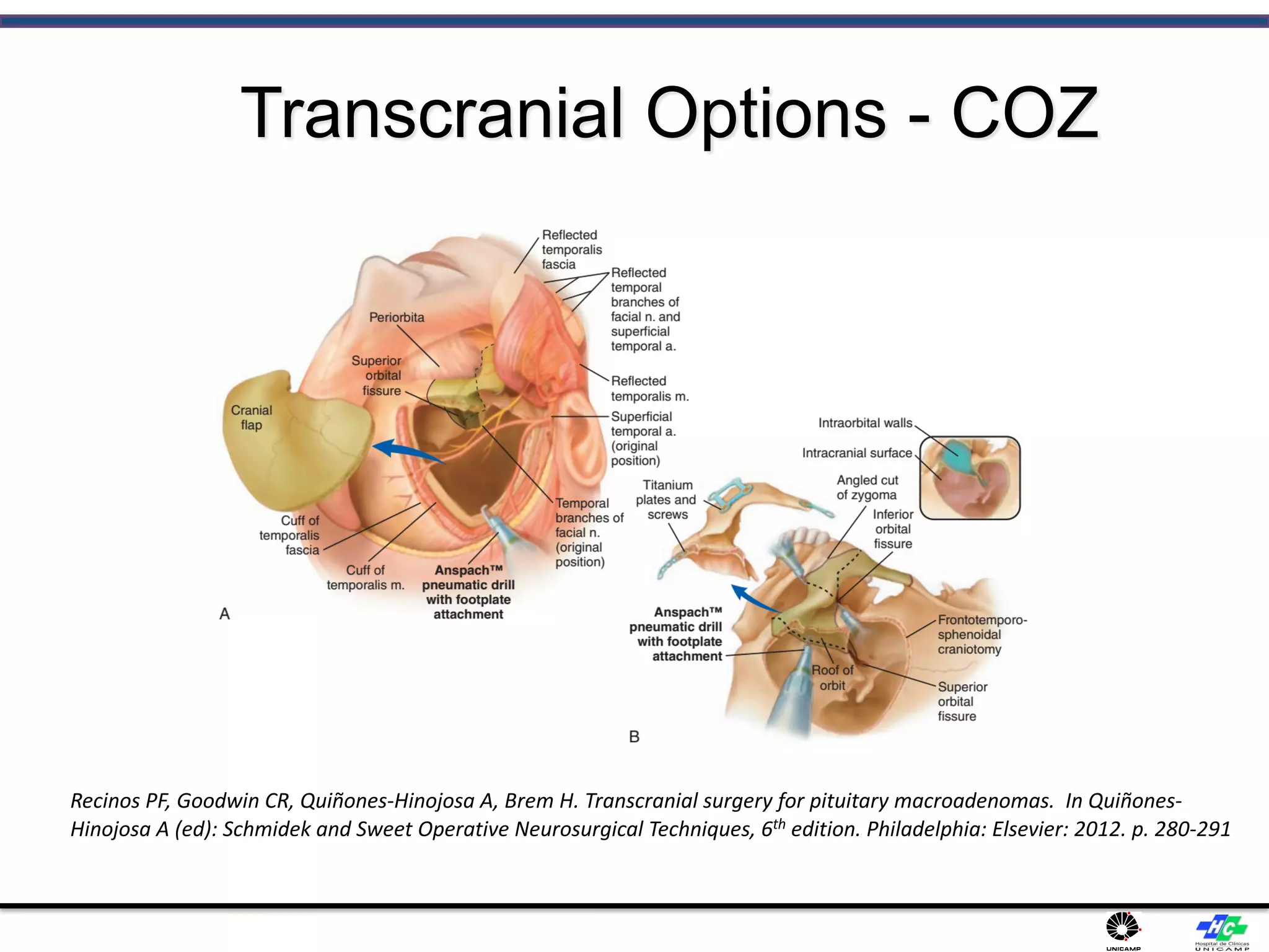
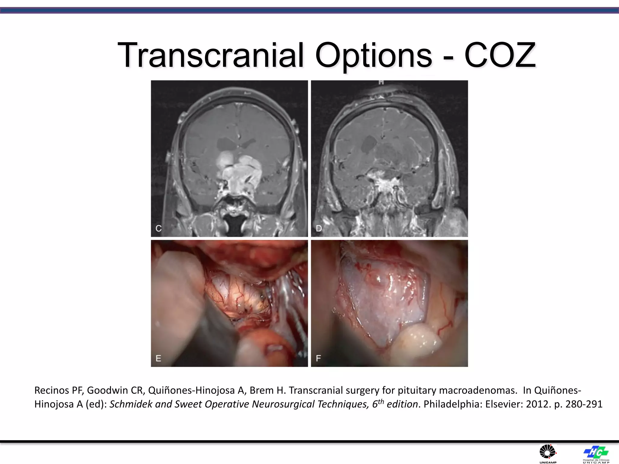
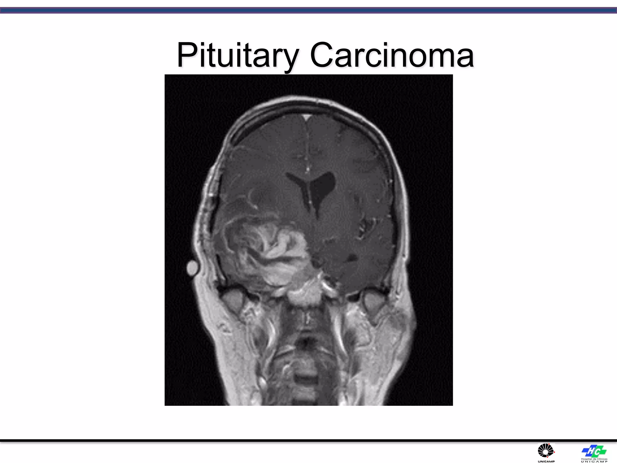
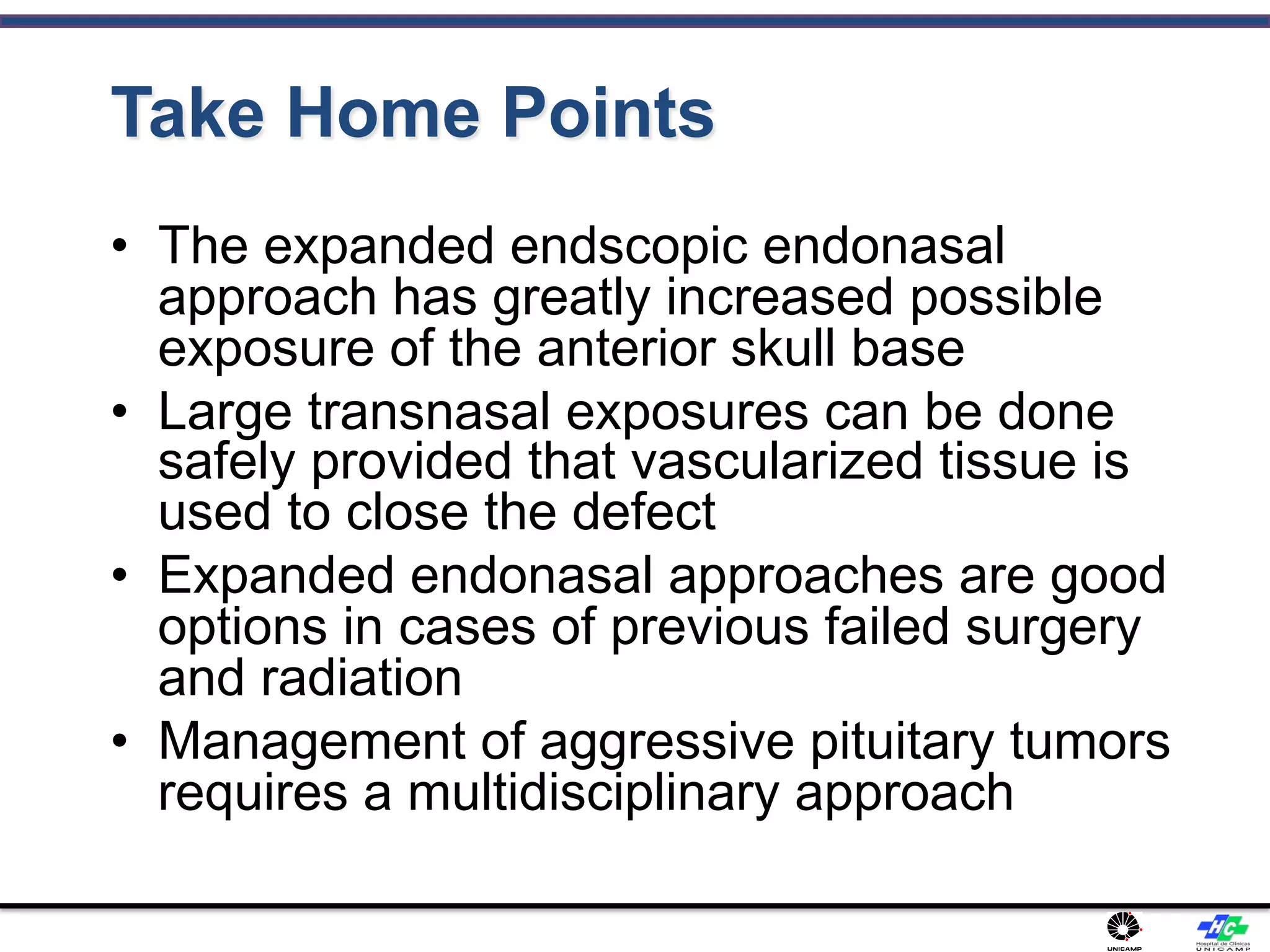
Erion Junior de Andrade
PGY 4 NEUROSUGERY RESIDENT
University of Campinas -UNICAMP
Pituitary Adenomas:
Contemporary Management
Introduction
• Neuroepithelial tumors of ADENOhypophysis
Epidemiology:
• Most common tumor in sella region (except
CRANIOPHARYNGIOMAS in childhood)
• Prevalence is 19-28 cases per 100,000 people.
• Meta-analysis of autopsy data and radiologic
studies in healthy volunteers: pituitary adenomas
are found in 14% of autopsies and 23% of CT/MRI
studies
Epidemiology:
• Mean prevalence of 17%
• 4-20 % of all CNS tumors.
• Young adults (peak 3-4th decades);
• men = women (symptomatic prolactinomas and
Cushing disease are found more frequently in
women)
Tumor development
• Adenomas grow slowly!
• Initially confined to sella turcica → may grow out
of sella and compress /encase / destroy:
a) optic chiasm
b) cavernous sinus and internal carotids (lateral
extension)
c) hypothalamus
d) surrounding bony structures (e.g. sphenoid sinus)
Tumor development
• Locally invasive adenomas nearly always are
histologically benign! CNS metastases and, rarely,
distant metastases can occur!
• Pituitary adenomas never have calcifications!?
(look at CT – if calcium is present, it is
craniopharyngioma)?
. Toshihiro Ogiwara et al. Acta Neurochirurgica 2017 August 19
Tumor development
Adenomas lack discrete capsule, but presence of
pseudocapsule facilitates surgical separation.
Microscopic Anatomy
• Routine staining:
a) chromophilic cells (acidophilic or basophilic)
b) chromophobic cells.
routine staining is meaningless - tumor can be
difficult to differentiate from normal tissue or
metastatic disease and immunohistochemical
staining and electron microscopy are essential!
.
Microscopic Anatomy
Adenoma - packeted arrangement of cells resembles that
of anterior pituitary, together with prominent vascular
network:
Macroscopic Anatomy
Clinical Features
HORMONAL FUNCTION CONTROL
A) hormonal hypersecretion (most commonly
prolactin!)
B) destruction of normal gland → hypopituitarism
(partial in 37-85% patients with nonsecretory
tumors, pan in 6-29% patients with nonsecretory
tumors)
• All MACROADENOMAS eventually cause
hypopituitarism.
Clinical Features
HORMONAL FUNCTION CONTROL
• If hypopituitarism occurs, hormone loss is
sequential: GH → gonadotropins → ACTH → TSH.
• Primary pituitary tumors rarely cause ADH
deficiency (except when induced by
hypophysectomy)
diabetes insipidus is more common in
CRANIOPHARYNGIOMAS.
Clinical Features
MASS EFFECT
1) Headache occurs in 20% (can be diffuse and
nonpulsatile and may be mistaken for daily
headaches; more often in females) – due to
stretching of diaphragma sellae and adjacent dural
structures; ICP is normal!!
Rizzoli P. et al “Headache in Patients With Pituitary
Lesions: A Longitudinal Cohort Study” Neurosurgery:
March 2016 - Volume 78 - Issue 3 - p 316–323
Clinical Features
2) crossing fibers in optic chiasm (superior
bitemporal quadrantanopia → full bitemporal
hemianopia - chief and earliest finding in most
patients!)
Clinical Features
• Relationship of pituitary and optic chiasm:
a) chiasm directly above pituitary (80%).
b) chiasm anteriorly to pituitary (9%) – PRE FIXED
c) chiasm behind pituitary (11%) – POST FIXED
Clinical Features
• further expansion compromises noncrossing
fibers - affects lower and finally upper nasal
quadrants.
any pattern of visual loss is possible, e.g.:
– asymmetrical loss results from chiasm ischemia
produced by vessel occlusion.
Clinical Features
3) Lateral extension into cavernous sinus → diplopia,
ophthalmoplegias, postganglionic Horner syndrome.
4) Hypothalamic compression (e.g. Stalk effect
leading to hyperprolactinemia, diabetes insipidus,
alterations in consciousness, memory, intake of food
and water).
5) Extension into sphenoid sinus → CSF rhinorrhea (≈
0.5% cases) - cortical bone separating sella from
sphenoid sinus is quite thin in normal individuals!
Clinical Features
6) Compression of 3rd ventricle → obstructive
hydrocephalus.
7) Basal forebrain abnormalities (personality
changes, dementia, anosmia).
8) Temporal lobe seizures.
Diagnosis – MRI :
• Gold standard, more sensitive method for tumor
identification (esp. 1-mm cuts and magnified views through
sella – pituitary protocol)
Normal NEUROHYPOPHYSIS
• on T1-MRI shows increased signal (representing
neurosecretory granules in ADH-containing axons).
Normal ADENOHYPOPHYSIS:
• isointense with grey matter on all MR sequences.
• circumventricular organ without an intact BBB - enhances
homogeneously
Diagnosis – MRI :
• MICROADENOMAS enhance later and/or lesser than
normal pituitary tissue!
• other indirect MRI signs:
1) gland height ↑ (normally < 10 mm)
2) gland upper margin contour alteration from concave or
straight to convex
3) erosion of sella turcica floor adjacent to hypointensity area
4) displacement of pituitary stalk (normally midline) away from
hypointensity area.
Diagnosis – MRI :
Diagnosis – MRI :
Diagnosis – SPECT:
• 99mTc(V)-DMSA is actively taken up by adenomas relative to
other sellar/suprasellar lesions.
• Radiolabeled somatostatin or dopamine can potentially
differentiate hormone producing from nonfunctioning
pituitary adenomas and identify patients who would benefit
from pharmacotherapy, although the clinical feasibility of this
is unclear.
Diagnosis– Laboratory:
Diagnosis – Laboratory:
• IPSS: Inferior petrosal sinus sampling is used to localize
tumors not seen radiographically (e.g. many ACTH-secreting
microadenomas are < 5 mm).
• Central hypothyroidism is typically confirmed by the
thyrotropin releasing hormone (TRH) stimulation test, in
which serum TSH is measured serially post-TRH at 20 and 60
minutes, with a normal response defined as the 20- minute
TSH value being higher than the 60-minute TSH value. A flat
response is seen in pituitary disease, and delayed response,
with the 60-minute value higher than the 20-minute value, is
seen in hypothalamic disease.
Diagnosis – Laboratory:
• PROLACTIN
• Nonsecreting tumors are commonly associated with slight
elevations of serum prolactin (< 150*).
• STALK SYNDROME (compression of pituitary stalk,
interrupting dopaminergic fibers that inhibit prolactin
release).
- Hook effect (s. prozone effect) - resulting in false negatives or
inaccurately low results – too much antigen (prolactin)
interferes with results (H: diluting blood sample; modern labs
do it automatically):
Diagnosis – Laboratory:
• Prolactin
Aggressive Pituitary Tumors
• 25-55% of pituitary tumors are invasive
• WHO Classification
– Typical adenomas
– Atypical adenomas
– Pituitary carcinomas
Di Ieva, A. et al. (2014) Aggressive pituitary adenomas—diagnosis and emerging treatments
Nat. Rev. Endocrinol. doi:10.1038/nrendo.2014.64
Atypical Adenomas
• Atypical morphological features
• Elevated mitotic index
• Ki-67 ≥ 3%
• Positive p53 staining
• 2.7% - 15% of all pituitary tumors
Di Ieva, A. et al. (2014) Aggressive pituitary adenomas—diagnosis and emerging treatments
Nat. Rev. Endocrinol. doi:10.1038/nrendo.2014.64
Pituitary Carcinomas
• Must have craniospinal dissemination or
systemic metastases
• 0.2% of all pituitary tumors
• Can share morphological features with
pituitary adenomasDi Ieva, A. et al. (2014) Aggressive pituitary adenomas—diagnosis and emerging treatments
Nat. Rev. Endocrinol. doi:10.1038/nrendo.2014.64
Aggressive vs Invasive
Aggressive
- Clinical behavior
Invasive
- Radiological, surgical, and histopathological
Recognizing Aggressive Tumors
Hardy Classification System
Di Ieva, A. et al. (2014) Aggressive pituitary adenomas—diagnosis and emerging treatments
Nat. Rev. Endocrinol. doi:10.1038/nrendo.2014.64
Knosp Classification System
Di Ieva, A. et al. (2014) Aggressive pituitary adenomas—diagnosis and emerging treatments
Nat. Rev. Endocrinol. doi:10.1038/nrendo.2014.64
Outline
• Evolution of Surgical Approaches to
Pituitary Tumors
“If I have seen further it is by standing
on the shoulders of Giants”
- Sir Isaac Newton
Presentation Title l October 20,
2019 l 36
Historical Approaches
Perneczky A, Müller-Forell W, van Lindert E, Fries G. Keyhole concept in neurosurgery.
New York: Thieme. 1999
Historical Approaches
Courtesy of Robert Wilkins, M.D.
Microscopic TSH
Microscopic TSH
Endoscopic TSH
The Right Plan: What does not change
• Goal of Surgery
– Same indications, end points, criteria of
success
• Basic techniques of tumor resection
– Suction, curettes
• Angle of approach and relevant
anatomy
The Right Plan: What does not change
• Major complications to avoid:
– Carotid injury
– Optic nerve damage, CSF leak, endocrine
complications, post-op hemorrhage
Picture from Johns Hopkins i-star lab website
The Right Plan: What does change
• Speculum use
• Nasal trauma
• Visual perspective
– 3-D at a distance = microscope
– 2-D plus proprioception up close =
endoscope
– Narrow vs wide visual working space
One surgeon vs Two surgeons
Visualization Difference
Simmen DB, Briner HR, Jones N. Endoscopically assisted bimanual operating technique. In Stamm AC(ed):
Transnasal endoscopic skull base and brain surgery. New York: Thieme. 2011. p 92-8.
Visualization Difference
Nomikos P, Fahlbusch R, Buchfelder M. Recent developments in transsphenoidal surgery
of pituitary surgery. Hormones (Athens). 3(2):85-91, 2004.
Looking Around Corners
Perneczky A, Müller-Forell W, van Lindert E, Fries G. Keyhole concept in
neurosurgery. New York: Thieme. 1999
Tools and Working Space
Simmen DB, Briner HR, Jones N. Endoscopically assisted bimanual operating technique. In
Stamm AC(ed): Transnasal endoscopic skull base and brain surgery. New York: Thieme.
2011. p 92-8.
Neuronavigation
Neuronavigation
TEAM APPROACH
§ Two surgeons
§ Simultaneous surgery
§ 2 monitors
§ Increased functional
room in sinus
§ Minimizes insertion
trauma
Learning to dance
Reconstruction
Hadad G, Bassagaisteguy L, Timperley D, Stamm AC. Management of skull base defects after extended
endoscopic skull base surgery: from free grafts to vascularized flaps. In Stamm AC(ed): Transnasal
endoscopic skull base and brain surgery. New York: Thieme. 2011. p 379-85.
Reconstruction
Hadad G, Bassagaisteguy L, Timperley D, Stamm AC. Management of skull base defects after extended
endoscopic skull base surgery: from free grafts to vascularized flaps. In Stamm AC(ed): Transnasal
endoscopic skull base and brain surgery. New York: Thieme. 2011. p 379-85.
- 39.1% pituitary adenomas, 11.8% meningiomas
- CSF leak most common complication (15.9%)
- <6% CSF leak after adoption of nasal-septal flap
Papers on rec outcomes
- Zanation
- Kassam
Outline
• Case Examples of
Persistent/Aggressive Tumors
Choosing the right approach
Firm Macroadenoma
Firm Macroadenoma
Firm Macroadenoma
Multiple prior surgeries + XRT
Aggressive Recurrent
Macroadenoma
Transcranial Options – Extended
Bifrontal
Recinos PF, Goodwin CR, Quiñones-Hinojosa A, Brem H. Transcranial surgery for pituitary macroadenomas. In Quiñones-
Hinojosa A (ed): Schmidek and Sweet Operative Neurosurgical Techniques, 6th edition. Philadelphia: Elsevier: 2012. p. 280-291
Transcranial Options – Extended
Bifrontal
Recinos PF, Goodwin CR, Quiñones-Hinojosa A, Brem H. Transcranial surgery for pituitary macroadenomas. In Quiñones-
Hinojosa A (ed): Schmidek and Sweet Operative Neurosurgical Techniques, 6th edition. Philadelphia: Elsevier: 2012. p. 280-291
Transcranial Options - COZ
Recinos PF, Goodwin CR, Quiñones-Hinojosa A, Brem H. Transcranial surgery for pituitary macroadenomas. In Quiñones-
Hinojosa A (ed): Schmidek and Sweet Operative Neurosurgical Techniques, 6th edition. Philadelphia: Elsevier: 2012. p. 280-291
Transcranial Options - COZ
Recinos PF, Goodwin CR, Quiñones-Hinojosa A, Brem H. Transcranial surgery for pituitary macroadenomas. In Quiñones-
Hinojosa A (ed): Schmidek and Sweet Operative Neurosurgical Techniques, 6th edition. Philadelphia: Elsevier: 2012. p. 280-291
Pituitary Carcinoma
Pituitary Carcinoma
Pituitary Carcinoma
Pituitary Carcinoma
Pituitary Carcinoma
Pituitary Carcinoma
Take Home Points
• The expanded endscopic endonasal
approach has greatly increased possible
exposure of the anterior skull base
• Large transnasal exposures can be done
safely provided that vascularized tissue is
used to close the defect
• Expanded endonasal approaches are good
options in cases of previous failed surgery
and radiation
• Management of aggressive pituitary tumors
requires a multidisciplinary approach
Pituitary adenomas

Pituitary adenomas

  • 1.
    Erion Junior deAndrade PGY 4 NEUROSUGERY RESIDENT University of Campinas -UNICAMP Pituitary Adenomas: Contemporary Management
  • 2.
  • 3.
    Epidemiology: • Most commontumor in sella region (except CRANIOPHARYNGIOMAS in childhood) • Prevalence is 19-28 cases per 100,000 people. • Meta-analysis of autopsy data and radiologic studies in healthy volunteers: pituitary adenomas are found in 14% of autopsies and 23% of CT/MRI studies
  • 4.
    Epidemiology: • Mean prevalenceof 17% • 4-20 % of all CNS tumors. • Young adults (peak 3-4th decades); • men = women (symptomatic prolactinomas and Cushing disease are found more frequently in women)
  • 5.
    Tumor development • Adenomasgrow slowly! • Initially confined to sella turcica → may grow out of sella and compress /encase / destroy: a) optic chiasm b) cavernous sinus and internal carotids (lateral extension) c) hypothalamus d) surrounding bony structures (e.g. sphenoid sinus)
  • 6.
    Tumor development • Locallyinvasive adenomas nearly always are histologically benign! CNS metastases and, rarely, distant metastases can occur! • Pituitary adenomas never have calcifications!? (look at CT – if calcium is present, it is craniopharyngioma)? . Toshihiro Ogiwara et al. Acta Neurochirurgica 2017 August 19
  • 7.
    Tumor development Adenomas lackdiscrete capsule, but presence of pseudocapsule facilitates surgical separation.
  • 8.
    Microscopic Anatomy • Routinestaining: a) chromophilic cells (acidophilic or basophilic) b) chromophobic cells. routine staining is meaningless - tumor can be difficult to differentiate from normal tissue or metastatic disease and immunohistochemical staining and electron microscopy are essential! .
  • 9.
    Microscopic Anatomy Adenoma -packeted arrangement of cells resembles that of anterior pituitary, together with prominent vascular network:
  • 10.
  • 11.
    Clinical Features HORMONAL FUNCTIONCONTROL A) hormonal hypersecretion (most commonly prolactin!) B) destruction of normal gland → hypopituitarism (partial in 37-85% patients with nonsecretory tumors, pan in 6-29% patients with nonsecretory tumors) • All MACROADENOMAS eventually cause hypopituitarism.
  • 12.
    Clinical Features HORMONAL FUNCTIONCONTROL • If hypopituitarism occurs, hormone loss is sequential: GH → gonadotropins → ACTH → TSH. • Primary pituitary tumors rarely cause ADH deficiency (except when induced by hypophysectomy) diabetes insipidus is more common in CRANIOPHARYNGIOMAS.
  • 13.
    Clinical Features MASS EFFECT 1)Headache occurs in 20% (can be diffuse and nonpulsatile and may be mistaken for daily headaches; more often in females) – due to stretching of diaphragma sellae and adjacent dural structures; ICP is normal!! Rizzoli P. et al “Headache in Patients With Pituitary Lesions: A Longitudinal Cohort Study” Neurosurgery: March 2016 - Volume 78 - Issue 3 - p 316–323
  • 14.
    Clinical Features 2) crossingfibers in optic chiasm (superior bitemporal quadrantanopia → full bitemporal hemianopia - chief and earliest finding in most patients!)
  • 15.
    Clinical Features • Relationshipof pituitary and optic chiasm: a) chiasm directly above pituitary (80%). b) chiasm anteriorly to pituitary (9%) – PRE FIXED c) chiasm behind pituitary (11%) – POST FIXED
  • 16.
    Clinical Features • furtherexpansion compromises noncrossing fibers - affects lower and finally upper nasal quadrants. any pattern of visual loss is possible, e.g.: – asymmetrical loss results from chiasm ischemia produced by vessel occlusion.
  • 17.
    Clinical Features 3) Lateralextension into cavernous sinus → diplopia, ophthalmoplegias, postganglionic Horner syndrome. 4) Hypothalamic compression (e.g. Stalk effect leading to hyperprolactinemia, diabetes insipidus, alterations in consciousness, memory, intake of food and water). 5) Extension into sphenoid sinus → CSF rhinorrhea (≈ 0.5% cases) - cortical bone separating sella from sphenoid sinus is quite thin in normal individuals!
  • 18.
    Clinical Features 6) Compressionof 3rd ventricle → obstructive hydrocephalus. 7) Basal forebrain abnormalities (personality changes, dementia, anosmia). 8) Temporal lobe seizures.
  • 19.
    Diagnosis – MRI: • Gold standard, more sensitive method for tumor identification (esp. 1-mm cuts and magnified views through sella – pituitary protocol) Normal NEUROHYPOPHYSIS • on T1-MRI shows increased signal (representing neurosecretory granules in ADH-containing axons). Normal ADENOHYPOPHYSIS: • isointense with grey matter on all MR sequences. • circumventricular organ without an intact BBB - enhances homogeneously
  • 20.
    Diagnosis – MRI: • MICROADENOMAS enhance later and/or lesser than normal pituitary tissue! • other indirect MRI signs: 1) gland height ↑ (normally < 10 mm) 2) gland upper margin contour alteration from concave or straight to convex 3) erosion of sella turcica floor adjacent to hypointensity area 4) displacement of pituitary stalk (normally midline) away from hypointensity area.
  • 21.
  • 22.
  • 23.
    Diagnosis – SPECT: •99mTc(V)-DMSA is actively taken up by adenomas relative to other sellar/suprasellar lesions. • Radiolabeled somatostatin or dopamine can potentially differentiate hormone producing from nonfunctioning pituitary adenomas and identify patients who would benefit from pharmacotherapy, although the clinical feasibility of this is unclear.
  • 24.
  • 25.
    Diagnosis – Laboratory: •IPSS: Inferior petrosal sinus sampling is used to localize tumors not seen radiographically (e.g. many ACTH-secreting microadenomas are < 5 mm). • Central hypothyroidism is typically confirmed by the thyrotropin releasing hormone (TRH) stimulation test, in which serum TSH is measured serially post-TRH at 20 and 60 minutes, with a normal response defined as the 20- minute TSH value being higher than the 60-minute TSH value. A flat response is seen in pituitary disease, and delayed response, with the 60-minute value higher than the 20-minute value, is seen in hypothalamic disease.
  • 26.
    Diagnosis – Laboratory: •PROLACTIN • Nonsecreting tumors are commonly associated with slight elevations of serum prolactin (< 150*). • STALK SYNDROME (compression of pituitary stalk, interrupting dopaminergic fibers that inhibit prolactin release). - Hook effect (s. prozone effect) - resulting in false negatives or inaccurately low results – too much antigen (prolactin) interferes with results (H: diluting blood sample; modern labs do it automatically):
  • 27.
  • 28.
    Aggressive Pituitary Tumors •25-55% of pituitary tumors are invasive • WHO Classification – Typical adenomas – Atypical adenomas – Pituitary carcinomas Di Ieva, A. et al. (2014) Aggressive pituitary adenomas—diagnosis and emerging treatments Nat. Rev. Endocrinol. doi:10.1038/nrendo.2014.64
  • 29.
    Atypical Adenomas • Atypicalmorphological features • Elevated mitotic index • Ki-67 ≥ 3% • Positive p53 staining • 2.7% - 15% of all pituitary tumors Di Ieva, A. et al. (2014) Aggressive pituitary adenomas—diagnosis and emerging treatments Nat. Rev. Endocrinol. doi:10.1038/nrendo.2014.64
  • 30.
    Pituitary Carcinomas • Musthave craniospinal dissemination or systemic metastases • 0.2% of all pituitary tumors • Can share morphological features with pituitary adenomasDi Ieva, A. et al. (2014) Aggressive pituitary adenomas—diagnosis and emerging treatments Nat. Rev. Endocrinol. doi:10.1038/nrendo.2014.64
  • 31.
    Aggressive vs Invasive Aggressive -Clinical behavior Invasive - Radiological, surgical, and histopathological
  • 32.
  • 33.
    Hardy Classification System DiIeva, A. et al. (2014) Aggressive pituitary adenomas—diagnosis and emerging treatments Nat. Rev. Endocrinol. doi:10.1038/nrendo.2014.64
  • 34.
    Knosp Classification System DiIeva, A. et al. (2014) Aggressive pituitary adenomas—diagnosis and emerging treatments Nat. Rev. Endocrinol. doi:10.1038/nrendo.2014.64
  • 35.
    Outline • Evolution ofSurgical Approaches to Pituitary Tumors
  • 36.
    “If I haveseen further it is by standing on the shoulders of Giants” - Sir Isaac Newton Presentation Title l October 20, 2019 l 36
  • 37.
    Historical Approaches Perneczky A,Müller-Forell W, van Lindert E, Fries G. Keyhole concept in neurosurgery. New York: Thieme. 1999
  • 38.
  • 39.
  • 40.
  • 41.
  • 42.
  • 43.
    The Right Plan:What does not change • Goal of Surgery – Same indications, end points, criteria of success • Basic techniques of tumor resection – Suction, curettes • Angle of approach and relevant anatomy
  • 44.
    The Right Plan:What does not change • Major complications to avoid: – Carotid injury – Optic nerve damage, CSF leak, endocrine complications, post-op hemorrhage Picture from Johns Hopkins i-star lab website
  • 45.
    The Right Plan:What does change • Speculum use • Nasal trauma • Visual perspective – 3-D at a distance = microscope – 2-D plus proprioception up close = endoscope – Narrow vs wide visual working space One surgeon vs Two surgeons
  • 46.
    Visualization Difference Simmen DB,Briner HR, Jones N. Endoscopically assisted bimanual operating technique. In Stamm AC(ed): Transnasal endoscopic skull base and brain surgery. New York: Thieme. 2011. p 92-8.
  • 47.
    Visualization Difference Nomikos P,Fahlbusch R, Buchfelder M. Recent developments in transsphenoidal surgery of pituitary surgery. Hormones (Athens). 3(2):85-91, 2004.
  • 48.
    Looking Around Corners PerneczkyA, Müller-Forell W, van Lindert E, Fries G. Keyhole concept in neurosurgery. New York: Thieme. 1999
  • 49.
    Tools and WorkingSpace Simmen DB, Briner HR, Jones N. Endoscopically assisted bimanual operating technique. In Stamm AC(ed): Transnasal endoscopic skull base and brain surgery. New York: Thieme. 2011. p 92-8.
  • 50.
  • 51.
  • 52.
    TEAM APPROACH § Twosurgeons § Simultaneous surgery § 2 monitors § Increased functional room in sinus § Minimizes insertion trauma
  • 53.
  • 55.
  • 56.
    Hadad G, BassagaisteguyL, Timperley D, Stamm AC. Management of skull base defects after extended endoscopic skull base surgery: from free grafts to vascularized flaps. In Stamm AC(ed): Transnasal endoscopic skull base and brain surgery. New York: Thieme. 2011. p 379-85.
  • 57.
    Reconstruction Hadad G, BassagaisteguyL, Timperley D, Stamm AC. Management of skull base defects after extended endoscopic skull base surgery: from free grafts to vascularized flaps. In Stamm AC(ed): Transnasal endoscopic skull base and brain surgery. New York: Thieme. 2011. p 379-85.
  • 58.
    - 39.1% pituitaryadenomas, 11.8% meningiomas - CSF leak most common complication (15.9%) - <6% CSF leak after adoption of nasal-septal flap
  • 59.
    Papers on recoutcomes - Zanation - Kassam
  • 61.
    Outline • Case Examplesof Persistent/Aggressive Tumors
  • 62.
  • 63.
  • 64.
  • 65.
  • 66.
  • 67.
  • 68.
  • 69.
  • 70.
  • 71.
  • 72.
    Transcranial Options –Extended Bifrontal Recinos PF, Goodwin CR, Quiñones-Hinojosa A, Brem H. Transcranial surgery for pituitary macroadenomas. In Quiñones- Hinojosa A (ed): Schmidek and Sweet Operative Neurosurgical Techniques, 6th edition. Philadelphia: Elsevier: 2012. p. 280-291
  • 73.
    Transcranial Options –Extended Bifrontal Recinos PF, Goodwin CR, Quiñones-Hinojosa A, Brem H. Transcranial surgery for pituitary macroadenomas. In Quiñones- Hinojosa A (ed): Schmidek and Sweet Operative Neurosurgical Techniques, 6th edition. Philadelphia: Elsevier: 2012. p. 280-291
  • 74.
    Transcranial Options -COZ Recinos PF, Goodwin CR, Quiñones-Hinojosa A, Brem H. Transcranial surgery for pituitary macroadenomas. In Quiñones- Hinojosa A (ed): Schmidek and Sweet Operative Neurosurgical Techniques, 6th edition. Philadelphia: Elsevier: 2012. p. 280-291
  • 75.
    Transcranial Options -COZ Recinos PF, Goodwin CR, Quiñones-Hinojosa A, Brem H. Transcranial surgery for pituitary macroadenomas. In Quiñones- Hinojosa A (ed): Schmidek and Sweet Operative Neurosurgical Techniques, 6th edition. Philadelphia: Elsevier: 2012. p. 280-291
  • 76.
  • 77.
  • 78.
  • 79.
  • 80.
  • 81.
  • 82.
    Take Home Points •The expanded endscopic endonasal approach has greatly increased possible exposure of the anterior skull base • Large transnasal exposures can be done safely provided that vascularized tissue is used to close the defect • Expanded endonasal approaches are good options in cases of previous failed surgery and radiation • Management of aggressive pituitary tumors requires a multidisciplinary approach