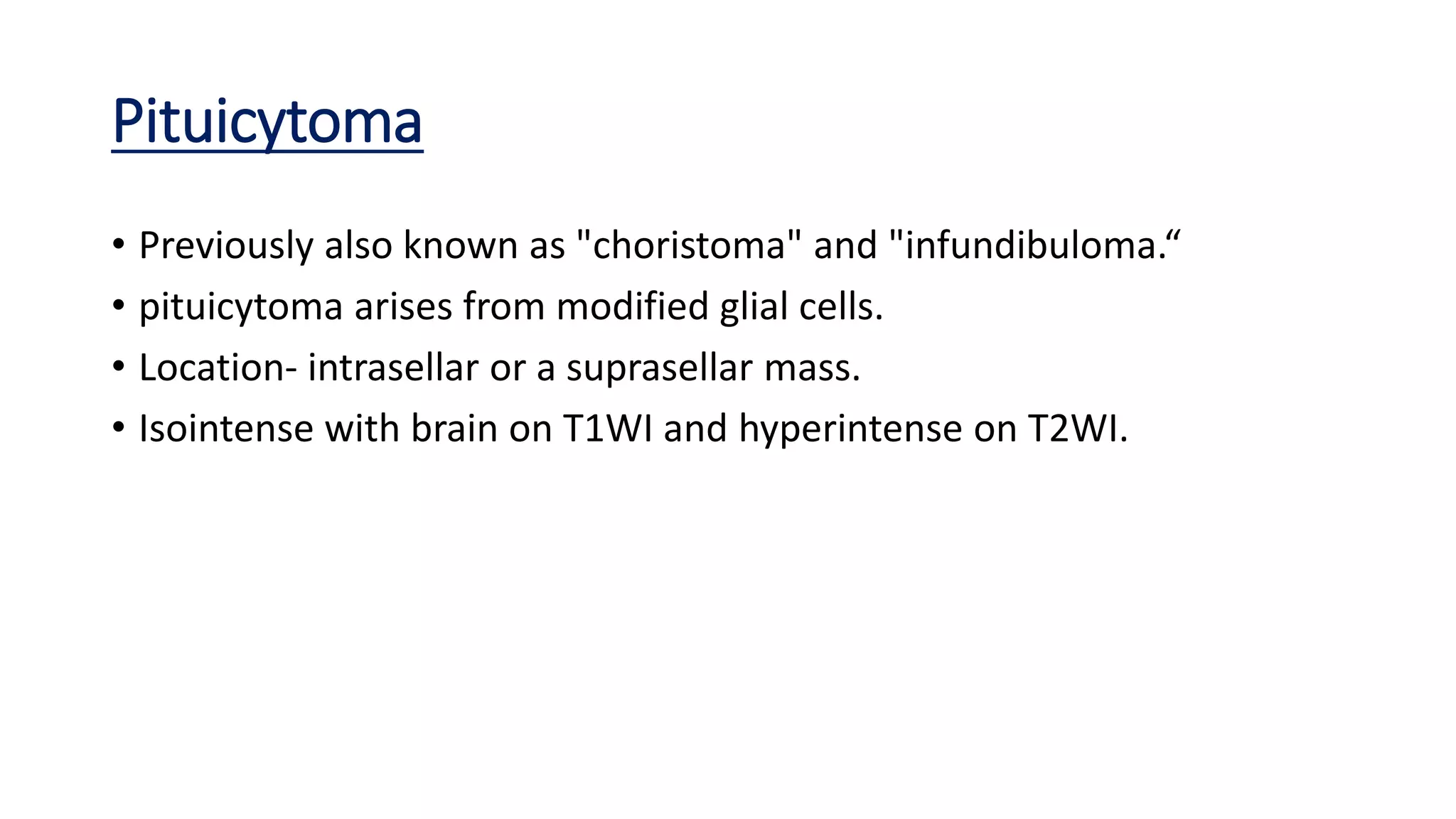
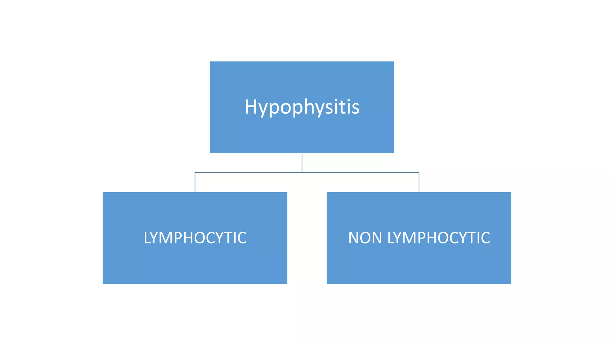
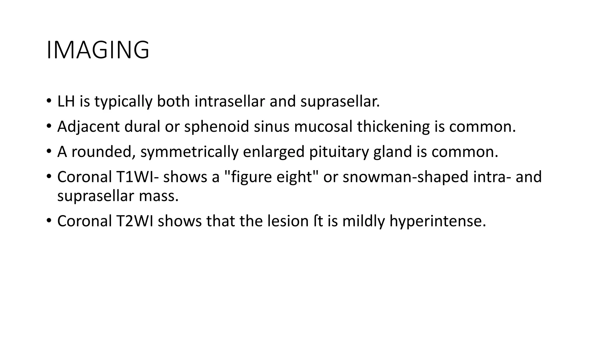
PITUITARY TUMORS
BY DR AVINASH DAHATRE
JR II RADIOLOGY
Spectrum of lesions that can occur in/around
the sella
S
•SARCOID
A
•ADENOMA
•ANEURYSM
T
•TUBERCULOSIS
•TERATOMA
C
•CYST
•CRANIOPHARYNGIOMA
H
•HYPOPHYSISTIS
•HAMARTOMA
M
•MENINGIOMA
•METASTASIS
O
•OPTIC GLIOMA
Diagnostic Considerations :-
• key to determining anatomic sublocation accurately is the
question, "Can I find the pituitary gland separate from the
mass?" If you can't, and the gland is the mass, the most likely
diagnosis is MACROADENOMA.
• If the mass is clearly separate from the pituitary gland, it is
extrapituitary and therefore not a macroadenoma.
• An enlarged pituitary gland in a child is almost always either
normal physiologic hypertrophy or nonphysiologic
nonneoplastic hyperplasia secondary to end-organ failure
(most commonly hypothyroidism).
• macroadenoma, meningioma, and aneurysm are generally
rare in children.
ANATOMY
• sella turcica ("Turkish saddle") is a midline concavity in the
basisphenoid of the central skull base that contains the pituitary
gland
• Anterior border:- Formed by the tuberculum sellae and anterior
clinoid processes of the lesser sphenoid wing.
• Posterior border:- Formed by the dorsum sellae.
• Floor:- is part of the sphenoid sinus roof.
• Roof :- Diaphragma sellae
• Dura covers the bony floor of the sella, separating it from the
pituitary gland.
VARIANTS:-
• Kissing" Carotid Arteries:-
• Cavernous internal carotid arteries (ICAs) normally lie lateral to the pituitary
gland in the parasellar carotid sulci but Occasionally, course inside the bony
sella, squeezing it upward and making it appear modestly enlarged.
•Pituitary Hyperplasia:-
Nonneoplastic increase in adenohypophysial cell number.
Physiologic Hyperplasia: Hypertrophy of puberty, Enlarged pituitary glands in
young menstruating female patients is very common secondary to prolactin.
Pathologic Hyperplasia: Most commonly occurs in response to end-organ failure.
Primary hypothyroidism is the most common cause of pathologic pituitary
hyperplasia.
Growth hormone (GH) cell hyperplasia.
ACTH cell hyperplasia in ACTH-dependent Cushing disease.
Mammosomatotroph hyperplasia occurs in Gigantism.
• NECT scans show that the superior margin of the gland is convex
upward
• MR demonstrates an enlarged gland that bulges upward and may
even contact the optic chiasm. The enlarged pituitary is isointense
with cortex on both T1- and T2WI.
SAGT1WI
SAGT1WI +Contrast
CORT2WI
Empty Sella
• It is an arachnoid-lined, CSF-filled protrusion that extends from the
suprasellar cistern through the diaphragma sellae into the sella turcica.
• ES is rarely completely "empty"; a small remnant of flattened pituitary
gland is almost always present at the bottom of the bony sella.
• CAUSES:-
• primary cause:-occurs due to opening in the diaphragma sellae allows
intrasellar herniation of arachnoid and CSF from the suprasellar cistern.
• Secondary cause:- occurs when pituitary volume is reduced with surgery,
bromocriptine therapy, or radiation treatment, Sheehans syndrome.
IMAGING:-
• CT Findings:- CSF-density fluid fills a sella that may be of normal size
or moderately enlarged
• The bony floor of the sella is intact in primary ES, but, in secondary
ES, it often shows a surgical defect caused by transsphenoidal
hypophysectomy
• MR Findings:- The intrasellar fluid behaves exactly like CSF on T1- and
T2WI and suppresses completely on FLAIR. DWI shows no diffusion
restriction.
PARTIALLY EMPTY
SELLA
EMPTY SELLA
Congenital Lesions Such As:-
Pituitary Anomalies :-
Pituitary Hypoplasia
Most frequent abnormality in
children with isolated growth
hormone deficiency.
Imaging abnormalities include a
small sella and anterior pituitary
lobe, hypoplasia or absence of
the stalk, and an "ectopic"
posterior pituitary "bright spot"
seen as displacement of the T1
hyperintense.
Pituitary duplication
• Pituitary duplication is a rare
anomaly in which two pituitary
stalks can be identified on the
coronal view.
• Female patients are more
commonly affected.
Pituitary hypoplasia with
displaced bright spot
Duplication of pituitary
stalk
Rathke Cleft Cyst
• Benign endodermal cyst of the sellar region.
• They are smoothly lobulated, sharply marginated cysts.
• Contents vary from clear and CSF-like to thick yellow inspissated
mucoid material.
• AGE of Onset:- occur at all ages, mean age at presentation is 45 years.
• Location:- 40% are completely intrasellar, whereas 60% are
suprasellar.
• They are 5-15 mm in diameter. Occasionally, an RCC becomes very
large and can compress the optic chiasm.
Imaging
• CT Findings:-
NECT scans show a well-delineated round or ovoid mass within or just
above the sella turcica.
RCCs are hypodense on NECT, whereas 20% are mixed hypo- and isodense
• MR Findings:-
Signal intensity varies with cyst contents. Half of all RCCs are hypointense on
T1WI, and half are hyperintense.
hyperintense on T2WI.
hyperintense on FLAIR
Enhancing rim ("claw" sign) of compressed pituitary gland can often be seen
surrounding the nonenhancing cyst.
hyperintense on T2WI.
hypointense on T1WI T1+C Enhancing rim
Neoplasms:-
PITUITARY
ADENOMAS
MACROADENOMA
MICROADENOMA
(<10mm)
• Adenohypophysial tumors composed of secretory cells that produce
pituitary hormones.
• Location:- Adenomas arise within the sella turcica. Reported ectopic
sites include the sphenoid sinus (the most common site),
nasopharynx, third ventricle, and suprasellar cistern.
• Size :- from microscopic lesions to giant tumors more than 5 cm.
GENETICS:-
• genetic defects are associated with pituitary adenomas are:
multiple endocrine neoplasia type 1 (MEN1):- Are often plurihormonal (most
commonly secreting prolactin and growth hormone)
Carney complex:- Associated with spotty skin pigmentation, myxomas, endocrine tumors,
and schwannomas.
McCune-Albright syndrome (MAS):-Defined by the triad of gonadotropin-
independent sexual precocity, café au lait skin lesions, and fibrous dysplasia. Caused by a
postzygotic mutation in the GNAS gene.
familial isolated pituitary adenoma (FIPA) syndrome:- It includes familial
pituitary tumors that are not associated with MEN1 and Carney complex
Imaging
• CT Findings :-
• Bone CT may show an enlarged, remodeled sella turcica.
• Macroadenomas are usually isodense with gray matter.
• Calcification is rare.
• Moderate but heterogeneous enhancement of macroadenomas is
typical on CECT.
MR Findings
• Macroadenomas are usually isointense with cortex
• T1WI- posterior pituitary "bright spot" is absent (20%) or displaced
into the supradiaphragmatic cistern (80%).
• T2WI- Adenomas are generally isointense with gray matter
• T2/FLAIR-Hyperintensity along the optic pathways.
• Microadenomas on T1WI post contrast- hypointense.
• Fast image acquisition during contrast administration can often
discriminate between the slowly enhancing microadenoma and
rapidly enhancing normal gland.
On T1WI-Normal bright spot believed to be from the
storage of vasopressin, which has a T1-shortening effect
T1WI- ‘Absent Bright spot’
FLAIR- Large homogeneously enhancing mass enlarges the pituitary fossa and
compresses the optic chiasm.
ANGIOGRAPHY
• macroadenoma may show the supraclinoid internal carotid and
anterior choroidal arteries displaced laterally.
Craniopharyngioma
• probably arises from epithelial remnants of Rathke pouch.
• Location-
CPs are primarily suprasellar tumors.
Completely intrasellar CPs are rare.
Size- Lesions larger than 5 cm are common.
Giant CPs may extend into both anterior and middle cranial fossa.
Age of onset- peak between 5 and 15 years and a second, smaller peak
at 45-60 years.
Gross Pathology.
• Two types of craniopharyngiomas are:-
• Adamantinomatous 90% - multilobulated, partially solid but mostly
cystic suprasellar mass. Cholesterol-rich "machinery oil" fluid.
• Papillary 10% - are often solid, with a cauliflower-like configuration.
CT Findings
• Adamantinomatous CPs follow a "rule of ninety," i.e.,
90% are mixed cystic/solid
90% are calcified
90% enhance
○ Can be giant (> 5 cm), involve multiple fossae
• Papillary CPs rarely calcify. They are often solid or mostly solid.
MRI
○ Variable signal on T1WI
○ Usually hyperintense on
T2/FLAIR
○ Enhancement (nodular or
rim) 90%
○ MRS: large lipid-lactate
peak
Differential Diagnosis
1. Rathke cleft cyst (RCC)-
RCCs do not calcify, appear to be much less heterogeneous, and do not
show nodular enhancement.
Nonadenomatous Pituitary Tumors
• Pituicytoma
• spindle cell oncocytoma (SCO)
• granular cell tumor
Pituicytoma
• Previously also known as "choristoma" and "infundibuloma.“
• pituicytoma arises from modified glial cells.
• Location- intrasellar or a suprasellar mass.
• Isointense with brain on T1WI and hyperintense on T2WI.
spindle cell oncocytoma (SCO)
• SCO, also previously known as folliculostellate cell tumor, consists of
"spindled" oncocytes containing granular, mitochondria-rich
cytoplasm.
granular cell tumor
• tumor of the neurohypophysis.
• Typically suprasellar masses.
• NECT- Hyperdense.
• MRI- isointense with brain on both T1- and T2WI.
Hypophysitis
LYMPHOCYTIC NON LYMPHOCYTIC
LYMPHOCYTIC HYPOPHYSITIS
• Uncommon autoimmune inflammatory disorder of the pituitary gland
that most often occurs in women of child-bearing age.
• Varying degrees of gland destruction and fibrosis may be present.
• pituitary gland and stalk in LH appear diffusely enlarged and firm.
IMAGING
• LH is typically both intrasellar and suprasellar.
• Adjacent dural or sphenoid sinus mucosal thickening is common.
• A rounded, symmetrically enlarged pituitary gland is common.
• Coronal T1WI- shows a "figure eight" or snowman-shaped intra- and
suprasellar mass.
• Coronal T2WI shows that the lesion ſt is mildly hyperintense.
T1-weighted MRI scans showing a sellar mass arising from the pituitary fossa and
extending into suprasellar region with heterogeneous contrast enhancement.
PITUITARY TUMORS

PITUITARY TUMORS

  • 1.
    PITUITARY TUMORS BY DRAVINASH DAHATRE JR II RADIOLOGY
  • 2.
    Spectrum of lesionsthat can occur in/around the sella S •SARCOID A •ADENOMA •ANEURYSM T •TUBERCULOSIS •TERATOMA C •CYST •CRANIOPHARYNGIOMA H •HYPOPHYSISTIS •HAMARTOMA M •MENINGIOMA •METASTASIS O •OPTIC GLIOMA Diagnostic Considerations :- • key to determining anatomic sublocation accurately is the question, "Can I find the pituitary gland separate from the mass?" If you can't, and the gland is the mass, the most likely diagnosis is MACROADENOMA. • If the mass is clearly separate from the pituitary gland, it is extrapituitary and therefore not a macroadenoma. • An enlarged pituitary gland in a child is almost always either normal physiologic hypertrophy or nonphysiologic nonneoplastic hyperplasia secondary to end-organ failure (most commonly hypothyroidism). • macroadenoma, meningioma, and aneurysm are generally rare in children.
  • 3.
    ANATOMY • sella turcica("Turkish saddle") is a midline concavity in the basisphenoid of the central skull base that contains the pituitary gland • Anterior border:- Formed by the tuberculum sellae and anterior clinoid processes of the lesser sphenoid wing. • Posterior border:- Formed by the dorsum sellae. • Floor:- is part of the sphenoid sinus roof. • Roof :- Diaphragma sellae • Dura covers the bony floor of the sella, separating it from the pituitary gland.
  • 5.
    VARIANTS:- • Kissing" CarotidArteries:- • Cavernous internal carotid arteries (ICAs) normally lie lateral to the pituitary gland in the parasellar carotid sulci but Occasionally, course inside the bony sella, squeezing it upward and making it appear modestly enlarged.
  • 6.
    •Pituitary Hyperplasia:- Nonneoplastic increasein adenohypophysial cell number. Physiologic Hyperplasia: Hypertrophy of puberty, Enlarged pituitary glands in young menstruating female patients is very common secondary to prolactin. Pathologic Hyperplasia: Most commonly occurs in response to end-organ failure. Primary hypothyroidism is the most common cause of pathologic pituitary hyperplasia. Growth hormone (GH) cell hyperplasia. ACTH cell hyperplasia in ACTH-dependent Cushing disease. Mammosomatotroph hyperplasia occurs in Gigantism.
  • 7.
    • NECT scansshow that the superior margin of the gland is convex upward • MR demonstrates an enlarged gland that bulges upward and may even contact the optic chiasm. The enlarged pituitary is isointense with cortex on both T1- and T2WI.
  • 8.
  • 9.
  • 10.
    Empty Sella • Itis an arachnoid-lined, CSF-filled protrusion that extends from the suprasellar cistern through the diaphragma sellae into the sella turcica. • ES is rarely completely "empty"; a small remnant of flattened pituitary gland is almost always present at the bottom of the bony sella. • CAUSES:- • primary cause:-occurs due to opening in the diaphragma sellae allows intrasellar herniation of arachnoid and CSF from the suprasellar cistern. • Secondary cause:- occurs when pituitary volume is reduced with surgery, bromocriptine therapy, or radiation treatment, Sheehans syndrome.
  • 11.
    IMAGING:- • CT Findings:-CSF-density fluid fills a sella that may be of normal size or moderately enlarged • The bony floor of the sella is intact in primary ES, but, in secondary ES, it often shows a surgical defect caused by transsphenoidal hypophysectomy • MR Findings:- The intrasellar fluid behaves exactly like CSF on T1- and T2WI and suppresses completely on FLAIR. DWI shows no diffusion restriction.
  • 12.
  • 13.
    Congenital Lesions SuchAs:- Pituitary Anomalies :- Pituitary Hypoplasia Most frequent abnormality in children with isolated growth hormone deficiency. Imaging abnormalities include a small sella and anterior pituitary lobe, hypoplasia or absence of the stalk, and an "ectopic" posterior pituitary "bright spot" seen as displacement of the T1 hyperintense. Pituitary duplication • Pituitary duplication is a rare anomaly in which two pituitary stalks can be identified on the coronal view. • Female patients are more commonly affected.
  • 14.
    Pituitary hypoplasia with displacedbright spot Duplication of pituitary stalk
  • 15.
    Rathke Cleft Cyst •Benign endodermal cyst of the sellar region. • They are smoothly lobulated, sharply marginated cysts. • Contents vary from clear and CSF-like to thick yellow inspissated mucoid material. • AGE of Onset:- occur at all ages, mean age at presentation is 45 years. • Location:- 40% are completely intrasellar, whereas 60% are suprasellar. • They are 5-15 mm in diameter. Occasionally, an RCC becomes very large and can compress the optic chiasm.
  • 16.
    Imaging • CT Findings:- NECTscans show a well-delineated round or ovoid mass within or just above the sella turcica. RCCs are hypodense on NECT, whereas 20% are mixed hypo- and isodense
  • 17.
    • MR Findings:- Signalintensity varies with cyst contents. Half of all RCCs are hypointense on T1WI, and half are hyperintense. hyperintense on T2WI. hyperintense on FLAIR Enhancing rim ("claw" sign) of compressed pituitary gland can often be seen surrounding the nonenhancing cyst. hyperintense on T2WI. hypointense on T1WI T1+C Enhancing rim
  • 18.
  • 19.
  • 20.
    • Adenohypophysial tumorscomposed of secretory cells that produce pituitary hormones. • Location:- Adenomas arise within the sella turcica. Reported ectopic sites include the sphenoid sinus (the most common site), nasopharynx, third ventricle, and suprasellar cistern. • Size :- from microscopic lesions to giant tumors more than 5 cm.
  • 21.
    GENETICS:- • genetic defectsare associated with pituitary adenomas are: multiple endocrine neoplasia type 1 (MEN1):- Are often plurihormonal (most commonly secreting prolactin and growth hormone) Carney complex:- Associated with spotty skin pigmentation, myxomas, endocrine tumors, and schwannomas. McCune-Albright syndrome (MAS):-Defined by the triad of gonadotropin- independent sexual precocity, café au lait skin lesions, and fibrous dysplasia. Caused by a postzygotic mutation in the GNAS gene. familial isolated pituitary adenoma (FIPA) syndrome:- It includes familial pituitary tumors that are not associated with MEN1 and Carney complex
  • 22.
    Imaging • CT Findings:- • Bone CT may show an enlarged, remodeled sella turcica. • Macroadenomas are usually isodense with gray matter. • Calcification is rare. • Moderate but heterogeneous enhancement of macroadenomas is typical on CECT.
  • 24.
    MR Findings • Macroadenomasare usually isointense with cortex • T1WI- posterior pituitary "bright spot" is absent (20%) or displaced into the supradiaphragmatic cistern (80%). • T2WI- Adenomas are generally isointense with gray matter • T2/FLAIR-Hyperintensity along the optic pathways. • Microadenomas on T1WI post contrast- hypointense. • Fast image acquisition during contrast administration can often discriminate between the slowly enhancing microadenoma and rapidly enhancing normal gland.
  • 25.
    On T1WI-Normal brightspot believed to be from the storage of vasopressin, which has a T1-shortening effect T1WI- ‘Absent Bright spot’
  • 26.
    FLAIR- Large homogeneouslyenhancing mass enlarges the pituitary fossa and compresses the optic chiasm.
  • 27.
    ANGIOGRAPHY • macroadenoma mayshow the supraclinoid internal carotid and anterior choroidal arteries displaced laterally.
  • 28.
    Craniopharyngioma • probably arisesfrom epithelial remnants of Rathke pouch. • Location- CPs are primarily suprasellar tumors. Completely intrasellar CPs are rare. Size- Lesions larger than 5 cm are common. Giant CPs may extend into both anterior and middle cranial fossa. Age of onset- peak between 5 and 15 years and a second, smaller peak at 45-60 years.
  • 30.
    Gross Pathology. • Twotypes of craniopharyngiomas are:- • Adamantinomatous 90% - multilobulated, partially solid but mostly cystic suprasellar mass. Cholesterol-rich "machinery oil" fluid. • Papillary 10% - are often solid, with a cauliflower-like configuration.
  • 31.
    CT Findings • AdamantinomatousCPs follow a "rule of ninety," i.e., 90% are mixed cystic/solid 90% are calcified 90% enhance ○ Can be giant (> 5 cm), involve multiple fossae • Papillary CPs rarely calcify. They are often solid or mostly solid.
  • 33.
    MRI ○ Variable signalon T1WI ○ Usually hyperintense on T2/FLAIR ○ Enhancement (nodular or rim) 90% ○ MRS: large lipid-lactate peak
  • 34.
    Differential Diagnosis 1. Rathkecleft cyst (RCC)- RCCs do not calcify, appear to be much less heterogeneous, and do not show nodular enhancement.
  • 35.
    Nonadenomatous Pituitary Tumors •Pituicytoma • spindle cell oncocytoma (SCO) • granular cell tumor
  • 36.
    Pituicytoma • Previously alsoknown as "choristoma" and "infundibuloma.“ • pituicytoma arises from modified glial cells. • Location- intrasellar or a suprasellar mass. • Isointense with brain on T1WI and hyperintense on T2WI.
  • 37.
    spindle cell oncocytoma(SCO) • SCO, also previously known as folliculostellate cell tumor, consists of "spindled" oncocytes containing granular, mitochondria-rich cytoplasm.
  • 38.
    granular cell tumor •tumor of the neurohypophysis. • Typically suprasellar masses. • NECT- Hyperdense. • MRI- isointense with brain on both T1- and T2WI.
  • 39.
  • 40.
    LYMPHOCYTIC HYPOPHYSITIS • Uncommonautoimmune inflammatory disorder of the pituitary gland that most often occurs in women of child-bearing age. • Varying degrees of gland destruction and fibrosis may be present. • pituitary gland and stalk in LH appear diffusely enlarged and firm.
  • 41.
    IMAGING • LH istypically both intrasellar and suprasellar. • Adjacent dural or sphenoid sinus mucosal thickening is common. • A rounded, symmetrically enlarged pituitary gland is common. • Coronal T1WI- shows a "figure eight" or snowman-shaped intra- and suprasellar mass. • Coronal T2WI shows that the lesion ſt is mildly hyperintense.
  • 42.
    T1-weighted MRI scansshowing a sellar mass arising from the pituitary fossa and extending into suprasellar region with heterogeneous contrast enhancement.