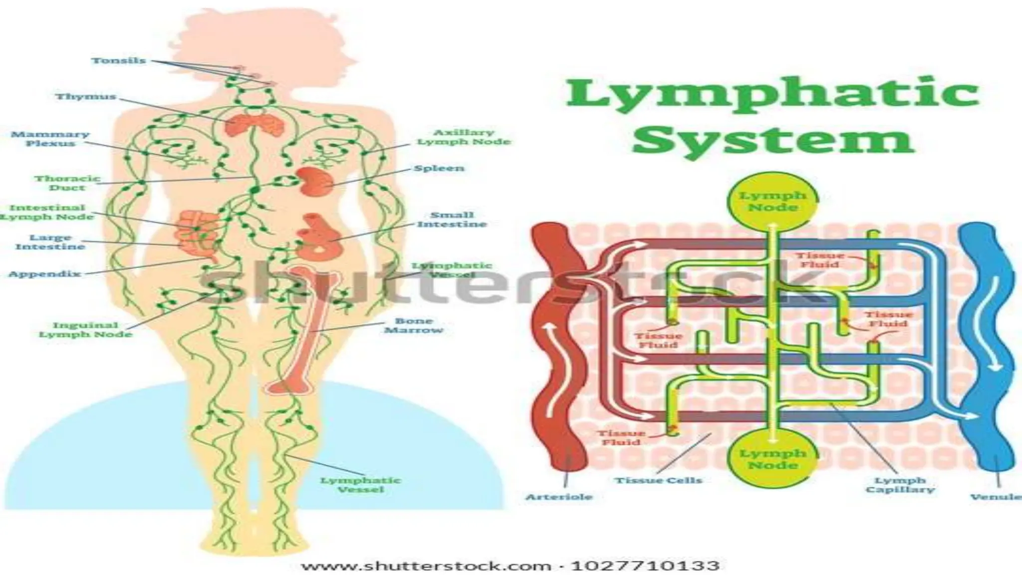
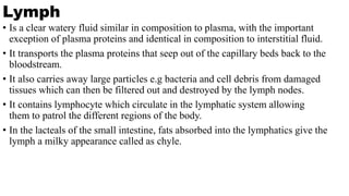
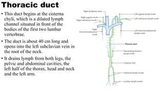
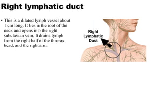
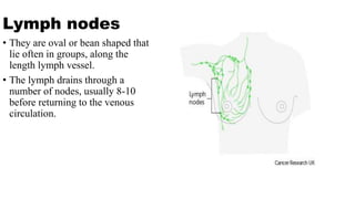
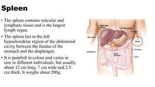
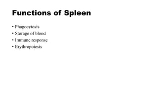
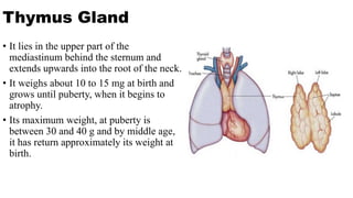
Best Rate (Patna ) Call Girls Patna ⟟ 8617370543 ⟟ High Class Call Girl In 5 ...
Presentation on lymphatic system.pptx
1.
2. Lymph
• Is a clear watery fluid similar in composition to plasma, with the important
exception of plasma proteins and identical in composition to interstitial fluid.
• It transports the plasma proteins that seep out of the capillary beds back to the
bloodstream.
• It also carries away large particles e.g bacteria and cell debris from damaged
tissues which can then be filtered out and destroyed by the lymph nodes.
• It contains lymphocyte which circulate in the lymphatic system allowing
them to patrol the different regions of the body.
• In the lacteals of the small intestine, fats absorbed into the lymphatics give the
lymph a milky appearance called as chyle.
3. Lymph Capillaries
• These originate as blind end tubes in
the interstitial spaces. They have the
same structures as blood capillaries,
i.e. a single layer of endothelial cells,
but their walls are more permeable to
all interstitial fluid constituents,
including proteins and cell debris.
• The tiny capillaries join up to form
larger lymph vessels.
4. Larger Lymph Vessels
• Their walls are about
the same thickness as
those of small veins
and have the same
layers of tissue i.e;
- Fibrous covering
- Middle layer smooth
muscle and elastic
tissue
- Inner lining of
endothelium.
• Lymph vessels have
numerous cup-shaped
valves to ensure that
lymph flows in one-
way system towards
the thorax.
• Lymph vessels
become larger as they
join together,
eventually forming
two large ducts, the
thoracic duct and
right lymphatic duct,
which empty lymph
into the subclavian
veins.
5. Thoracic duct
• This duct begins at the cisterna
chyli, which is a dilated lymph
channel situated in front of the
bodies of the first two lumbar
verterbrae.
• The duct is about 40 cm long and
opens into the left subclavian vein in
the root of the neck.
• It drains lymph from both legs, the
pelvic and abdominal cavities, the
left half of the thorax, head and neck
and the left arm.
6. Right lymphatic duct
• This is a dilated lymph vessel about
1 cm long. It lies in the root of the
neck and opens into the right
subclavian vein. It drains lymph
from the right half of the throrax,
head, and the right arm.
7. Lymph nodes
• They are oval or bean shaped that
lie often in groups, along the
length lymph vessel.
• The lymph drains through a
number of nodes, usually 8-10
before returning to the venous
circulation.
8. Structure of lymph nodes
• They have an outer
capsule of fibrous tissue
that dips down into the
node substance forming
partitions or trabeculae.
• The main substance of
the node consists of
reticular and lymphatic
tissue containing many
lymphocytes and
macrophages.
• Each node has a concave
surface called the hilum.
• Lymph from the head
and neck passes through
deep and superficial
cervical nodes..
• Lymph from the upper
limb passes through
nodes situated in the
elbow region, then
through the deep and
superficial axillary
nodes.
• The lymph from the
lower limbs drains
through deep and
superficial nodes
including groups of
nodes behind the knee
and in the groin(
Inguinal nodes)
10. Spleen
• The spleen contains reticular and
lymphatic tissue and is the largest
lymph organ.
• The spleen lies in the left
hypochondriac region of the abdominal
cavity between the fundus of the
stomach and the diaphragm.
• It is purplish in colour and varies in
size in different individuals, but usually
about 12 cm long, 7 cm wide and 2.5
cm thick. It weighs about 200g.
11. Structure of spleen
• The spleen is slightly
oval in shape with the
hilum on the lower
medial border.
• The anterior surface is
covered with
peritoneum. It is
enclosed in a
fibroelastic capsule that
dips into the organ,
forming trabeculae.
• The cellular material,
consisting of
lymphocytes and
macrophages is called
splenic pulp, and lies
between the trabeculae.
• Red pulp is the part
suffused with blood
and white pulp consists
of areas of lymphatic
tissue and where there
are sleeves of
lymphocytes and
macrophages around
blood vessels.
12. Structures leaving and entering
the spleen at the hilum
• Splenic artery, a branch of the coeliac aretery
• Splenic vein, branch of the portal vein
• Lymph vessel( efferent only)
• nerves
13. Functions of Spleen
• Phagocytosis
• Storage of blood
• Immune response
• Erythropoiesis
14. Thymus Gland
• It lies in the upper part of the
mediastinum behind the sternum and
extends upwards into the root of the neck.
• It weighs about 10 to 15 mg at birth and
grows until puberty, when it begins to
atrophy.
• Its maximum weight, at puberty is
between 30 and 40 g and by middle age,
it has return approximately its weight at
birth.
15. Structure
• The thymus consists of two lobes
joined by areolar tissue.
• The lobes are enclosed by a
fibrous capsule which dips into
their substance, dividing them
into lobules that consists of an
irregular branching framework of
epithelial cells and lymphocytes.
16. Functions of Thymus Gland
• Lymphocytes originate from the stem cells in red bone marrow.
• Those enter the thymus develop into the activated T- Lymphocytes.
• Thymic processing produces mature T- lymphocytes that can distinguish ‘self”
tissue from foreign tissue, and also provides each t- lymphocytes with the ability
to react to only one specific antigen from the millions it will encounter.
• T- lymphocytes then leaves the thymus and enter the blood.
• Some enter the lymphoid tissues and others circulate in the bloodstream.
• T- lymphocyte production, although most prolific in youth, probably continues
throughout the from a resident population of thymic stem cells.