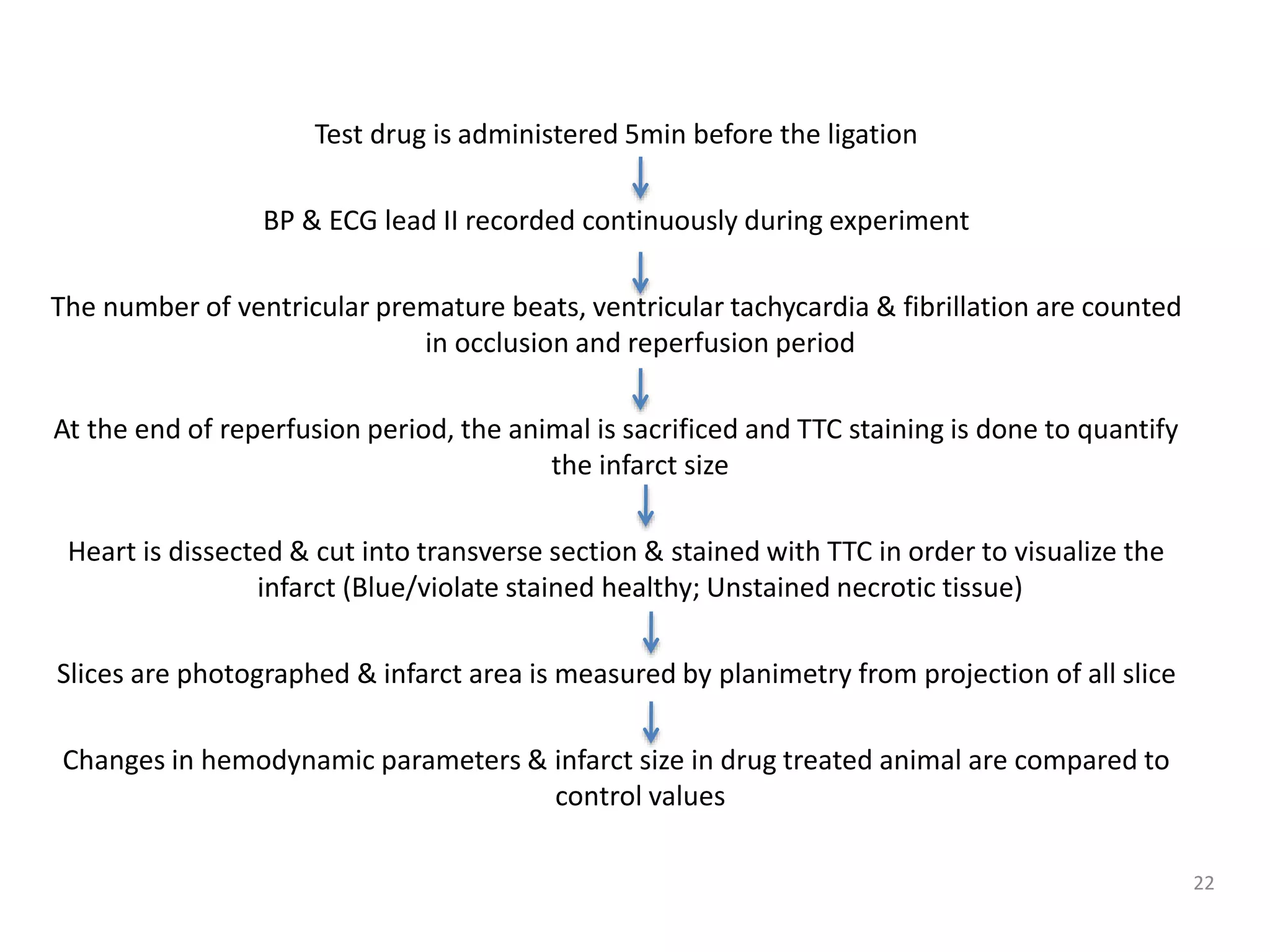
Downloaded 500 times


![Test compound administered through perfusion medium either before or after occlusion.
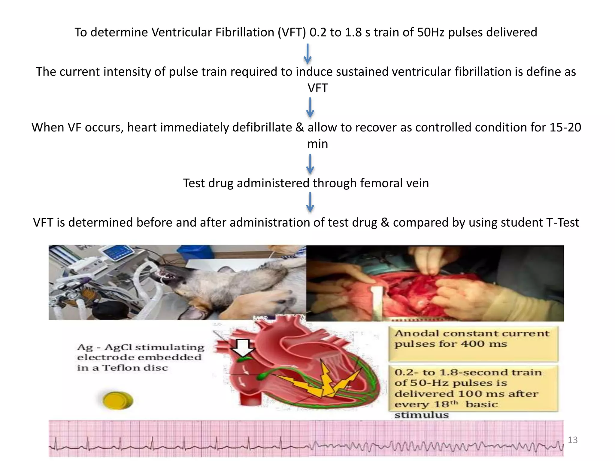
Epicardial ECG electrode is used for pulsatile stimulation and induction of arrhythmia
[Rectengular pulces 0.75 msec duration, 10 V; 400-1800 shocks/min]
Small steel hook with string attached to apex of heart and HR measured with chronometer
Incidence & duration of VF or VT is recorded in control & test group
5](https://image.slidesharecdn.com/dsmarrhythmias-170212150841/75/Drug-screening-methods-for-antiarrhythmic-agents-5-2048.jpg)

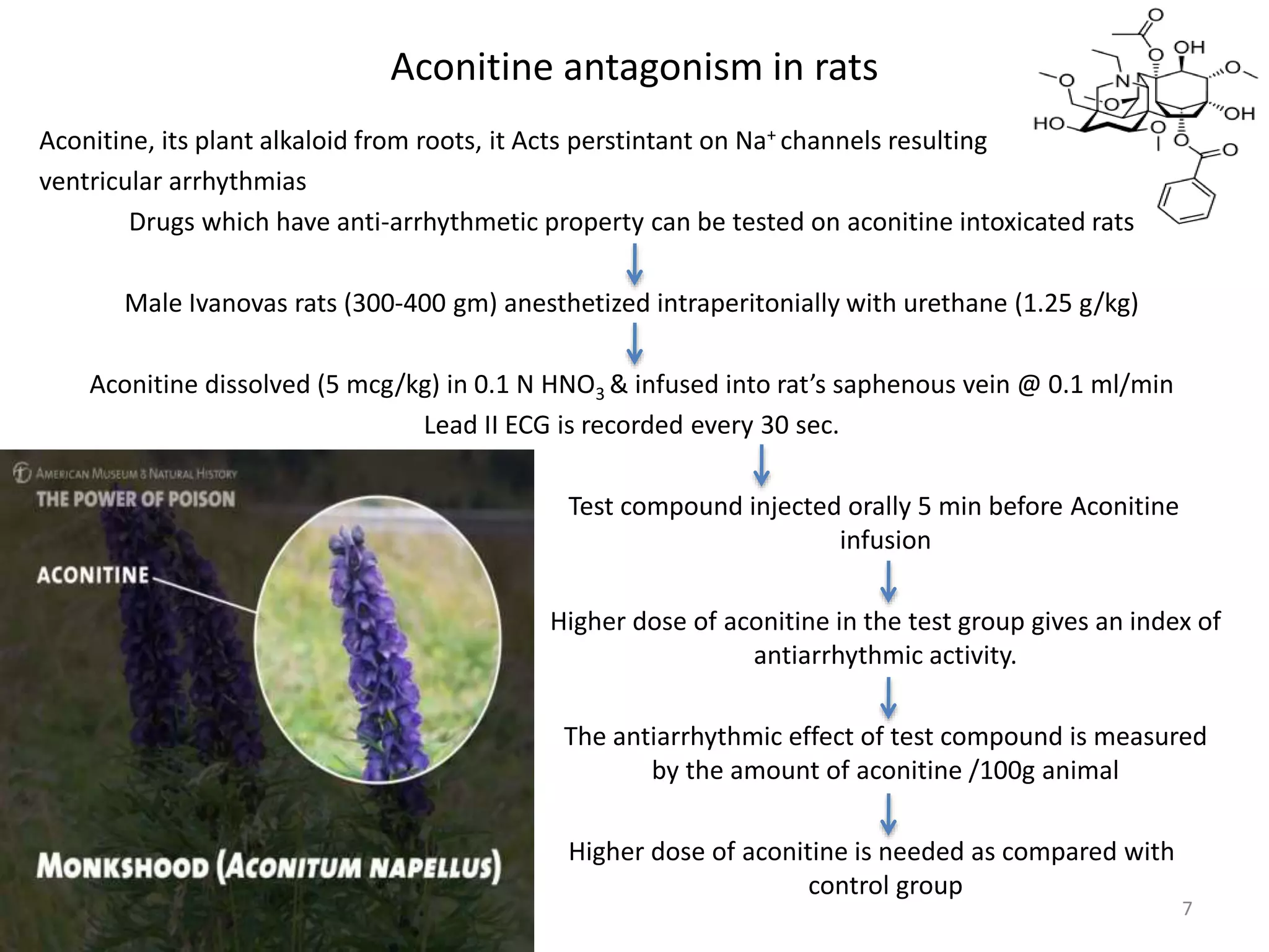


This document describes various in vitro and in vivo drug screening methods for evaluating potential antiarrhythmic agents. In vitro methods include using acetylcholine or potassium to induce arrhythmias in isolated rabbit atria and the Langendorff technique of perfusing isolated guinea pig hearts. In vivo methods involve chemically inducing arrhythmias using drugs like aconitine, digoxin, strophanthin, and adrenaline in rats and dogs. Other methods include electrically inducing arrhythmias by determining ventricular fibrillation threshold and using programmed stimulation in dogs with induced myocardial ischemia. A canine model of sudden coronary death is also described.


