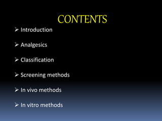
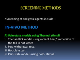
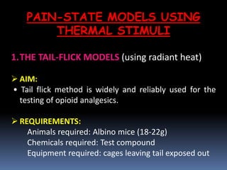
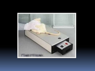
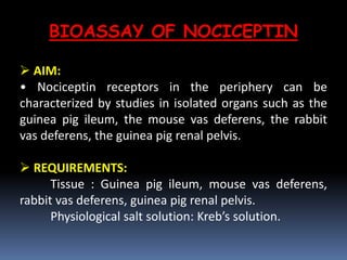
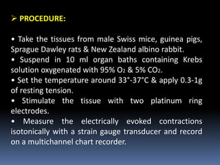
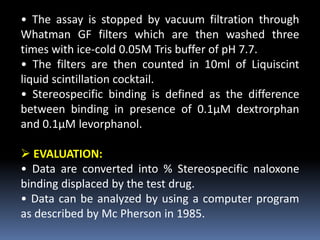
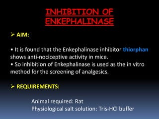
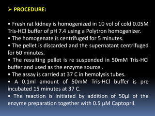
SCREENING OF
ANALGESICS
 BY: FOZIYA KHAN
 PHARMACOLOGY BRANCH
 SEM I
CONTENTS
 Introduction
 Analgesics
 Classification
 Screening methods
 In vivo methods
 In vitro methods
INTRODUCTION
• Pain is an unpleasant sensory and emotional
experience associated with actual and potential tissue
damage.
• Various types of pains seen in humans are:
1. Somatic pain (arising from skin, muscles, joints,
ligaments and bones)
2. Visceral pain
3. Referred pain
4. Neuropathic pain
5. Cancer pain etc.
• The pain sensing neurons “nociceptors” are
stimulated by physical (heat, cold and pressure) and
chemical stimuli.
• Pain can be classified as acute or chronic,
 Acute pain: it is of soft tissue damage, infection or
inflammation and will be short of duration.
 Chronic pain: it lasts for 6 months or larger than that
period (E.g. cancer pain, neuropathic pain and arthritic
pain).
ANALGESICS
 An analgesic or pain killer is any member of the
group of drug used to achieve analgesia, relief from
pain.
 These can be sold as an over the counter (OTC) or
prescription drug.
 The medicines are commonly used to treat pain
due to arthritis, surgery, injury, toothache, headache,
menstrual cramps, sore muscles or other causes.
Narcotic Analgesics Non Narcotic Analgesics
Act centrally Act peripherally
Cause addiction Do not cause addiction
Produce CNS depression Do not produce CNS
depression
Do not produce gastric
irritation
Produce gastric irritation
Show no anti-
inflammatory effect
Show anti- inflammatory
effect
Ex. Morphine, tremadol,
pethidine etc.
Ex. Diclofenac, ibuprofen,
aspirin etc.
CLASSIFICATION
SCREENING METHODS
• Screening of analgesic agents include –
IN-VIVO METHOD
A) Pain-state models using Thermal stimuli
i. The tail-flick model using radiant heat/ Immersion of
the tail in hot water.
ii. Paw-withdrawal test.
iii. Hot-plate test.
iv. Pain-state models using Cold- stimuli
B) Pain-state models using Mechanical stimuli
I. Strain gauges.
i. Von-Frey filaments.
ii. Inclined-plane test.
C) Pain-state models using Electrical stimuli
i. Electrical stimulation of the tail.
ii. Grid-shock test.
iii. Stimulation of the tooth pulp.
iv. Monkey-shock titration test.
v. Stimulation of the limbs.
D) Pain-state models using Chemical stimuli
i. Formalin test.
ii. Acetic acid induced writhing test.
iii. Stimulation of hollow organs.
IN-VITRO METHOD
A. Bioassay of nociceptin.
B. Receptor binding of nociceptin.
C. 3H- naloxone binding assay.
D. 3H- bremazocine binding assay.
E. Cannabinoids binding assay.
F. Inhibition of Enkephalinase.
PAIN-STATE MODELS USING
THERMAL STIMULI
1.THE TAIL-FLICK MODELS (using radiant heat)
 AIM:
• Tail flick method is widely and reliably used for the
testing of opioid analgesics.
 REQUIREMENTS:
Animals required: Albino mice (18-22g)
Chemicals required: Test compound
Equipment required: cages leaving tail exposed out
 PRINCIPLE AND ROCEDURE:
• The application of thermal radiation to the tail of an
animal provokes the withdrawal of the tail by a brief
vigorous movement.
• The withdraw of the tail from the heat source is
referred to as tail-flick latency.
• In this model a timer is started at the same time as the
application of the heat source and time taken by the rat
to withdraw its tail is recorded.
• Usually withdrawal time is within 2 to 10s.
• The lengthening of this reaction time by the animal
seen after the administration of a drug is interpreted as
an analgesic action.
• It is advisable not to prolong the exposure to radiant
heat beyond 20s because the skin of the tail may be burnt.
• A rheostat is inserted in the apparatus so as to control
the intensity of the current passing through the filament,
which further controls the intensity of radiant heat.
• This test is more sensitive to morphine.
 EVALUATION:
• At each time interval those animals that show higher
reaction time than the time before drug administration
are regarded as positive.
• Percentage of positive animals are counted for each time
interval and each dose and ED50 values of test compound
can be calculated
2. HOT PLATE MODEL
 AIM:
• Hot plate method has been widely used to evaluate
opioid analgesics.
 REQUIREMENTS:
Animals required: Albino mice
Chemicals required: Test compound
Equipment requirement: Hot plate at 55-66 C
(electrically heated plate)
 PRINCIPLE AND ROCEDURE:
• This test consists of introducing a rat or mouse into an
open-ended cylindrical space with a floor consisting of a
metallic plate that is heated by a thermode or a boiling
liquid.
• A plate heated to a constant temperature produces two
behavioral components that can be measured in terms of
their reaction times, namely paw licking and jumping.
• Animals are placed on the hot plate, which consist of
electrically heated surface.
• Temperature on the hot plate is maintained at 55-66
degree Celsius.
• Responses such as jumping ,withdrawal of the paws and
licking of the paws are seen
• Both are considered to be supra-spinally integrated
responses.
• The time period (latency period), when animals are
placed and until responses occur, is recorded by a
stopwatch.
• After administration of analgesic substances, the paw
licking behavior time and the jumping reaction time is
increased.
 EVALUATION:
• Test compounds are administered orally or
subcutaneously and latency or latency period is recorded
after 20,60 and 90 min.
• These values are compared with the values before
administration of the drug by using T-test.
PAIN-STATE MODELS USING
CHEMICAL STIMULI
3. ACETIC ACID INDUCED WRITHING TEST
 AIM:
• This test is used to screen analgesics by using
chemical stimulus.
 REQUIREMENTS:
Animals required: Albino mice
Chemicals required: Acetic acid 0.7% v/v
(1ml/100g) Aspirin (100mg/kg)
 PRINCIPLE AND PROCEDURE:
• Pain is often induced in rats or mice by injecting certain irritants
such as phenyl quinone or acetic acid into the peritoneal cavity.
• The animal reacts with a characteristic stretching behavior which is
called writhing.
• The intra-peritoneal administration of agents that irritate serous
membranes elicits a stereotyped behavior in the mouse/rat, which is
characterized by abdominal contractions, movements of the body as
a whole (particularly of the hind paws), twisting of dorso-abdominal
muscles, and motor in- coordination.
• Divide the animals into 2 groups each consisting of 5 animals.
• The first group serves as control and the second group is the test
group
• Administer the appropriate volume of acetic acid
solution to the first group and place individually under
glass jar for observation.
• Note the onset of writhing and number of writhing for
a period of 10 minutes.
• Administer Aspirin 100mg/kg orally to the second
group of animals and 30 minutes later administer acetic
acid solution.
• Note the onset and number of writhing responses.
 EVALUATION:
• Calculating the mean writhing response of control and
test group.
• From this value the percentage protection of
analgesics can be calculated.
BIOASSAY OF NOCICEPTIN
 AIM:
• Nociceptin receptors in the periphery can be
characterized by studies in isolated organs such as the
guinea pig ileum, the mouse vas deferens, the rabbit
vas deferens, the guinea pig renal pelvis.
 REQUIREMENTS:
Tissue : Guinea pig ileum, mouse vas deferens,
rabbit vas deferens, guinea pig renal pelvis.
Physiological salt solution: Kreb’s solution.
 PROCEDURE:
• Take the tissues from male Swiss mice, guinea pigs,
Sprague Dawley rats & New Zealand albino rabbit.
• Suspend in 10 ml organ baths containing Krebs
solution oxygenated with 95% O2 & 5% CO2.
• Set the temperature around 33°-37°C & apply 0.3-1g
of resting tension.
• Stimulate the tissue with two platinum ring
electrodes.
• Measure the electrically evoked contractions
isotonically with a strain gauge transducer and record
on a multichannel chart recorder.
• After equilibration period of about 60 min the
contractions induced by electrical field stimulation
become stable; at this time, perform the cumulative
concentration response curves to nociceptin or opioid
peptides.
• Perform four electrical field stimulation with each
tissue at 30 min intervals.
• Add Agonists & Antagonists to the bath.
• The biological effects of the application of agonists or
antagonists are expressed as % inhibition of electrical
filed stimulation-induced contraction.
• Contractile responses to electrical field stimulation are
expressed as % increment to the spontaneous activity of
the tissue.
 EVALUATION:
• Data are expressed as means of ‘n’ experiments and
statistically analyzed and recorded.
3H- NALOXONE BINDING ASSAY
AIM:
•This test is used for the study of opiate agonist and
antagonist.
 REQUIREMENTS:
Animal required: Wistar rat
Physiological salt solution: Tris buffer
 PROCEDURE:
• Decapitate the male Wistar rat and rapidly remove
their brain.
• Weigh whole brain except cerebella and homogenize
in 50ml of 0.05M Tris buffer with a Tekmar tissue
homogenizer.
• Centrifuge the homogenate for 15 minutes, decant
the supernatant and re-suspend the pellet in fresh
buffer and centrifuge.
• Then suspend the final pellet in original volume of
fresh 0.05M Tris buffer.
• Incubate the tubes for 30 minutes at 37 degree
Celsius.
• The assay is stopped by vacuum filtration through
Whatman GF filters which are then washed three
times with ice-cold 0.05M Tris buffer of pH 7.7.
• The filters are then counted in 10ml of Liquiscint
liquid scintillation cocktail.
• Stereospecific binding is defined as the difference
between binding in presence of 0.1μM dextrorphan
and 0.1μM levorphanol.
 EVALUATION:
• Data are converted into % Stereospecific naloxone
binding displaced by the test drug.
• Data can be analyzed by using a computer program
as described by Mc Pherson in 1985.
INHIBITION OF
ENKEPHALINASE
 AIM:
• It is found that the Enkephalinase inhibitor thiorphan
shows anti-nociceptive activity in mice.
• So inhibition of Enkephalinase is used as the in vitro
method for the screening of analgesics.
 REQUIREMENTS:
Animal required: Rat
Physiological salt solution: Tris-HCl buffer
 PROCEDURE:
• Fresh rat kidney is homogenized in 10 vol of cold 0.05M
Tris-HCl buffer of pH 7.4 using a Polytron homogenizer.
• The homogenate is centrifuged for 5 minutes.
• The pellet is discarded and the supernatant centrifuged
for 60 minutes.
• The resulting pellet is re suspended in 50mM Tris-HCl
buffer and used as the enzyme source .
• The assay is carried at 37 C in hemolysis tubes.
• A 0.1ml amount of 50mM Tris-HCl buffer is pre
incubated 15 minutes at 37 C.
• The reaction is initiated by addition of 50μl of the
enzyme preparation together with 0.5 μM Captopril.
•The tubes are incubated for 30 minutes in a water bath
with constant shaking
• The enzymatic reaction is stopped by boiling at 100 C
for 5 minutes.
• The samples are then diluted with 1.35ml of Tris-HCl
buffer and centrifuged for 30 minutes.
• 1ml of supernatant is transferred to thermostat cells
of a spectro fluorometer.
• Reading are performed at 562nm with an excitation
wavelength of 342nm.
• A calibration curve is prepared.
 EVALUATION:
• The inhibitory potencies of test compounds are
compared with the standard.
Screening methods for Analgesics

Screening methods for Analgesics

  • 1.
    SCREENING OF ANALGESICS  BY:FOZIYA KHAN  PHARMACOLOGY BRANCH  SEM I
  • 2.
    CONTENTS  Introduction  Analgesics Classification  Screening methods  In vivo methods  In vitro methods
  • 3.
    INTRODUCTION • Pain isan unpleasant sensory and emotional experience associated with actual and potential tissue damage. • Various types of pains seen in humans are: 1. Somatic pain (arising from skin, muscles, joints, ligaments and bones) 2. Visceral pain 3. Referred pain 4. Neuropathic pain 5. Cancer pain etc.
  • 4.
    • The painsensing neurons “nociceptors” are stimulated by physical (heat, cold and pressure) and chemical stimuli. • Pain can be classified as acute or chronic,  Acute pain: it is of soft tissue damage, infection or inflammation and will be short of duration.  Chronic pain: it lasts for 6 months or larger than that period (E.g. cancer pain, neuropathic pain and arthritic pain).
  • 5.
    ANALGESICS  An analgesicor pain killer is any member of the group of drug used to achieve analgesia, relief from pain.  These can be sold as an over the counter (OTC) or prescription drug.  The medicines are commonly used to treat pain due to arthritis, surgery, injury, toothache, headache, menstrual cramps, sore muscles or other causes.
  • 6.
    Narcotic Analgesics NonNarcotic Analgesics Act centrally Act peripherally Cause addiction Do not cause addiction Produce CNS depression Do not produce CNS depression Do not produce gastric irritation Produce gastric irritation Show no anti- inflammatory effect Show anti- inflammatory effect Ex. Morphine, tremadol, pethidine etc. Ex. Diclofenac, ibuprofen, aspirin etc. CLASSIFICATION
  • 7.
    SCREENING METHODS • Screeningof analgesic agents include – IN-VIVO METHOD A) Pain-state models using Thermal stimuli i. The tail-flick model using radiant heat/ Immersion of the tail in hot water. ii. Paw-withdrawal test. iii. Hot-plate test. iv. Pain-state models using Cold- stimuli
  • 8.
    B) Pain-state modelsusing Mechanical stimuli I. Strain gauges. i. Von-Frey filaments. ii. Inclined-plane test. C) Pain-state models using Electrical stimuli i. Electrical stimulation of the tail. ii. Grid-shock test. iii. Stimulation of the tooth pulp. iv. Monkey-shock titration test. v. Stimulation of the limbs.
  • 9.
    D) Pain-state modelsusing Chemical stimuli i. Formalin test. ii. Acetic acid induced writhing test. iii. Stimulation of hollow organs. IN-VITRO METHOD A. Bioassay of nociceptin. B. Receptor binding of nociceptin. C. 3H- naloxone binding assay. D. 3H- bremazocine binding assay. E. Cannabinoids binding assay. F. Inhibition of Enkephalinase.
  • 11.
    PAIN-STATE MODELS USING THERMALSTIMULI 1.THE TAIL-FLICK MODELS (using radiant heat)  AIM: • Tail flick method is widely and reliably used for the testing of opioid analgesics.  REQUIREMENTS: Animals required: Albino mice (18-22g) Chemicals required: Test compound Equipment required: cages leaving tail exposed out
  • 13.
     PRINCIPLE ANDROCEDURE: • The application of thermal radiation to the tail of an animal provokes the withdrawal of the tail by a brief vigorous movement. • The withdraw of the tail from the heat source is referred to as tail-flick latency. • In this model a timer is started at the same time as the application of the heat source and time taken by the rat to withdraw its tail is recorded. • Usually withdrawal time is within 2 to 10s. • The lengthening of this reaction time by the animal seen after the administration of a drug is interpreted as an analgesic action.
  • 14.
    • It isadvisable not to prolong the exposure to radiant heat beyond 20s because the skin of the tail may be burnt. • A rheostat is inserted in the apparatus so as to control the intensity of the current passing through the filament, which further controls the intensity of radiant heat. • This test is more sensitive to morphine.  EVALUATION: • At each time interval those animals that show higher reaction time than the time before drug administration are regarded as positive. • Percentage of positive animals are counted for each time interval and each dose and ED50 values of test compound can be calculated
  • 15.
    2. HOT PLATEMODEL  AIM: • Hot plate method has been widely used to evaluate opioid analgesics.  REQUIREMENTS: Animals required: Albino mice Chemicals required: Test compound Equipment requirement: Hot plate at 55-66 C (electrically heated plate)
  • 17.
     PRINCIPLE ANDROCEDURE: • This test consists of introducing a rat or mouse into an open-ended cylindrical space with a floor consisting of a metallic plate that is heated by a thermode or a boiling liquid. • A plate heated to a constant temperature produces two behavioral components that can be measured in terms of their reaction times, namely paw licking and jumping. • Animals are placed on the hot plate, which consist of electrically heated surface. • Temperature on the hot plate is maintained at 55-66 degree Celsius. • Responses such as jumping ,withdrawal of the paws and licking of the paws are seen
  • 18.
    • Both areconsidered to be supra-spinally integrated responses. • The time period (latency period), when animals are placed and until responses occur, is recorded by a stopwatch. • After administration of analgesic substances, the paw licking behavior time and the jumping reaction time is increased.  EVALUATION: • Test compounds are administered orally or subcutaneously and latency or latency period is recorded after 20,60 and 90 min. • These values are compared with the values before administration of the drug by using T-test.
  • 19.
    PAIN-STATE MODELS USING CHEMICALSTIMULI 3. ACETIC ACID INDUCED WRITHING TEST  AIM: • This test is used to screen analgesics by using chemical stimulus.  REQUIREMENTS: Animals required: Albino mice Chemicals required: Acetic acid 0.7% v/v (1ml/100g) Aspirin (100mg/kg)
  • 21.
     PRINCIPLE ANDPROCEDURE: • Pain is often induced in rats or mice by injecting certain irritants such as phenyl quinone or acetic acid into the peritoneal cavity. • The animal reacts with a characteristic stretching behavior which is called writhing. • The intra-peritoneal administration of agents that irritate serous membranes elicits a stereotyped behavior in the mouse/rat, which is characterized by abdominal contractions, movements of the body as a whole (particularly of the hind paws), twisting of dorso-abdominal muscles, and motor in- coordination. • Divide the animals into 2 groups each consisting of 5 animals. • The first group serves as control and the second group is the test group
  • 22.
    • Administer theappropriate volume of acetic acid solution to the first group and place individually under glass jar for observation. • Note the onset of writhing and number of writhing for a period of 10 minutes. • Administer Aspirin 100mg/kg orally to the second group of animals and 30 minutes later administer acetic acid solution. • Note the onset and number of writhing responses.  EVALUATION: • Calculating the mean writhing response of control and test group. • From this value the percentage protection of analgesics can be calculated.
  • 24.
    BIOASSAY OF NOCICEPTIN AIM: • Nociceptin receptors in the periphery can be characterized by studies in isolated organs such as the guinea pig ileum, the mouse vas deferens, the rabbit vas deferens, the guinea pig renal pelvis.  REQUIREMENTS: Tissue : Guinea pig ileum, mouse vas deferens, rabbit vas deferens, guinea pig renal pelvis. Physiological salt solution: Kreb’s solution.
  • 25.
     PROCEDURE: • Takethe tissues from male Swiss mice, guinea pigs, Sprague Dawley rats & New Zealand albino rabbit. • Suspend in 10 ml organ baths containing Krebs solution oxygenated with 95% O2 & 5% CO2. • Set the temperature around 33°-37°C & apply 0.3-1g of resting tension. • Stimulate the tissue with two platinum ring electrodes. • Measure the electrically evoked contractions isotonically with a strain gauge transducer and record on a multichannel chart recorder.
  • 26.
    • After equilibrationperiod of about 60 min the contractions induced by electrical field stimulation become stable; at this time, perform the cumulative concentration response curves to nociceptin or opioid peptides. • Perform four electrical field stimulation with each tissue at 30 min intervals. • Add Agonists & Antagonists to the bath. • The biological effects of the application of agonists or antagonists are expressed as % inhibition of electrical filed stimulation-induced contraction. • Contractile responses to electrical field stimulation are expressed as % increment to the spontaneous activity of the tissue.
  • 27.
     EVALUATION: • Dataare expressed as means of ‘n’ experiments and statistically analyzed and recorded.
  • 28.
    3H- NALOXONE BINDINGASSAY AIM: •This test is used for the study of opiate agonist and antagonist.  REQUIREMENTS: Animal required: Wistar rat Physiological salt solution: Tris buffer
  • 29.
     PROCEDURE: • Decapitatethe male Wistar rat and rapidly remove their brain. • Weigh whole brain except cerebella and homogenize in 50ml of 0.05M Tris buffer with a Tekmar tissue homogenizer. • Centrifuge the homogenate for 15 minutes, decant the supernatant and re-suspend the pellet in fresh buffer and centrifuge. • Then suspend the final pellet in original volume of fresh 0.05M Tris buffer. • Incubate the tubes for 30 minutes at 37 degree Celsius.
  • 30.
    • The assayis stopped by vacuum filtration through Whatman GF filters which are then washed three times with ice-cold 0.05M Tris buffer of pH 7.7. • The filters are then counted in 10ml of Liquiscint liquid scintillation cocktail. • Stereospecific binding is defined as the difference between binding in presence of 0.1μM dextrorphan and 0.1μM levorphanol.  EVALUATION: • Data are converted into % Stereospecific naloxone binding displaced by the test drug. • Data can be analyzed by using a computer program as described by Mc Pherson in 1985.
  • 31.
    INHIBITION OF ENKEPHALINASE  AIM: •It is found that the Enkephalinase inhibitor thiorphan shows anti-nociceptive activity in mice. • So inhibition of Enkephalinase is used as the in vitro method for the screening of analgesics.  REQUIREMENTS: Animal required: Rat Physiological salt solution: Tris-HCl buffer
  • 32.
     PROCEDURE: • Freshrat kidney is homogenized in 10 vol of cold 0.05M Tris-HCl buffer of pH 7.4 using a Polytron homogenizer. • The homogenate is centrifuged for 5 minutes. • The pellet is discarded and the supernatant centrifuged for 60 minutes. • The resulting pellet is re suspended in 50mM Tris-HCl buffer and used as the enzyme source . • The assay is carried at 37 C in hemolysis tubes. • A 0.1ml amount of 50mM Tris-HCl buffer is pre incubated 15 minutes at 37 C. • The reaction is initiated by addition of 50μl of the enzyme preparation together with 0.5 μM Captopril.
  • 33.
    •The tubes areincubated for 30 minutes in a water bath with constant shaking • The enzymatic reaction is stopped by boiling at 100 C for 5 minutes. • The samples are then diluted with 1.35ml of Tris-HCl buffer and centrifuged for 30 minutes. • 1ml of supernatant is transferred to thermostat cells of a spectro fluorometer. • Reading are performed at 562nm with an excitation wavelength of 342nm. • A calibration curve is prepared.  EVALUATION: • The inhibitory potencies of test compounds are compared with the standard.