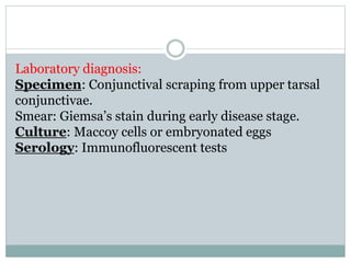
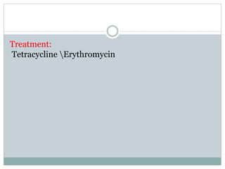
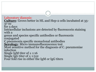
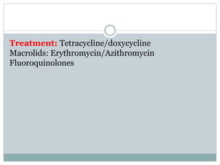
OMAR WALEED
MASTER STUDENT
GENUS: MYCOPLASMA
Characteristics
Part of normal flora of human genital tract or oral cavity of
healthy
adults
Formerly named as pleuropneumonia-like organism
(PPLO).
The smallest living micro-organism capable of free living in
nature
self-replicating on laboratory media
Highly pleomorphic due to absence of rigid cell wall, instead
bounded by a triple-layered “unit membrane”
Completely resistant to penicillin and cephalosporin.
Can reproduce in complex cell-free media.
Have an affinity to mammalian cell membrane
14 species of mycoplasma is identified in humans
and
classification of species is based on biochemical
reaction and
serological tests
Antigenic structure
The species are classified by biochemical and serologic
features. The complement fixation (CF) antigens of mycoplasmas
are glycolipids. Antigens for enzyme-linked immunoassay
(ELISA) tests are proteins. Some species have more
than one serotype
Mycoplasma species of medical importance
Mycoplasma pneumoniae
Mycoplasma hominis
Ureaplasma urealyticum
Mycoplasma pneumoniae
Clinical features:
Route of transmission: Infected respiratory secretion
Infection is initiated after adherence of bacterial
polar tip
adhesion protein to respiratory epithelial cells
is a major cause of pneumonia in young age groups
(5-20 yrs.)
Extra pulmonary manifestations:
Hemolytic anemia
Skin rashes/lesions
Meningoencephalitis
Myelitis
Neuritis
Myopericarditis
Arthritis
Laboratory diagnosis:
Specimen: consist of throat swabs; sputum; inflammatory
exudates; and respiratory, urethral, or genital secretions
Culture: The material is inoculated into broth and onto special solid
media depending on the organism sought. Agar media are
best incubated at 37°C with 5–10% CO2 (under microaerophilic
conditions or even anaerobic conditions). Broths
require incubation at 37°C under atmospheric (aerobic) conditions
Identification: Observe for “fried-egg” colonies embedded into the
surface of the medium or inhibition of growth around discs
impregnated with specific antisera
Serology:
Complement fixation test
Indirect Immunofluorescent test
Haemagglutination inhibition test
NB: Cold hem agglutinins titer ≥ 1:64 suggests M. pneumonia infection
Treatment:
Erythromycin
Tetracycline
Mycoplasma are resistant to penicillin, cephalosporin's and
vancomycin
Mycoplasma hominis and Ureaplasma urealyticum
Found as a normal flora in the lower genital tract.
Mycoplasma hominis causes genital infection and
post partum sepsis.
Ureaplasma urealyticum causes non-gonococcal
urethritis
Treatment is the same as pneumoniae
GENUS: CHLAMYDIA
Characteristics:
Obligate intracellular gram-negative bacteria.
Reproduce by binary fission.
Posses both DNA and RNA.
Have cell wall and ribosome's.
Sensitive to anti-microbial agents.
Have enzyme systems and make their own proteins,
lipids,
nucleic acids and vitamins
Three species of medical importance
C. tracomatis This species produces compact intracytoplasmic
inclusions
that contain glycogen; it is usually inhibited by sulfonamides.
It includes agents of human disorders such as trachoma,
inclusion conjunctivitis, nongonococcal urethritis, salpingitis
, cervicitis, pneumonitis of infants, and LGV
C. pneumoniae:
This species produces intracytoplasmic inclusions that lack
glycogen; it is usually resistant to sulfonamides. It causes
respiratory tract infections in humans
C. psittacii: This species produces diffuse intracytoplasmic inclusions
that lack glycogen; it is usually resistant to sulfonamides. It
includes agents of psittacosis in humans, ornithosis in birds,
feline pneumonitis, and other animal diseases
Developmental cycle of Chlamydia
The infectious environmental stable particle, named as
elementary
body is ingested by a host cell. The elementary body is
reorganized
into reticulate body in the host cell which is specifically
adapted for
intracellular growth. The reticulate body grows and divides
many
times to form inclusions in the host cell cytoplasm.
With in 24-48 hours of developmental cycle, the reticulate
bodies
rearrange them selves into infective elementary bodies and
released
after host cell rupture
Chlamydia trachomatis
Stained with giamsa’s and iodine stain.
Appearance in giemsa’s stain
Elementary body -------- Purple
Reticulate body---------- Blue
Host cell cytoplasm----- Blue
Appearance in iodine stain
Brown inclusions in host cell cytoplasm because of glycogen
matrix surrounding the particle.
. There are 15 serotypes of C. trachomatis.
C. trachomatis: serotype A, B, C causes trachoma.
C. trachomatis: serotype D-K causes genital infection.
C. trachomatis: serotype L1-L 3 causes lymphogranuloma
venereum(LGV)
C. trachomatis serotype A, B, C causes trachoma.
Incubation period is 3-10 days
Route of transmission is through indirect contact like eye-
to eye by infected fingers or sharing towels.
It manifests as a chronic keratoconjunctivitis producing
scarring and deformity of the eyelids, corneal
vascularization
and opacities which may lead to blindness.
It is associated with a low standard of living and poor
personal hygiene
Laboratory diagnosis:
Specimen: Conjunctival scraping from upper tarsal
conjunctivae.
Smear: Giemsa’s stain during early disease stage.
Culture: Maccoy cells or embryonated eggs
Serology: Immunofluorescent tests
Treatment:
Tetracycline Erythromycin
C.trachomatis serotype D-K causes
1. Genital infection.
. Male ---------- non-gonococcal urethritis
Epididymitis
Females------ Urethritis
Cervicitis
Pelvic inflamatory diseases
If complicated in females, it causes infertility and ectopic pregnancy.
2. Inclusion conjunctivitis resembling trachoma.
Transmission is by self-inoculation of the eye with infected genital
secretion.
3. Neonatal inclusion conjunctivitis and neonatal pneumonia.
Transmission is during passage through the infected birth canal
Laboratory diagnosis:
Specimen: Endocervical scraping
Culture: maccoy cells
Serology: Enzyme immunoassay for group-specific antigen.
Direct fluorescent antibody test for species- specific antigen.
C. trachomatis serotype L1-L 3 causes lymphogranuloma venereum
(LGV).
It is a sexually transmitted disease (STD) which is characterized by
suppurative ingunal adenitis.
Complication: . . Elephantiasis of penis, scrotum or vulva due to
lymphatic obstruction.
Sinus formation at site of lesion
Chlamydia pneumoniae
Humans are the only known host
Produces sulfonamide-reisistant, round, dense, glycogen
negative
inclusions
Only one serovar has been demonstrated
Route of transmission: person –to-person transmission via
Air borne
Clinical features:
Most infection are asymptomatic to mildly symptomatic
Symptomatic cases present with
Chlamydial pneumonia
Pharyngitis
Sinusitis
Otitis media
Laboratory dianosis:
Culture: Grows better in HL and Hep-2 cells incubated at 35-
37 0c
for 3 days
Intracellular inclusions are detected by fluoresccein staining
with a
genus and species specific antibodies or fluorescein
conjugated
C.pneumonia specific monoclonal antibodies
Serology: Micro immunofluorescence test
Most sensitive method for the diagnosis of C. pneumoniae
infection
Single IgM titer of ≥ 1:16
Single IgG titer of ≥ 1:512
Four fold rise in either the IgM or IgG titers
Treatment: Tetracycline/doxycycline
Macrolids: Erythromycin/Azithromycin
Fluoroquinolones
تشخيص بكتريا.pptx

تشخيص بكتريا.pptx

  • 1.
  • 2.
    Characteristics Part of normalflora of human genital tract or oral cavity of healthy adults Formerly named as pleuropneumonia-like organism (PPLO). The smallest living micro-organism capable of free living in nature self-replicating on laboratory media Highly pleomorphic due to absence of rigid cell wall, instead bounded by a triple-layered “unit membrane”
  • 3.
    Completely resistant topenicillin and cephalosporin. Can reproduce in complex cell-free media. Have an affinity to mammalian cell membrane 14 species of mycoplasma is identified in humans and classification of species is based on biochemical reaction and serological tests
  • 4.
    Antigenic structure The speciesare classified by biochemical and serologic features. The complement fixation (CF) antigens of mycoplasmas are glycolipids. Antigens for enzyme-linked immunoassay (ELISA) tests are proteins. Some species have more than one serotype Mycoplasma species of medical importance Mycoplasma pneumoniae Mycoplasma hominis Ureaplasma urealyticum
  • 5.
    Mycoplasma pneumoniae Clinical features: Routeof transmission: Infected respiratory secretion Infection is initiated after adherence of bacterial polar tip adhesion protein to respiratory epithelial cells
  • 6.
    is a majorcause of pneumonia in young age groups (5-20 yrs.) Extra pulmonary manifestations: Hemolytic anemia Skin rashes/lesions Meningoencephalitis Myelitis Neuritis Myopericarditis Arthritis
  • 7.
    Laboratory diagnosis: Specimen: consistof throat swabs; sputum; inflammatory exudates; and respiratory, urethral, or genital secretions Culture: The material is inoculated into broth and onto special solid media depending on the organism sought. Agar media are best incubated at 37°C with 5–10% CO2 (under microaerophilic conditions or even anaerobic conditions). Broths require incubation at 37°C under atmospheric (aerobic) conditions Identification: Observe for “fried-egg” colonies embedded into the surface of the medium or inhibition of growth around discs impregnated with specific antisera
  • 10.
    Serology: Complement fixation test IndirectImmunofluorescent test Haemagglutination inhibition test NB: Cold hem agglutinins titer ≥ 1:64 suggests M. pneumonia infection Treatment: Erythromycin Tetracycline Mycoplasma are resistant to penicillin, cephalosporin's and vancomycin
  • 11.
    Mycoplasma hominis andUreaplasma urealyticum Found as a normal flora in the lower genital tract. Mycoplasma hominis causes genital infection and post partum sepsis. Ureaplasma urealyticum causes non-gonococcal urethritis Treatment is the same as pneumoniae
  • 12.
    GENUS: CHLAMYDIA Characteristics: Obligate intracellulargram-negative bacteria. Reproduce by binary fission. Posses both DNA and RNA. Have cell wall and ribosome's. Sensitive to anti-microbial agents. Have enzyme systems and make their own proteins, lipids, nucleic acids and vitamins
  • 13.
    Three species ofmedical importance C. tracomatis This species produces compact intracytoplasmic inclusions that contain glycogen; it is usually inhibited by sulfonamides. It includes agents of human disorders such as trachoma, inclusion conjunctivitis, nongonococcal urethritis, salpingitis , cervicitis, pneumonitis of infants, and LGV C. pneumoniae: This species produces intracytoplasmic inclusions that lack glycogen; it is usually resistant to sulfonamides. It causes respiratory tract infections in humans
  • 14.
    C. psittacii: Thisspecies produces diffuse intracytoplasmic inclusions that lack glycogen; it is usually resistant to sulfonamides. It includes agents of psittacosis in humans, ornithosis in birds, feline pneumonitis, and other animal diseases
  • 16.
    Developmental cycle ofChlamydia The infectious environmental stable particle, named as elementary body is ingested by a host cell. The elementary body is reorganized into reticulate body in the host cell which is specifically adapted for intracellular growth. The reticulate body grows and divides many times to form inclusions in the host cell cytoplasm. With in 24-48 hours of developmental cycle, the reticulate bodies rearrange them selves into infective elementary bodies and released after host cell rupture
  • 18.
    Chlamydia trachomatis Stained withgiamsa’s and iodine stain. Appearance in giemsa’s stain Elementary body -------- Purple Reticulate body---------- Blue Host cell cytoplasm----- Blue Appearance in iodine stain Brown inclusions in host cell cytoplasm because of glycogen matrix surrounding the particle. . There are 15 serotypes of C. trachomatis. C. trachomatis: serotype A, B, C causes trachoma. C. trachomatis: serotype D-K causes genital infection. C. trachomatis: serotype L1-L 3 causes lymphogranuloma venereum(LGV)
  • 19.
    C. trachomatis serotypeA, B, C causes trachoma. Incubation period is 3-10 days Route of transmission is through indirect contact like eye- to eye by infected fingers or sharing towels. It manifests as a chronic keratoconjunctivitis producing scarring and deformity of the eyelids, corneal vascularization and opacities which may lead to blindness. It is associated with a low standard of living and poor personal hygiene
  • 20.
    Laboratory diagnosis: Specimen: Conjunctivalscraping from upper tarsal conjunctivae. Smear: Giemsa’s stain during early disease stage. Culture: Maccoy cells or embryonated eggs Serology: Immunofluorescent tests
  • 22.
  • 23.
    C.trachomatis serotype D-Kcauses 1. Genital infection. . Male ---------- non-gonococcal urethritis Epididymitis Females------ Urethritis Cervicitis Pelvic inflamatory diseases If complicated in females, it causes infertility and ectopic pregnancy. 2. Inclusion conjunctivitis resembling trachoma. Transmission is by self-inoculation of the eye with infected genital secretion. 3. Neonatal inclusion conjunctivitis and neonatal pneumonia. Transmission is during passage through the infected birth canal
  • 24.
    Laboratory diagnosis: Specimen: Endocervicalscraping Culture: maccoy cells Serology: Enzyme immunoassay for group-specific antigen. Direct fluorescent antibody test for species- specific antigen. C. trachomatis serotype L1-L 3 causes lymphogranuloma venereum (LGV). It is a sexually transmitted disease (STD) which is characterized by suppurative ingunal adenitis. Complication: . . Elephantiasis of penis, scrotum or vulva due to lymphatic obstruction. Sinus formation at site of lesion
  • 25.
    Chlamydia pneumoniae Humans arethe only known host Produces sulfonamide-reisistant, round, dense, glycogen negative inclusions Only one serovar has been demonstrated Route of transmission: person –to-person transmission via Air borne Clinical features: Most infection are asymptomatic to mildly symptomatic Symptomatic cases present with Chlamydial pneumonia Pharyngitis Sinusitis Otitis media
  • 26.
    Laboratory dianosis: Culture: Growsbetter in HL and Hep-2 cells incubated at 35- 37 0c for 3 days Intracellular inclusions are detected by fluoresccein staining with a genus and species specific antibodies or fluorescein conjugated C.pneumonia specific monoclonal antibodies Serology: Micro immunofluorescence test Most sensitive method for the diagnosis of C. pneumoniae infection Single IgM titer of ≥ 1:16 Single IgG titer of ≥ 1:512 Four fold rise in either the IgM or IgG titers
  • 27.