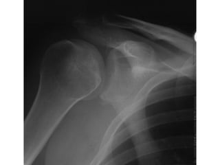
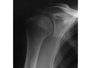
A 22 year old footballer is “smashed” in a tackle and presents with
right shoulder pain. He has no other injuries. An AP x-ray is taken
of the shoulder.
•What injury does the x-ray show ?
•How should this injury be managed ?
This patient has a posterior dislocation of the shoulder. The give away on the x-ray
is a so called “light bulb” sign. The humeral head in the normal patient is lopsided
extending substantially further medially than laterally. In this x-ray the humeral head
looks symmetrical in shape looking much like a light bulb. The x-ray also suggests
that the gleno-humeral joint space is widened. In many posterior dislocations the
widening is so obvious that the posterior rim of the glenoid can be seen (normally
this is hidden behind the humeral head on an AP film), the so called “rim sign”. To
see how different the joint looks once relocated compare it to the post reduction film
which is underneath this sheet. The x-ray probably also shows a fracture fragment at
the inferior rim of the glenoid reflecting the force involved.
Posterior dislocations are unusual (anterior dislocations are perhaps 20 times as
common) so seek advice if uncertain how to deal with them. They can be reduced
under procedural sedation using a combination of:
• Traction
• Abduction
• External Rotation
This is the components of the “Milch” method that is also very effective in anterior
dislocations.
The other technique used is the DePalma….which is traction, ADDuction, and
internal rotation, applying lateral force to the humerus
Posterior shoulder dislocation 2019

Posterior shoulder dislocation 2019

  • 1.
    A 22 yearold footballer is “smashed” in a tackle and presents with right shoulder pain. He has no other injuries. An AP x-ray is taken of the shoulder. •What injury does the x-ray show ? •How should this injury be managed ?
  • 3.
    This patient hasa posterior dislocation of the shoulder. The give away on the x-ray is a so called “light bulb” sign. The humeral head in the normal patient is lopsided extending substantially further medially than laterally. In this x-ray the humeral head looks symmetrical in shape looking much like a light bulb. The x-ray also suggests that the gleno-humeral joint space is widened. In many posterior dislocations the widening is so obvious that the posterior rim of the glenoid can be seen (normally this is hidden behind the humeral head on an AP film), the so called “rim sign”. To see how different the joint looks once relocated compare it to the post reduction film which is underneath this sheet. The x-ray probably also shows a fracture fragment at the inferior rim of the glenoid reflecting the force involved. Posterior dislocations are unusual (anterior dislocations are perhaps 20 times as common) so seek advice if uncertain how to deal with them. They can be reduced under procedural sedation using a combination of: • Traction • Abduction • External Rotation This is the components of the “Milch” method that is also very effective in anterior dislocations. The other technique used is the DePalma….which is traction, ADDuction, and internal rotation, applying lateral force to the humerus