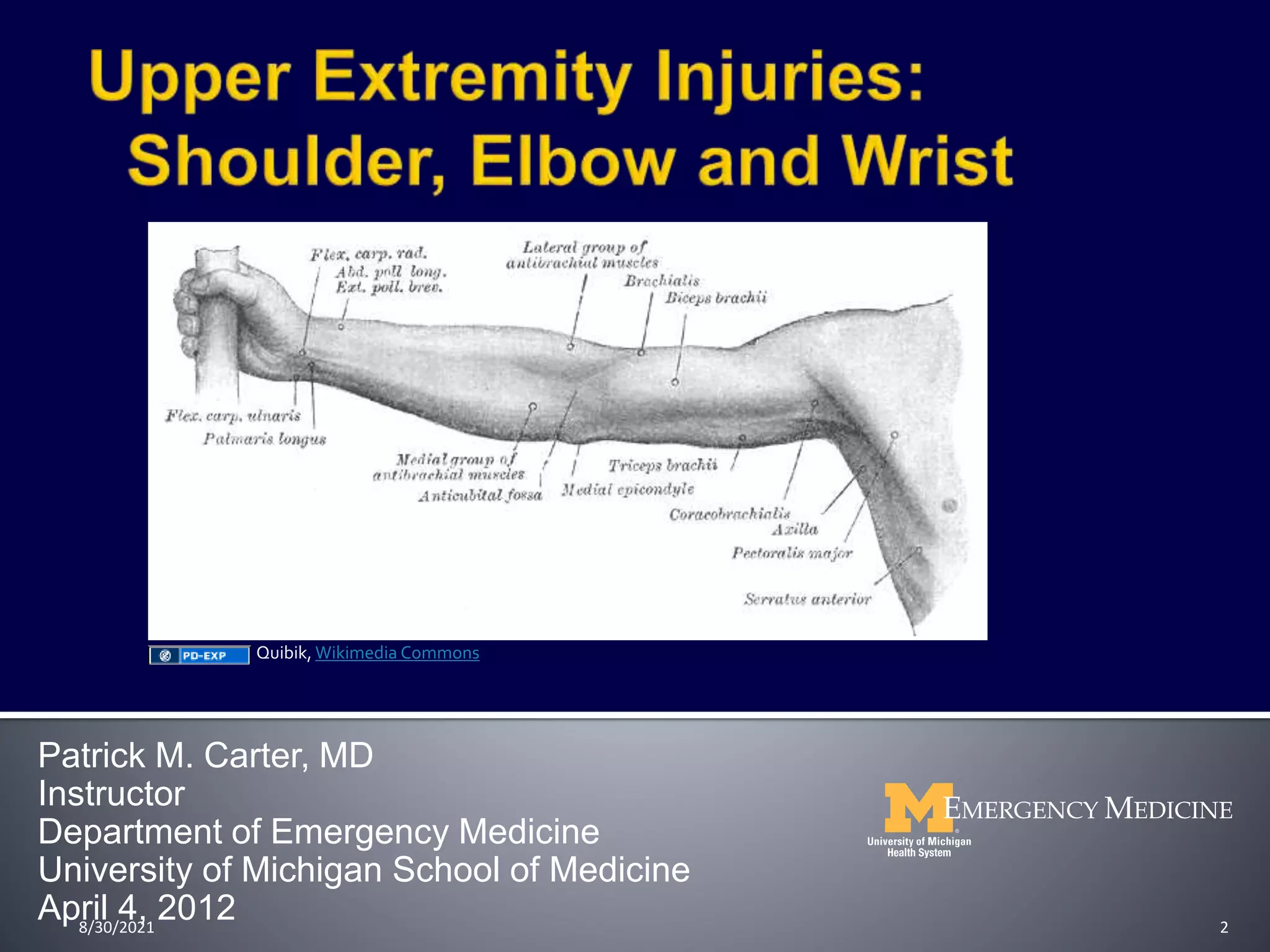
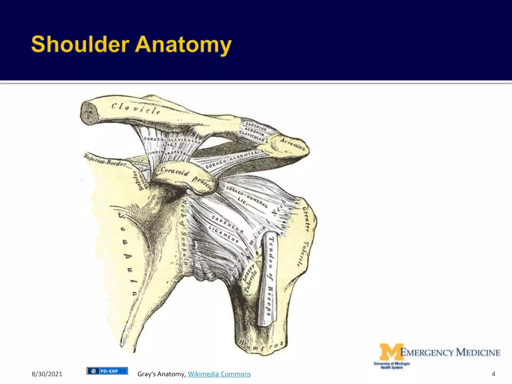
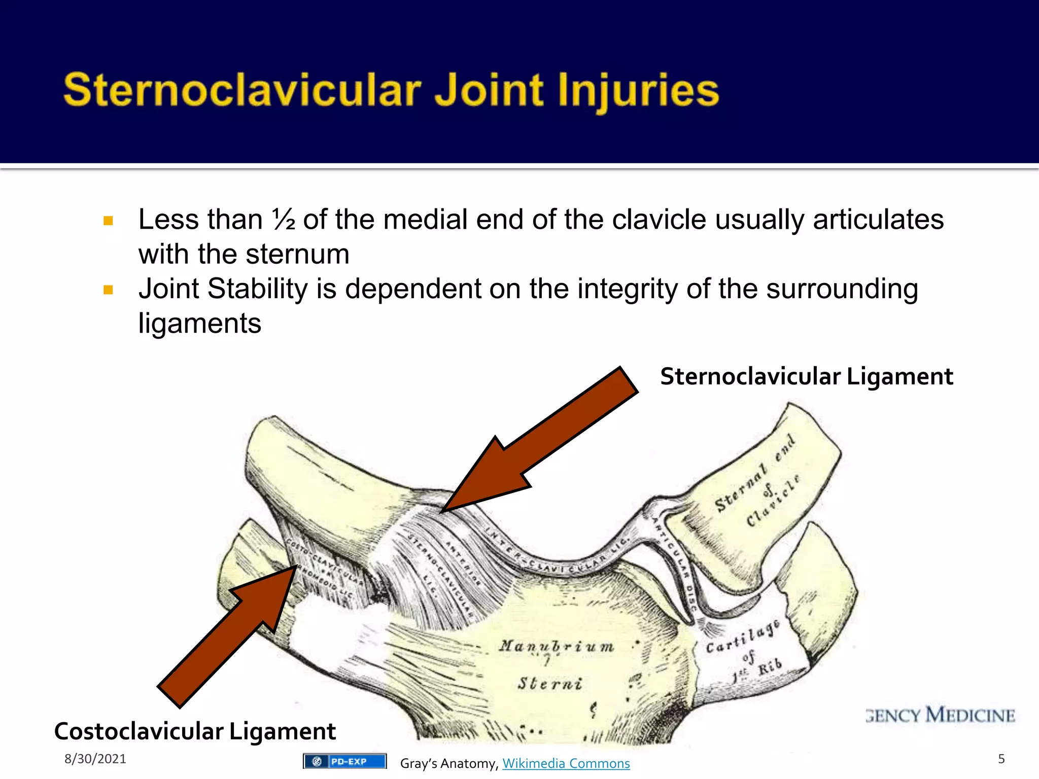
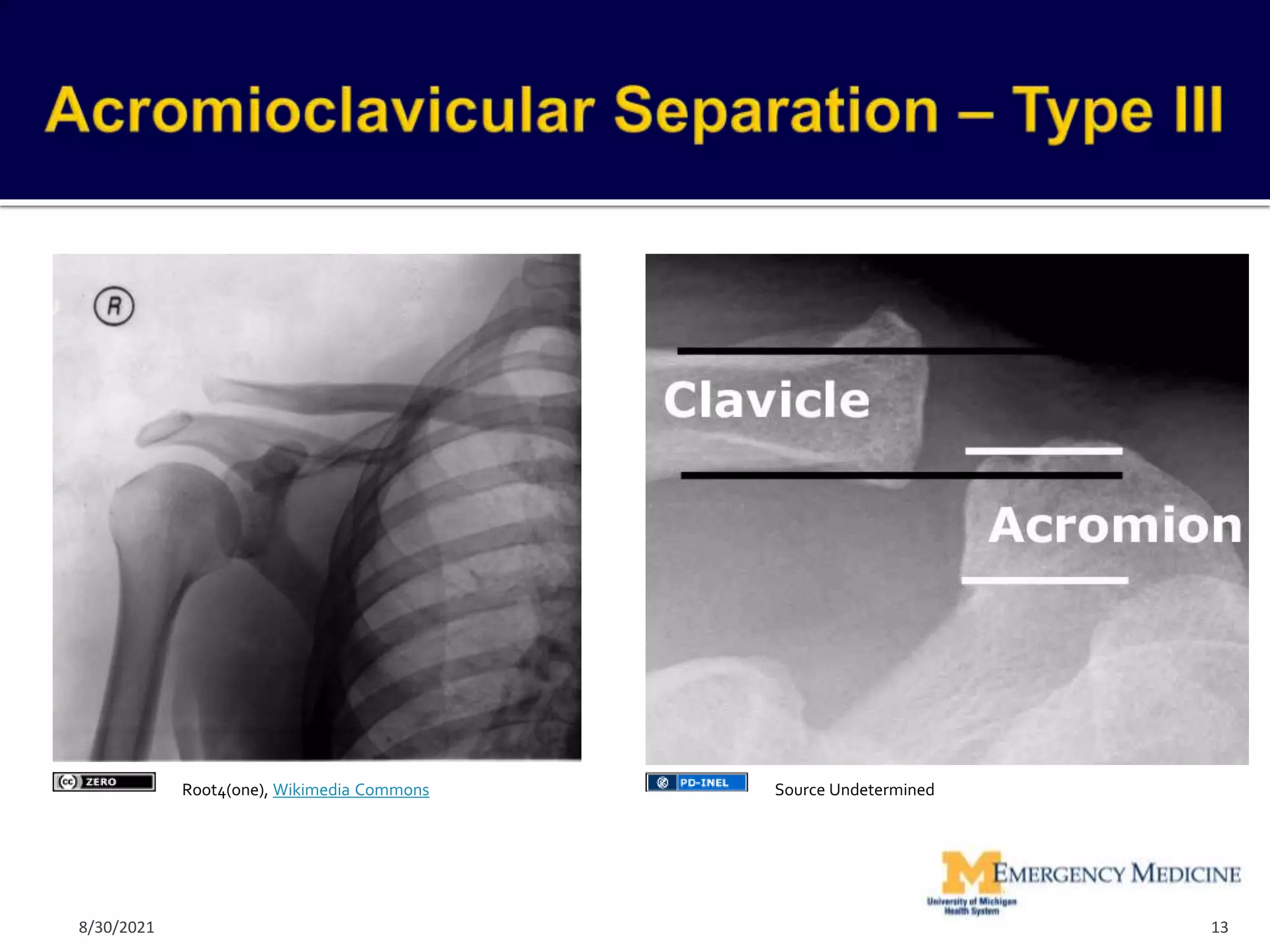
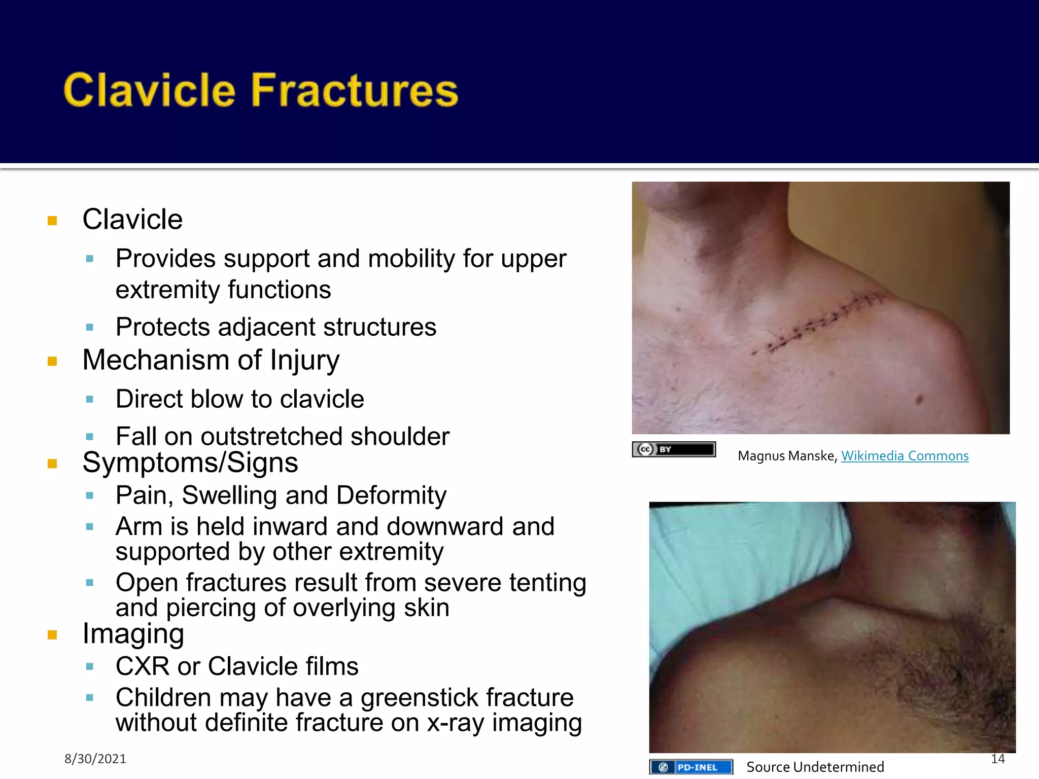
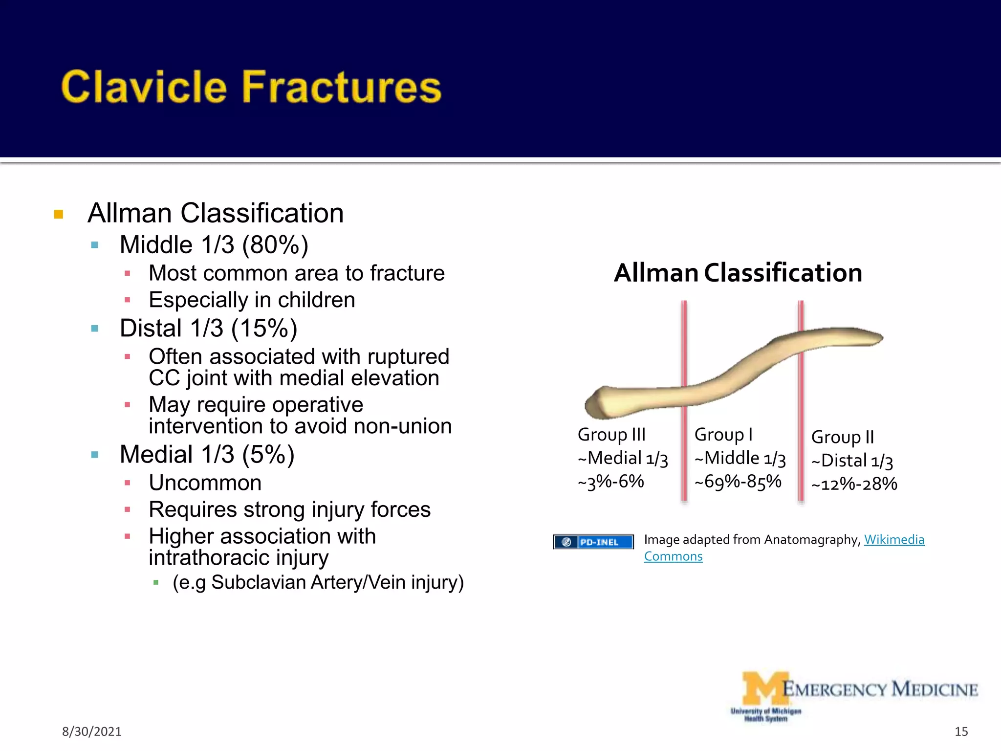
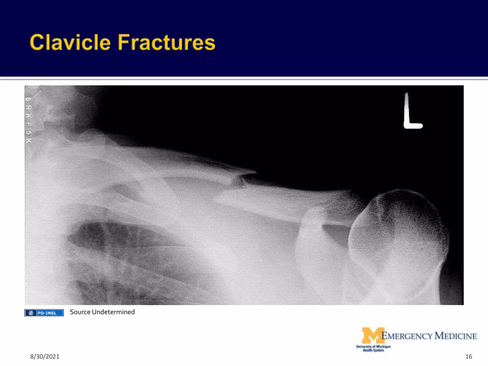
The document provides an overview of orthopedic injuries of the upper extremity, including the shoulder, arm, elbow, forearm, and wrist. It reviews common fractures and dislocations, their presentations, diagnostic approaches, and treatment options. Key injuries discussed include sternoclavicular dislocations, acromioclavicular separations, clavicle and scapula fractures, and shoulder dislocations.
Tractions in orthopaedics by Dr O.O. AfuyeAlade Olubunmi
Traction is an act of drawing or exerting a pulling force on bones or other tissues to offer realignment. It is very important in the management of fractures in other to prevent unwanted complications.
Tractions in orthopaedics by Dr O.O. AfuyeAlade Olubunmi
Traction is an act of drawing or exerting a pulling force on bones or other tissues to offer realignment. It is very important in the management of fractures in other to prevent unwanted complications.
As a general term, traction means pulling on part of the body.
Most often, traction uses mechanical force (sometimes generated by weights and pulleys) to put tension on a displaced bone or joint, such as a dislocated shoulder, to put it back in position and keep it still. In the medical field, traction refers to the practice of slowly and gently pulling on a fractured or dislocated body part. It’s often done using ropes, pulleys, and weights. These tools help apply force to the tissues surrounding the damaged area.
Traction: a basic physiotherapy modality used for inducing space between the joints. this slideshow deals with various types of traction and its application to cervical, thoracic and lumbar spine.
Acromioclavicular (AC) joint injury is a term used to describe an injury to the top of the shoulder, where the front of the shoulder blade (acromion) attaches to the collarbone (clavicle).
How to Make a Field invisible in Odoo 17Celine George
It is possible to hide or invisible some fields in odoo. Commonly using “invisible” attribute in the field definition to invisible the fields. This slide will show how to make a field invisible in odoo 17.
A review of the growth of the Israel Genealogy Research Association Database Collection for the last 12 months. Our collection is now passed the 3 million mark and still growing. See which archives have contributed the most. See the different types of records we have, and which years have had records added. You can also see what we have for the future.
Unit 8 - Information and Communication Technology (Paper I).pdfThiyagu K
This slides describes the basic concepts of ICT, basics of Email, Emerging Technology and Digital Initiatives in Education. This presentations aligns with the UGC Paper I syllabus.
Biological screening of herbal drugs: Introduction and Need for
Phyto-Pharmacological Screening, New Strategies for evaluating
Natural Products, In vitro evaluation techniques for Antioxidants, Antimicrobial and Anticancer drugs. In vivo evaluation techniques
for Anti-inflammatory, Antiulcer, Anticancer, Wound healing, Antidiabetic, Hepatoprotective, Cardio protective, Diuretics and
Antifertility, Toxicity studies as per OECD guidelines
This slide is special for master students (MIBS & MIFB) in UUM. Also useful for readers who are interested in the topic of contemporary Islamic banking.
Model Attribute Check Company Auto PropertyCeline George
In Odoo, the multi-company feature allows you to manage multiple companies within a single Odoo database instance. Each company can have its own configurations while still sharing common resources such as products, customers, and suppliers.
Exploiting Artificial Intelligence for Empowering Researchers and Faculty, In...Dr. Vinod Kumar Kanvaria
Exploiting Artificial Intelligence for Empowering Researchers and Faculty,
International FDP on Fundamentals of Research in Social Sciences
at Integral University, Lucknow, 06.06.2024
By Dr. Vinod Kumar Kanvaria
Safalta Digital marketing institute in Noida, provide complete applications that encompass a huge range of virtual advertising and marketing additives, which includes search engine optimization, virtual communication advertising, pay-per-click on marketing, content material advertising, internet analytics, and greater. These university courses are designed for students who possess a comprehensive understanding of virtual marketing strategies and attributes.Safalta Digital Marketing Institute in Noida is a first choice for young individuals or students who are looking to start their careers in the field of digital advertising. The institute gives specialized courses designed and certification.
for beginners, providing thorough training in areas such as SEO, digital communication marketing, and PPC training in Noida. After finishing the program, students receive the certifications recognised by top different universitie, setting a strong foundation for a successful career in digital marketing.
Macroeconomics- Movie Location
This will be used as part of your Personal Professional Portfolio once graded.
Objective:
Prepare a presentation or a paper using research, basic comparative analysis, data organization and application of economic information. You will make an informed assessment of an economic climate outside of the United States to accomplish an entertainment industry objective.
2. Patrick M. Carter, MD
Instructor
Department of Emergency Medicine
University of Michigan School of Medicine
April 4, 2012 2
Quibik, Wikimedia Commons
8/30/2021
3. Review key orthopedic injuries of the shoulder, upper arm, elbow,
forearm and wrist
Fractures
Dislocations
Ligamentous Injuries
Identify key x-ray findings
Review treatment options for orthopedic disorders of upper
extremity
Review key complications of upper extremity disorders
Not a complete review of all upper extremity injuries
3
8/30/2021
5. Sternoclavicular Ligament
Costoclavicular Ligament
Less than ½ of the medial end of the clavicle usually articulates
with the sternum
Joint Stability is dependent on the integrity of the surrounding
ligaments
5
Gray’s Anatomy, Wikimedia Commons
8/30/2021
6. Classification
1st Degree = Sprain
▪ Partial tear of SC and CC ligaments with mild subluxation
2nd Degree = Subluxation
▪ Complete tear of SC ligament with partial tear of CC
ligament
▪ Clavicle subluxates from the manubrium on x-ray
3rd Degree = Dislocation
▪ Complete tear of SC and CC ligaments
▪ Complete dislocation of clavicle from the manubrium
▪ Anterior > Posterior
▪ Posterior = True Emergency – 25% will have concurrent life-
threatening injuries to adjacent mediastinal structures
6
8/30/2021
7. Mechanism of Injury
Direct force applied to the medial end of the clavicle
Indirect force to the shoulder with the shoulder rolled either forward or
backward that tears medial ligaments
Symptoms/Signs
Pain and swelling over the SC joint
Pain with movement of shoulder
Anterior Dislocation = Prominent medial clavicle anterior to sternum
Posterior Dislocation = Clavicle may not be palpable, may be subtle
Diagnosis
X-ray
CT scan (Diagnostic Study of Choice if concern for underlying
structures)
7
8/30/2021
8. Treatment
1st Degree = Sling, Analgesia, Ice
2nd Degree
▪ Sling or Figure of Eight Clavicular Strap, Orthopedic Follow-up
3rd Degree
▪ Anterior Dislocation
▪ Uncomplicated anterior dislocations often don’t require reduction
▪ Sling or Figure of Eight, Analgesia and outpatient follow-up
▪ Posterior Dislocation
▪ Reduction often necessary due to underlying injury
▪ Closed reduction in OR
▪ Reduction
Towel roll between scapula
Traction applied to arm
Towel clip on clavicle with traction to reduce
8
8/30/2021
9. AC Joint Anatomy
Mechanism of Injury
Fall on outstretched arm with
transmission to AC joint
Fall on shoulder with arm
adducted (most common)
Scapula and Shoulder girdle
driven inferiorly with clavicle in
normal position
Signs/Symptoms
Joint Tenderness
Swelling over the joint
Pain with movement of affected
extremity
Displacement of clavicle Coracoclavicular Ligaments
- Coracoacromial ligament
-Trapezoid Coracoclavicular ligament
- Conoid Coracoclavicular ligament
Acromioclavicular
Ligament
9
Gray’s Anatomy, Wikimedia Commons
8/30/2021
10. AC Joint Injury Classification
Tossy and Allman Classification (Types 1-3)
Rockwood Classification (Types 4-6)
Classification
Type 1 = Sprain = Partial tear of AC ligament, No CC ligament
injury
Type 2 = Subluxation = Complete tear of AC ligament, CC
ligament stretched or incompletely torn
Type 3 = Dislocation = Complete tears of AC and CC ligaments
with displacement of clavicle
Direction of displacement defines types 4-6
▪ Type IV = Posterior displacement in or through trapezius
▪ Type V = Superior displacement (more serious type 3 injury)
▪ Type VI = Inferior displacement of clavicle behind biceps tendon
10
8/30/2021
12. X-rays
AP views of clavicle usually sufficient
Stress views not commonly used anymore and do not alter course of
treatment
Axillary views necessary for posterior dislocation identification (Type 4)
Findings
▪ Type 1 = Radiographically normal
▪ Type 2 = Increased distance between clavicle and acromion (< 1 cm)
▪ Type 3 = Increased distance between the clavicle and acromion (> 1 cm)
▪ Type 4-6 = Defined by displacement
Treatment
Type 1-2 = Sling x 1-2 weeks, Rest, Ice, Analgesia, Early ROM 7-14
days
Type 3 = Immobilize in sling, Prompt orthopedic referral
▪ Controversy regarding operative vs. conservative treatment options
▪ Shift towards conservative treatment
Type 4-6 = Sling, Prompt orthopedic referral, Likely will require surgical
management
12
8/30/2021
14. Clavicle
Provides support and mobility for upper
extremity functions
Protects adjacent structures
Mechanism of Injury
Direct blow to clavicle
Fall on outstretched shoulder
Symptoms/Signs
Pain, Swelling and Deformity
Arm is held inward and downward and
supported by other extremity
Open fractures result from severe tenting
and piercing of overlying skin
Imaging
CXR or Clavicle films
Children may have a greenstick fracture
without definite fracture on x-ray imaging
14
Magnus Manske, Wikimedia Commons
Source Undetermined
8/30/2021
15. Allman Classification
Middle 1/3 (80%)
▪ Most common area to fracture
▪ Especially in children
Distal 1/3 (15%)
▪ Often associated with ruptured
CC joint with medial elevation
▪ May require operative
intervention to avoid non-union
Medial 1/3 (5%)
▪ Uncommon
▪ Requires strong injury forces
▪ Higher association with
intrathoracic injury
▪ (e.g Subclavian Artery/Vein injury)
15
Image adapted from Anatomagraphy, Wikimedia
Commons
Group III
~Medial 1/3
~3%-6%
Group I
~Middle 1/3
~69%-85%
Group II
~Distal 1/3
~12%-28%
Allman Classification
8/30/2021
17. Emergency Orthopedic Consultation
Open Fractures
Fractures with neurovascular injuries
Fractures with significant tenting at high risk for converting to open
Indications for Surgical Repair
Displaced distal third
Open
Bilateral
Neurovascular injury
Treatment = Sling, Orthopedic Follow-up
Non-operative management is successful in 90%
Middle 1/3 Clavicle Non-union risk factors
Shortening > 2 cm
Comminuted fracture
Elderly female
Displaced fracture
Significant associated trauma
17
8/30/2021
18. Scapula
Links the axial skeleton to the upper extremity
Stabilizing platform for the motion of the arm
1% cases of blunt trauma have scapular fracture
3-5% of shoulder injuries
Mechanism of Injury
Direct blow to the scapula
Trauma to the shoulder
Fall on an outstretched arm
Clinical Presentation
Localized pain over the scapula
Ipsilateral arm held in adduction
Any movement of arm exacerbates pain
High association with other intrathoracic injuries (>75%)
Due to high degree of energy required for fracture
Pulmonary contusion > 50% of cases
Pneumothorax, Rib fractures commonly associated
Glenoid
Body Neck
18
Gray’s Anatomy, Wikimedia Commons
8/30/2021
19. Classification
Anatomic Location
Body = 50-60%
Neck = 25%
Imaging
Shoulder/Dedicated
Scapular Series
▪ AP/Lateral/Axillary
Axillary views help identify
fractures:
▪ Glenoid fossa
▪ Acromion
▪ Coracoid Process
Consider CXR/Chest CT to
rule out associated injuries
19
Gray’s Anatomy, Wikimedia
Commons
8/30/2021
20. Treatment
Sling, Ice, Analgesia
Immobilization
Early ROM exercises
Orthopedic Referral for ORIF
▪ Glenoid articular surface fractures
with displacement
▪ Scapular neck fractures with
angulation
▪ Acromial fractures associated with
rotator cuff injuries
20
Source Undetermined
8/30/2021
21. Shoulder dislocation = Most common dislocation in the ED
Classification
Anterior (95-97%)
▪ Subcoricoid, Subglenoid, Subclavicular, Intrathroracic
Posterior (2-3%)
▪ Most commonly missed dislocation in the ED
▪ Association with Seizure, Electric Shock/lightening injuries
Inferior (Luxatio Erecta)
Superior (Very Rare)
Mechanism of Injury
Anterior = Abduction, Extension and External Rotation with force applied
to shoulder
Posterior = Indirect force with forceful internal rotation and adduction
21
8/30/2021
22. Clinical Presentation
Severe pain
“Squared off” Shoulder
Patient resists abduction and internal
rotation
Humeral head palpable anteriorly
Must test axillary nerve
function/sensation
Quebec Decision Rule
Radiographs needed for:
▪ Age > 40 and humeral ecchymosis
▪ Age > 40 and 1st dislocation
▪ Age < 40 and mechanism other than fall
from standing height or lower
Failed to be validated due to low
sensitivity (CJEM 2011)
Recurrent Shoulder dislocations
Radiographs
AP/Lateral/Y-view
22
Source Undetermined
Source Undetermined
8/30/2021
23. Clinical Presentation
Medial rotation of the arm.
Prominence of posterior shoulder
Anterior flatness
Unable to externally rotate or abduct the
affected arm
Radiography
AP Radiograph
▪ “Light Bulb Sign”
▪ Internal rotation of the humerus
Y view
▪ Diagnostic for posterior dislocation
23
Source Undetermined
Source Undetermined
8/30/2021
24. Inferior Shoulder Dislocation
Hyperabduction force
Levers humerus against the acromion
tearing inferior capsule
Forces humeral head out inferiorly
Clinical Presentation
Humerus is fully abducted, elbow
flexed, hand behind the head
Humeral head palpated on lateral chest
wall
Frequently associated with:
Soft tissue injuries/rotator cuff tears
Fractures of humeral head
Neurovascular compression injury is
common
24
Source Undetermined
8/30/2021
25. Treatment
Reduction using a variety of techniques
▪ Success rate = 70-96% regardless of
technique
Shoulder dislocation with associated
humeral head fracture typically require
orthopedic consultation and may require
operative repair
Neurovascular exam pre- and post
reduction
Procedural Sedation if initial attempts
unsuccessful
Intra-articular injection of 10-20 cc
lidocaine alternative to procedural
sedation
After reduction, patient should be placed
in shoulder immobilizer and orthopedic
follow-up arranged
25
Nevit Dilman, Wikimedia Commons
8/30/2021
26. External Rotation
Hennepin Technique
Gentle external rotation
Followed by slow
abduction of arm
Reduction typically
complete prior to reaching
coronal plane
78% success rate
Procedural sedation rarely
needed
26
Source: University of Hawaii School of Medicine
8/30/2021
30. Scapular Manipulation
Technique
▪ Seated Position
▪ Steady forward traction on wrist
parallel to floor
▪ Rotate inferior tip of scapula
medially and superior aspect
laterally
96% Success rate
Requires two people
Borders of scapula can be
difficult to identify in obese
patients
Rarely requires sedation
30
Source: University of Hawaii School of Medicine
8/30/2021
31. Stimpson or Hanging Weight Technique
31
Source: University of Hawaii School of Medicine
8/30/2021
32. Complications
Recurrent dislocation (Most Common)
▪ < 20 years old: > 90%
▪ > 40 years old: 10-15%
Bony Injuries
▪ Hill-Sachs Deformity
▪ Compression fracture or groove of posterolateral aspect of humeral head
▪ Results from impact of humeral head on the anterior glenoid rim as it dislocates or reduces
▪ Avulsion of greater tuberosity (Higher incidence > 45 years old)
▪ Bankart’s Fracture = Fracture of the anterior glenoid lip
Nerve Injuries (10-25% dislocations)
▪ Most often are traction related neuropraxias and resolve spontaneously
▪ Axillary nerve (most common) or Musculocutaneous nerve
Rotator Cuff Tears
▪ 86% of patients > 40 years will have associated rotator cuff tear
Axillary Artery Injury (rare)
▪ Elderly patients with weak pulse
▪ Rapidly expanding hematoma
32
8/30/2021
34. Rotator cuff = 4 muscles that insert tendons into the greater and lesser tuberosity
SITS MUSCLES = Subscapularis, Supraspinatous, Infraspinatous, Teres minor
Mechanisms of Injury
Acute tear = Forceful abduction of the arm against resistance (e.g. fall on outstretched arm)
Chronic teat = 90% = Results from subacromial impingement and decreased blood supply to
the tendons (worsens as patient ages)
Clinical Picture
Typically affects males at 40 y/o or later
Pain over anterior aspect of shoulder, tearing quality to pain, typically worse at night
PE with weak and painful abduction or inability to initiate abduction (if complete tear)
Tenderness on palpation of supraspinatous over greater tuberosity
Imaging
In ED, plain film x-rays indicated to exclude fracture and may show degenerative changes
and superior displacement of humeral head
MRI is diagnostic (not typically done in ED setting)
Treatment
Sling Immobilization, Analgesia, Ortho Referral
Complete tears require early surgical repair (< 3 weeks)
Chronic tears are managed with immobilization, analgesia and orthopedic follow-up for
rehabilitation exercises and possible steroid injection
34
8/30/2021
35. Proximal Humerus Fractures
Common in elderly patients with osteoporosis
Mechanism of Injury = Fall on outstretched hand with elbow extended
Clinical Presentation
▪ Pain, swelling and tenderness around the shoulder
▪ Brachial plexus and axillary arteries injuries
▪ Higher incidence (>50%) in displaced fractures
Neer Classification guides treatment
▪ Fractures separate humerus into 4 fragments by epiphyseal lines
▪ Displacement > 1 cm or angulation > 45 degrees defines a fragment as a
“separate part” when fractures occur
▪ If none of fragments are displaced > 1cm, fracture is termed 1 part
Treatment
▪ One part fractures (85%) = immobilization in sling/swathe, ice, analgesics,
orthopedic referral
▪ Two/Three/Four part fractures = Orthopedic Consultation
35
8/30/2021
36. Fragments of Humerus Head
Articular surface of humeral head
Greater tubercle
Lesser tubercle
Shaft of humerus
36
3
1
2
James Heilman, MD, Wikimedia Commons
Gray’s Anatomy,
Wikimedia Commons
8/30/2021
39. Typically involve middle 1/3 of the humeral shaft
Mechanism of Injury
Direct Blow (Most common)
Fall on outstretched arm or elbow
Pathologic Fracture (e.g. breast cancer)
Clinical Presentation
Pain and deformity over affected region
Associated Injuries
▪ Radial Nerve injury = Wrist Drop (10-20%)
▪ Neuropraxia will often resolve spontaneously
▪ Nerve palsy after manipulation or splinting is due to nerve entrapment and must be
immediately explored by orthopedic surgery
▪ Ulnar and Median nerve injury (less common)
▪ Brachial Artery Injury
39
8/30/2021
40. Most of the time is Conservative
Closed Reduction in upright position followed by application of U
shaped Slab of POP or Cylinder cast
Few weeks later or initially in stable fractures Functional Brace
may be used
http://ptjournal.apta.org/cgi/collection/injuries_and_conditions_upper_extremity
47. Proximal or distal biceps tendon rupture
Mechanism of Injury = Sudden or prolonged
contraction against resistance in middle aged
or elderly patients
Clinical Presentation
“Snap” or “Pop” typically described
Pain, swelling, tenderness over site of tendon
rupture
Flexion of elbow = Mid-arm ball
Loss of strength sometimes minimal
X-rays to exclude avulsion fracture
ED Treatment
Sling, Ice, Analgesia, Orthopedic referral
Surgical repair for young, active patients
47
Patenthalse, Wikimedia Commons
Gray’s Anatomy, Wikimedia
Commons
8/30/2021
49. Anterior Fat Pad
“Sail Sign”
Posterior Fat Pad
(Never normal)
Anterior Humeral Line
• Normal = Middle of capitellum
• Abnormal = Anterior 1/3 of
capitellum or completely anterior
Radial-Capitellar Line
•Normal =Transects
middle of capitellum
49
Hellerhoff, Wikimedia Commons
Source Undetermined
Source Undetermined
8/30/2021
50. Supracondylar Extension Fractures
Most Common Type
Mechanism of injury
▪ Fall on outstretched arm with elbow in extension
Imaging
▪ Distal humerus fractures and humeral fragment displaced posteriorly
▪ Sharp fracture fragments displaced anteriorly with potential for injury
of brachial artery and median nerve
Treatment
▪ Non-displaced fracture (Rare) = Immobilization in posterior splint
▪ May be discharged home with close follow-up
▪ Displaced fracture
▪ Orthopedic Consultation and reduction
▪ Patients with displaced fractures or significant soft tissue swelling require
admission for observation
50
8/30/2021
53. Supracondylar Flexion Fractures (rare)
Mechanism of Injury
▪ Direct blow to posterior aspect of flexed elbow
Fractures are frequently open
Imaging = Distal humerus fracture displaced anteriorly
Treatment
▪ Non-displaced fractures
▪ Splint immobilization and early orthopedic follow-up
▪ Displaced fractures
▪ Orthopedic consultation for reduction
▪ Patients with displacement and soft tissue swelling require admission
53
8/30/2021
54. Absolute Emergency
Should de done under G A by experienced doctor as soon as
possible
In the past the arm was held in flexed elbow position in back-slab
POP after reduction
At present time Percutaneous K wire fixation is ALWAYS carried
out after reduction
http://ptjournal.apta.org/cgi/collection/injuries_and_conditions_upper_extremity
56. Early Complications
Neurologic (7%)
▪ Results from traction, direct trauma or nerve ischemia
▪ Radial Nerve (Posterior-medial displacement)
▪ Median Nerve (Posterior-lateral displacement)
▪ Ulnar Nerve (Uncommon)
▪ Anterior Interosseous Nerve Injuries
▪ High incidence with supracondylar fractures
▪ No sensory component, Motor component must be tested (“OK sign”)
Vascular Entrapment (Brachial Artery)
Late Complications
Non-union/Mal-union
Loss of mobility
56
8/30/2021
57. A. Early= Compartment syndrome
Brachial Artery injury ( Acute
Volkmann's Ischemia )
Nerve Injury : Median, Ulnar or Radial
B. Late= Stiffness
Volkmann's Ischemic contracture
Heterotopic Calcification
Mal-Union ( Cubitus Valgus or varus)
http://ptjournal.apta.org/cgi/collection/injuries_and_conditions_upper_extremity
58. Compartment syndrome of the forearm
Complication of elbow/forearm fractures
Increased compartment pressure results in ischemia of muscles of
forearm, typically flexor compartment
Patient complains of pain out of proportion of injury, digit swelling
and paresthesias
Also consider in any patient presenting with pain and numbness in
hand after casting has been performed
Irreversible damage in 6 hours (see image)
Treatment
Removal of cast
Surgical decompression with fasciotomy
58
Source Undetermined
8/30/2021
60. Most common fractures of the elbow
Mechanism of Injury = Fall on outstretched hand
Clinical Finding = Tenderness and swelling over the radial head
Imaging
May not be seen on initial x-ray or may be subtle on x-ray
Evaluate for anterior or posterior fat pad which suggests diagnosis
Associated Injuries
Essex-Lopresti Lesion
▪ Disruption of fibrocartilage of the wrist and interosseus membrane
▪ Distal radial-ulnar dissociation
Articular surface of capitellum frequently also injured
Treatment
Non-displaced = Sling, Ortho follow-up
Comminuted/Displaced Fractures require urgent orthopedic referral
within 24 hours
60
Source Undetermined
8/30/2021
61. Nursemaid’s elbow = Subluxation of
radial head beneath the annular
ligament
Mechanism of injury = Longitudinal
traction on hand or forearm with
arm in pronation
X-rays not necessary
Treatment = Reduction
Thumb over radial head with
concurrent supination of forearm and
flexion of elbow
Extension and pronation (another
option for reduction)
61
David Tan, Flickr
8/30/2021
63. Third most common joint dislocation
Posterolateral (90%)
Mechanism of Injury = Fall on outstretched hand
Clinical Findings
▪ Marked swelling with loss of landmarks
▪ Posterior prominence of olecranon
Immediate consideration must be given to neurovascular status
▪ Ulnar or Median Nerve injury common (8-21%)
▪ Brachial artery injury (5-13%)
Associated fractures (30-60%) of coronoid process and radial head
Terrible triad injury = elbow dislocation + radial head and coronoid
fracture (unstable)
Anterior (Uncommon)
Mechanism of Injury = Blow to Olecranon with elbow in flexion
Associated Injuries = Much higher incidence of vascular impingement
63
8/30/2021
65. Elbow Reduction
Immobilize humerus
Apply traction at wrist
Slight flexion of the elbow
Posterior pressure on olecranon
Post-Reduction
Long Term Complications
Post-traumatic arthritis
Joint instability
65
8/30/2021
66. Fracture of both ulnar and radius
Usually displaced fracture
Mechanism of Injury
Direct blow to forearm
Associated Injury
Peripheral Nerve Deficits
▪ Uncommon in most closed injuries
▪ More common with open fractures
Development of compartment syndrome
Treatment
Displaced – ORIF
Complications
Compartment Syndrome
Malunion
66
Source Undetermined
8/30/2021
67. Isolated fracture of ulnar
shaft
Mechanism
Direct blow to ulna
Patient raising forearm to
protect face
Treatment
Non-displaced
▪ Immobilization in splint
Displaced
▪ >10 degrees angulation
▪ Displacement > 50% of ulna
▪ Orthopedic consultation - ORIF
67
Source Undetermined
8/30/2021
68. Distal Radius Fracture
Distal radio-ulnar
dislocation
Reverse Monteggia’s fx
Mechanism of Injury
Direct blow to back of wrist
Fall on outstretched hand
Complication = Ulnar
nerve injury
Treatment = ORIF
http://www.learningradiology.com/caseofweek/ca
seoftheweekpix2/cow157lg.jpg
68
Th. Zimmermann, Wikimedia Commons
8/30/2021
69. Proximal 1/3 Ulnar Fracture
Dislocation of radial head
Mechanism of Injury = Direct blow
to posterior aspect of ulna
Fall on outstretched hand
Imaging
Elbow/Forearm x-rays
Radial head dislocation missed in
25% of cases
Carefully examine the alignment of
radial head
Associated Injury = Radial Nerve
Injury
Treatment
ORIF
Closed Reduction/Splinting
69
Jane Agnes, Wikimedia Commons
8/30/2021
71. Transverse fracture of distal radius with dorsal displacement of distal
fragment
Mechanism = Fall on outstretched hand
Most common fracture in adults > 50 years old
Exam = Classic Dinner Fork Deformity
Associated Injuries
Ulnar styloid fracture
Median Nerve Injury
Unstable Fractures
>20 degrees angulation, intra-articular involvement, comminuted fractures or
> 1 cm of shortening
Treatment
Non-displaced Fracture
▪ Sugar Tong Splint, Referral to Orthopedic Surgery
Displaced Fracture
▪ Reduction – Finger traps and manipulation under procedural sedation or with
hematoma block
▪ Immobilization in Sugar tong splint
▪ Referral to Orthopedic Surgery
71
8/30/2021
72. Transverse fracture of distal
radius with volar displacement
Mechanism = Fall on
outstretched arm with forearm
in supination
Associated Injury = Median
Nerve Injury
Treatment
Reduction with finger traps and
manipulation
Immobilization in sugar tong or
long arm splint
Orthopedic referral
72
8/30/2021
74. Carpal tunnel (CTS)
result from repetitive stress
to tissue
64% of work injuries
Compressive neuropathy
Wrist flexion/ext and finger
movements
Risk factors
exertion
repetitive stress
posture
localized contact
cold
Markiewitz AD, Andrish JT. Hand and wrist injuries in the preadolescent and adolescent athlete. Clin in Sport Med. 11(1):203-25,
1992.
76. Carpal fractures
compressive loads to
hyperextended wrist
hyper flexion
rotation loading
against a fixed wrist
Scaphoid
▪ 60-70%
Lunate
Markiewitz AD, Andrish JT. Hand and wrist injuries in the preadolescent and adolescent athlete. Clin in Sport Med. 11(1):203-25,
1992.
77. Thumb: essential to
prehension
Sprain: skiers thumb
fall with thumb in abducted
position
tensile loads on MCL
Hyperextension
Bennets fracture (fighting)
Bowler’s thumb: ulnar digital
nerve trauma
tingling, sensitivity
Markiewitz AD, Andrish JT. Hand and wrist injuries in the preadolescent and adolescent athlete. Clin in Sport Med. 11(1):203-25,
1992.
78. Metacarpal &
phalangeal injuries
Fractures
Boxers
Dislocations
Markiewitz AD, Andrish JT. Hand and wrist injuries in the preadolescent and adolescent athlete. Clin in Sport Med. 11(1):203-25,
1992.
79. Scaphoid Fracture
Most common carpal bone fracture
Mechanism = fall on outstretched hand or axial load to thumb
2/3 of fracture in waist of scaphoid
Imaging – Initial x-rays may fail to demonstrate fracture
▪ > 10% of cases
▪ Repeat Imaging in 2 weeks will often show fracture
Clinical findings = tenderness in anatomical snuff box
Treatment
▪ Non-displaced or clinically suspected fracture
▪ Thumb spica Splint
▪ Displaced fractures will require ORIF
▪ Complications
▪ Avascular necrosis of proximal fragment -> arthritis
▪ Delayed union or malunion
79
8/30/2021
81. Triquetrum Fracture (2nd most common)
Mechanism = Fall on outstretched hand
Body fracture or avulsion chip fractures
Exam = Tenderness on palpation distal to ulnar styloid on dorsal aspect of
wrist, painful flexion
Avulsion fracture best visualized on lateral or oblique view of wrist
Treatment = Volar splint, Orthopedic referral
Lunate Fracture
Mechanism = Fall on outstretched hand
Exam = Pain over mid-dorsum of wrist increased with axial loading of 3rd
digit
Vascular supply is through distal end of bone -> high risk for avascular
necrosis of the proximal portion
Plain x-rays are often normal
Treatment = Immobilization in thumb spica splint, orthopedic referral
Complications
▪ Kienbock’s disease = Avascular necrosis of proximal segment
▪ Chronic pain, decreased grip strength, osteoarthritis
81
8/30/2021
83. Lunate is at the center of the carpal bones
Majority of ligamentous injuries are centered on the lunate
Injuries are from forceful dorsiflexion of wrist
Degree of force determines severity of injury
▪ Spectrum from isolated tear to dislocations
Spectrum of ligamentous injuries
Scapholunate ligament instability
Triquetrolunate ligament instability
Perilunate and Lunate dislocations
83
8/30/2021
84. Scapholunate ligament binds the scaphoid and lunate together
Most common ligamentous injury of hand
Commonly missed
Pain with wrist hyperextension, snapping or clicking sensation with
radial/ulnar deviation
Radiographic signs
Scaphoid is foreshortened and has a dense ring shaped image around
its distal edge (signet or cortical ring sign)
Widening of space between the lunate/scaphoid
▪ > 3 mm, Terry Thomas sign
Treatment
Thumb spica or radial gutter splint
Orthopedic Referral
84
8/30/2021
85. Terry Thomas and Signet Ring Sign
85
Source Undetermined
8/30/2021
86. Perilunate and lunate dislocations are the result of the most severe
carpal ligamentous injury
Mechanism of Injury = Violent Hyperextension usually combined with a
fall from height or motor vehicle crash
Clinical examination
Generalized swelling, pain and tenderness over wrist
May be deceiving with no evidence of gross deformity
Radiographic evaluation is key to diagnosis
Treatment = Orthopedic Consultation
Treatment is dependent on severity of injury
Closed reduction and long-arm immobilization if possible
Open, unstable and irreducible dislocations require OR
Some orthopedists take all dislocations to OR
Complications
Degenerative Arthritis
Delayed union/Malunion/Non-union
Avascular necrosis
86
8/30/2021
87. 4 C’s Need to line up on normal x-ray
Lunate
87
Source Undetermined
8/30/2021
88. Lunate Dislocation
Capitate is centered over the
radius and the lunate is tilted out
Spilled Tea cup deformity
Peri-lunate Dislocation
Lunate is centered over the
radius and capitate is tilted out
Associated with scaphoid fx
88
Source: Radiology
Assistant
Source: Radiology
Assistant
8/30/2021
89. Carpal Tunnel Syndrome
Entrapment of Median nerve
Tinel’s sign = Tapping over volar wrist produces paresthesias
Phalen’s sign = Hyperflexion of wrist = Paresthesias
Risk Factors = Pregnancy, Hypothyroid, DM, RA
Treatment = Splinting, Rest, Surgical Decompression
DeQuervain’s Tenosynovitis
Overuse syndrome with inflammation of extensor tendons of thumb
Characterized by pain along radial aspect of wrist that is exacerbated with
use of thumb
Finkelstein’s test = Ulnar deviation of fisted hand produces pain
Treatment = NSAIDS, Splint, Rest
Guyon’s Canal Syndrome
Ulnar nerve entrapment syndrome
Numbness and tingling in ring and small finger
Causes = repetitive trauma (handle bar neuropathy), cyst
Treatment = Splint, Surgical Decompression
89
8/30/2021
Editor's Notes
Shoulder is the most mobile joint in the human body
Due to the increased functional abilities of the shoulder, it is at high risk for injury
Shoulder Anatomy
Sternoclavicular Joint
Clavicle
Acromioclavicular Joint
Scapula
Humerus
Glenohumeral joint
Muscles/ligaments that attach the shoulder bones, including special rotator cuff muscles
Sternoclavicular Injuries
SC joint is most frequently moved, non-axial joint in the body
SC joint has the least amount of stability of any major joint because less than ½ of the medial end of the clavicle articulates with the sternum
Thus, Joint stability depends on the integrity of surrounding ligaments
SC = Sternoclavicular
CC = Costoclavicular
SC Sprains
Mechanism of Injury = forcing the shoulder forward suddenly or applying medial directed force to the shoulder Sprain
Symptoms = Pain and swelling localized to the joint
Treatment = Ice, Sling, Analgesics
****Remember to think about possible septic arthritis in injection drug users****
SC Dislocations
Uncommon injuries requiring significant force to the shoulder usually from motor vehicle crashes
Anterior dislocations > Posterior Dislocations
Mechanism of injury
Anterior Dislocation = Usually the result of either direct or indirect force applied directly to the joint with the should rolled backward at the time of impact
Posterior Dislocation = If the shoulder is rolled forward at the time of impact, a posterior dislocation can result from a direct impact or indirect force to the shoulder
Symptoms = Severe pain that is exacerbated arm motion
Exam
Anterior Dislocations = Prominent medial clavicle that is palpable anterior to the sternum
Posterior Dislocations = clavicle may not be palpable
X-rays views may not be diagnostic and special views or comparison views may be necessary. CT is the diagnostic tool of choice. If concerns for underlying vascular structures, consider IV contrast
Treatment
Anterior SC Dislocations = Uncomplicated anterior dislocations do not require attempted reduction as injury has little or no impact on function
Injury is often unstable, so attempts at reduction will usually be unsuccessful, but reduction can be attempted by placing the patient supine with a towel roll under the scapula - - - traction is applied to the arm with pressure on the medial end of the clavicle
Sling, Ice, analgesics and orthopedic referral are recommended
Posterior SC Dislocations = May be associated with lifethreatening injuries = = Pneumothorax, compression or laceration of vessels, trachea or esophagus
ORTHOPEDIC CONSULTATION
Closed reduction should be attempted in the operating room with vascular surgery present
If immediate reduction is needed, position the patient the same as for anterior DC dislocations, towel clip is applied to clavicle and pulled upward
**** Figure of 8 clavicular strap is older method of immobilizing. Simple sling is good enough for support, tends to cause less discomfort and the results are equal in studies comparing two techniques. Figure of 8 might be used in places without commercially available slings (e.g. Ghana)
AC = Acromioclavicular
CC = Corococlavicular
> 1 cm displacement defines type 3
Body of scapula fracture
Associated rib fractures
Mechanism of Injury
Anterior = Abduction, Extension and External Rotation
Posterior = Seizure or Electric Shock
Fall on forward-flexed, adducted and internally rotated arm
(1) age 40 and humeral ecchymosis, (2) age 40 and first dislocation, and (3) age <40 and injury mechanism other than nontrauma or a fall from standing height or lower.
Procedural sedation often necessary
If in seated position, have an assistant stand, facing the patient, and use one arm to firmly grasp the wrist of the dislocated arm. The assistant should then apply steady forward traction parallel to the floor while applying countertraction with the other arm, which is outstretched and resting on the patient's clavicle
Use both hands to rotate the inferior tip of the scapula medially and the superior aspect laterally with slight dorsal displacement. The goal is to move the glenoid fossa back into anatomical position.
Hill-Sachs Deformity
Compression fracture of posterolateral aspect of humeral head
Results from impact of humeral head on the anterior glenoid rim as it dislocates or reduces
Bankart’s Fracture = Fracture of the anterior glenoid lip
Displacement of a fracture fragment by 1 cm, or angulation between fracture fragments of 45° or greater, is what defines a fragment as being a "separate" part. Hence, a proximal humerus fractures may be called 2-part, 3-part, or 4-part according to the Neer classification system, depending upon the amount of displacement and angulation seen on x-ray.
Radial Nerve injury = Wrist Drop = Inability of extend wrist, fingers, thumb, Loss of sensation over dorsal web space of 1st digit
Elbow radiographic evaluation can be difficult
True Lateral X-ray = Hourglass or Figure of 8 at distal Humerus
Fat Pad Signs
Posterior Fat Pad Sign = Never seen on normal x-ray imaging
Indicates distension of joint capsule by effusion with likely occult fracture
Often associated with occult radial head fracture
Anterior Fat Pad Sign = Small one may be present on normal x-rays
Increased anterior fat pad (sail sign) is abnormal and may indicate fracture
Anterior Humeral Line
Line drawn along anterior surface of humerus and extending through the elbow
Normally, transects the middle of the capitellum but with Supracondylar fractures, transects the anterior 1/3 of the capitellum or passes completely anterior to the capitellum
Radial-Capitellar Line
Line drawn through the middle of the radius
Normally, transects the middle of the capitellum
Abnormal line may indicate radial head dislocation or subtle fracture
Radial Head Evaluation
Carefully inspect the radial head.
Fracture may be subtle and only clue may be slight cortical irregularity
Distal Humerus Evaluation
Careful inspection and evaluation of anterior humeral head line
Posterior Fat Pad Sign = Never seen on normal x-ray imaging Anterior Fat Pad Sign = Small one may be present on normal x-rays
Anterior Humeral Line = Line drawn along anterior surface of humerus and extending through the elbow
Normally, transects the middle of the capitellum but with Supracondylar fractures, transects the anterior 1/3 of the capitellum or passes completely anterior to the capitellum
Radial-Capitellar Line
Line drawn through the middle of the radius
Normally, transects the middle of the capitellum
Abnormal line may indicate radial head dislocation or subtle fracture
Traction distally at wrist with assistant immobilizing the humerus
While maintaining traction, flex the elbow and apply posterior pressure to the humerus
Post red= Reassess ROM of elbow and neurovascular status
Immobilize in long-arm posterior splint in 120 degrees of flexion (i.e. full flexion)
Observe for delayed vascular compromise
ORIF = majority, closed red/splinting = may be possible, more commonly in kids