SlideShare a Scribd company logo
Page 1
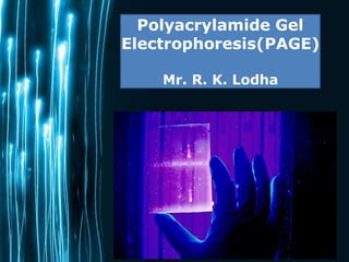
Polyacrylamide Gel
Electrophoresis(PAGE)
Mr. R. K. Lodha
Why Use
Polyacrylamide
Gels to
Separate
Proteins?
• Polyacrylamide gel has a tight matrix
• Ideal for protein separation
• Smaller pore size than agarose
• Proteins much smaller than DNA
– Average amino acid = 110 daltons
– Average nucleotide pair = 649 daltons
– 1 kilobase of DNA = 650 kD
– 1 kilobase of DNA encodes 333 amino acids =
36 kD
Why polyacrylamide used for
a gel?
⚫Chemically inert
⚫Electrically neutral
⚫Hydrophillic
⚫Transparent for optical detection
Acrylamide
Page 4
•
•
Acrylamide CF- C3H5NO
White odourless crystalline solid, soluble in water,
ethanol,ether & chloroform
•
•
•
Prepared on industrial scale by the hydrolysis
of acrylonitrile by nitrile hydratase
carcinogenic as well as Neurotoxic compound.
used in the manufacture of dyes, Waste water
treatment and other monomers
Polyacrylamide
Page 5
• Also called Cross-linked Polyacrylamide
• Polyacrylamide is not toxic
• Polyacrylamide is a cross-linked polymer of Acrylamide
• It is recommended to handle it with caution
• It is highly water-absorbent, forming a soft gel when hydrated
• Used in-
- Flocculate or coagulate solids in a liquid
- A subdermal filler for aesthetic facial surgery
- Polyacrylamide gel electrophoresis
- In soft contact lenses etc.
Polyacrylamide gel
Page 6
• It is a white odorless gel, soluble in water
•
•
•
After polymerization of acrylamide it get cross-linked
structure
TEMED stabilizes free radicals and improves
polymerization
Here, the toxic affect of acrylamide get vanish (95%)
• Amount of polyacrylamide salt dissolved (conc.) is
directly proportion to cross –linked nature of gel
▶Polyacrylamide gels are
characterized by two parameters:
1. total monomer concentration
(%T, in g/100 ml)
2. weight percentage of cross
linker (%C).
▶ Higher %T - smaller pores.
▶The practical ranges for
monomer concentration are
stock solutions of 30-40%
Gel Types
Page 8
•
•
•
•
•
•
•
•
•
•
•
Agarose
Polysaccharide extracted
from sea weed.
Gel casted horizontally
Non-toxic.
Separate large molecules
Commonly used for DNA
separations.
Staining can be done before
or pouring the gel.
Polyacrylamide Gel
• Cross-linked polymer of
acrylamide.
Gel casted vertically.
Potent neuro-toxic.
Separate small molecules.
Used for DNA or protein
separations.
Staining can be done after
pouring the gel.
A. On The Basis of Supporting Media
1.Slab Gel
A.Horizontal
B.Vertical
2. Tube Gel
B. On The Basis of Types of Separation
1. No-Denaturing/native (Separation by size and charge;
charge/mass and shape)
2. Denaturing/Non-Native (separation by size)
3. Other Types (IEF, 2-D-GEL)
TYPES OF GEL ELECTROPHORESIS
PAGE
Page 10
Poly Acrylamide Gel
Electrophoresis
Page 11
• It is a subtype of the gel electrophoresis
whereby the normal gel is replaced with
polyacrylamide gels used as support matrix.
• Gels are made by free radical-induced
polymerization of acrylamide and N,N’-
Methylenebisacrylamide.
• It is the most widely used technique of
electrophoresis.
Types Of PAGE
Page 12
•
•
•
No denaturing agents
Proteins separated based on size, charge and
shape.
Used when want to keep protein active to study
conformation, self-association or aggregation, and the
binding of other proteins
Native PAGE
Page 13
Principle
Electrophoretic migration occurs because most proteins
carry a net negative charge in alkaline running buffers.
The higher the negative charge density (more charges
per molecule mass), the faster a protein will migrate.
At the same time, the frictional force of the gel matrix
creates a sieving effect, retarding the movement of
proteins according to their size and three-dimensional
shape.
Small proteins face only a small frictional force while
large proteins face a larger frictional force.
Thus native PAGE separates proteins based upon both
their charge and mass.
Because no denaturants are used in native PAGE,
subunit interactions within a multimeric protein are
generally retained and information can be gained about
the quaternary structure.
In addition, some proteins retain their enzymatic
activity (function) following separation by native
PAGE. Thus, it may be used for preparation of purified,
active proteins.
Type of Native PAGE
Native or Non-Denaturing
1. Continuous System: Both gels and
electrophoresis tank have same buffer
composition (Single phase gel; a resolving
gel)….e.g DNAAgarose gel electrophoresis
2. Discontinuous System: Both tank and gels have
different buffers (two phase gel; a stacking gel
and separating or resolving gel) e.g. Polyacryl
amide gel electrophoresis (PAGE) for proteins
SDS - PAGE
Page 17
• It is a modified version of PAGE whereby
Sodium-dodecyl-sulphate (SDS) is used.
• SDS is an amphipathic surfactant.
• It denatures proteins by binding to the protein
chain with its hydrocarbon ‘tail’, exposing
normally buried regions and ‘coating’ the
protein chain with surfactant molecules.
• The polar ‘head’ group of SDS adds an
additional benefit to the use of this
denaturant.
Structure of SDS
Page 18
(NaC12H25SO4)
also called sodium lauril sulfate or sodium lauryl sulfate
⚫ Mercaptoethanol will break the disulphide bridges.
⚫ SDS binds strongly to and denatures the protein.
⚫ Each protein is fully denatured and open into rod-
shape with series of negatively charged SDS
molecule on polypeptide chain.
SDS is an
anionic
detergent.
The sample is
first boiled for
5min in buffer
containing
20
• Beta-
Mercaptoethanol
• SDS
⚫On average, One SDS molecule bind for every
two amino acid residue.
⚫Hence original native charge is completely
swamped by the negative charge of SDS
molecule.
⚫Also referred as Discontinuous gel
electrophoresis.
21
SDS-PAGE
Page 22
WHY ? ? ?
• In their native form, proteins fold into a variety
of shapes, some compact, some elongated.
• The rate of migration of native proteins
through a sieving medium is therefore more a
reflection of their relative compactness, and
less an accurate measure of molecular weight.
• Denaturing the proteins nullifies structural
effects on mobility, allowing separation on a
true charge/mass ratio basis.
• It also separates subunits in multimeric
proteins, allowing analysis of large, complex
aggregates.
BEFORE SDS
AFTER SDS
Principle
• In denaturing PAGE protein samples are heated with
SDS before electrophoresis so that the charge-density
of all proteins is made roughly equal. Heating in SDS, an
anionic detergent, denatures proteins in the sample and
binds tightly to the uncoiled molecule.
• Usually, a reducing agent such as dithiothreitol (DTT) or
2-mercaptoethanol is also added to cleave protein
disulfide bonds and ensure that no quaternary or
tertiary protein structure remains.
• Consequently, when these samples are
electrophoresed, proteins separate according to mass
alone, with very little effect from compositional
differences.
(2S,3S)-1,4-
bis(sulfanyl)butane-2,3-diol
(DTT)
Differences
Page 28
Native PAGE
• Separation is based
upon charge, size,
and shape of
macromolecules.
• Useful for separation
and/or purification of
mixture of proteins
• This was the original
mode of
electrophoresis.
SDS PAGE
• Separation is based
upon the molecular
weight of proteins.
• The most common
method for
determining MW of
proteins
• Very useful for
checking purity of
protein samples
Two gel system
 PAGE is widely used to analyze the proteins in complex
extracts.
 The system actually consists of two gels - a resolving
(aka running) gel in which proteins are resolved on the
basis of their molecular weights (MWs) and a stacking
gel in which proteins are concentrated prior to entering
the resolving gel.
 Gel matrices are permeated with networks of pores
through which the molecules move. The amount of
resistance that the matrix presents to the movement of a
molecule depends on the diameter of the pore as well as
the size and geometry of the molecule
Stacking gel: ordering/arranging and conc the
macromolecule before entering the field of
separation. (4% of acrylamide) pH 6.7
• Purpose is to concentrate protein sample in sharp
band before enters main separating gel.
Running gel: the actual zone of separation of
the particle/molecules based on their mobility.
(15% of acrylamide) pH 8.9
Pore size: routinely used as 3% to 30% which
is of pore size 0.2nm to 0.5nm resp.
30
31
Movement of particle
[Cl] > [protein-SDS] > [Glycinate]
Chemistry of acrylamide polymerization
• The polyacrylamide gels used to separate proteins are formed
by the chemical polymerization of acrylamide and a cross-
linking reagent, N,N’methylenebisacrylamide.
• Polymerization occurs because of free oxygen radicals that
react with the vinyl groups in acrylamide and bisacrylamide.
• The oxygen radicals are generated from the catalyst,
ammonium persulfate (APS), when it reacts with a second
catalyst, N,N,N’,N’-tetramethylethylenediamine (TEMED).
ELECTROPHORESIS
INSTRUMENTATION
Page 36
•
•
A typical setup consists of a gel slab sandwiched between
two glass plates, with the ends enclosed in upper and
lower reservoirs of buffer
Samples to be run are loaded in wells at the top of the
gel, in conjunction with tracking dye. An electrical voltage
is applied between the upper and lower reservoirs,
causing the samples to migrate down through the gel.
Page 13
Procedure of SDS- PAGE
Procedure
Set up Gel Load the Buffer
Load Sample
Page 39
Assembling the glass plates:
•Assemble the glass plate on a clean surface. Lay the longer glass
plate (the one with spacer) down first, then place the shorter glass
plate on top of it.
•Embed them into the casting frame and clamp them properly Make
sure that the that the bottom ends of the glass plates are properly
aligned.
•Then place it on the casting stand.
Page 40
Casting the gels
Page 41
2. Prepare 10%of resolving gel and 4.5% of stacking gel.
•Prepare the separating gel solution by combining all reagents. Do not
add Ammonium persulfate and TEMED.
•Add APS and TEMED to the monomer solution (just before pouring)
and mix well by swirling gently. Pour the solution till the mark. (It is ok if
you introduce air bubbles, add a layer of isopropanol or distilled water
on top of the gel so as to level the poured gel.)
•Allow the gel to polymerize for 20-30 minutes .
•Prepare stacking gel. Mix all reagents except APS and TEMED. Drain
the isopropanol with strips of filter paper .
•Add APS and TEMED to the monomer solution (just before pouring)
and mix well by swirling gently. (Make sure you keep the comb ready by
the side.)
•Place a comb in the stacking gel sandwich. Allow it to polymerize for 10
minutes.
Contents of Gel
Page 42
Preparation of samples
3.Mix your protein in the ratio 4:1 with the
sample buffer. Heat your sample by either:
a) Boiling for 5-10 minutes. (works for most proteins)
b) 65°C for 10 minutes.
c) 37°C for 30 minutes.
Page 43
Running the gel
•To assemble, take out the gels from the casting frame and clamp
them in the gel apparatus. (Make sure that the short plate always
faces inside and if you have got only one gel to run use the dummy
plate that is available to balance).
•When the plates are secured, place them in the cassette and then
lock it.
•Place them in the gel running tank.
Page 44
•
•
Fill the inner chamber of the tank with buffer.(Now it
is easy to remove the comb, since it is lubricated).
Remove the comb CAREFULLY (without breaking the
well).
[Now the gel is ready to load the samples]
Page 45
•
•
Rinse the loading tip a few times with distilled water.
(Make sure that all the water is poured out before
loading the samples.)
Insert the loading tip to a few mm from the well bottom
and deliver the samples into the well. Rinse the syringe
with distilled water after loading for a few times .
Page 46
• Attach the power supply by putting the lid (Make sure
that the connection is in correct way i.e., black - black
and red - red). Set the voltage up to 180 V and run for
1 hour.(Don't allow the dye front to go out of the gel).
Page 47
Staining the gel
•After running, switch off the power supply and take out
the gel plates, remove the gel. Place the gel in the staining
solution for 30 minutes.
Page 48
Staining solution
Page 49
•Weigh 0.25g of Coomassie Brilliant Blue R250
in a beaker.
•Add 90 ml methanol : water (1:1 v/v) and
10ml of Glacial acetic acid ,mix properly using
a magnetic stirrer.
•Filter through a Whatman No. 1 filter & store
in bottles.
Destaining the Gel
• Destain the gel until the bands are properly seen.
Determine the approximate molecular weight of the
visualised protein bands by comparing them with
the molecular weight ladders(markers).
Page 50
Destaining solution
Page 51
•Mix 90 ml methanol: water (1:1 v/v)
and 10ml of Glacial acetic acid using a
magnetic stirrer and store in
appropriate bottles.
•
•
•
Staining
Coomassie blue-sensitive to 0.1ug of protein
Silver- sensitive to 0.002ug of protein, based on ppt of silver ions
producing brown stain.
• greater sensitivity, radioactive samples can be used, allowing for
exposure over time to produce images on photographic film, as
seen in the sequencing gel on the right
• To calibrate the relative migrations of molecules of different size, a
marker lane is often added, where samples of known size wPiallge27
migrate to reference positions
Page 53
Relative Mobility
where,
Z = charge on the molécule
E = Voltage applied
and ,
Rf is measured by: f = frictional resistance
Direction of movement is
determined from Z: -
if Z < 0, then →+
if Z > 0, then →-
if Z = 0, then no movement
Page 54
Significance of SDS
Page 55
• SDS is a anionic detergent (soap) that can dissolve
hydrophobic molecules but also has a negative charge
• For uniform distribution of charge per unit area(surface)
(q/A)
• For getting the uniform direction of motion of molecules
• If a cell is incubated with SDS, the membranes will be
dissolved and the proteins will be solubilized by the
detergent
Advantages of PAGE
Page 56
Applications
Page 57
•
•
•
•
Used for estimation of molecular weight of proteins and
nucleic acids.
Determination of subunit structure of proteins.
Purification of isolated proteins.
Monitoring changes of protein content in body fluids.
a) To identify whether a particular protein is pure or not.
b) Separation of proteins, prior to Western Blot transfer.
c) Species identification.
d) Antigen preparation.
e) To measure genetic diversity
Potential problems with Polyacrylamide gels
Page 58
– Under loaded (bands invisible)
– Sloppy loading or to little concentration of protein
– Bent bands
– Tearing
– frowning
1. Over loaded 3. Frowning (run too hot)
4. Bent bands. Tearing
2. Tearing
5. Sloppy loading or
too low conc.
Page 59
POLYACRYLAMIDE GEL ELECTROPHORESIS
Page 60
References
Page 61
• BIOCHEMISTRY by Donald Voet & Judith Voet, Wiley publications.
• BIOCHEMISTRY by Satyanarayana & Chakrapani.
• Biochemistry by Upadhyay , Upadhyay and Nath.
•
•
•
•
http://amrita.vlab.co.in/?sub=3&brch=186&sim=319&cnt=2
http://www.protocolonline.org/prot/Molecular_Biology/Elec
trophoresis/Polyacrylamide_Gel_Electrophoresis PAGE_/in
dex.html
http://www.sciencedirect.com/science/article/pii/00032697
60900361
http://www.nature.com/nature
physci/journal/v230/n12/abs/physci230092a0.html
https://slideplayer.com/slide/3408337/
<div style="width:708px"> <strong
style="display:block;margin:12px 0 4px"><a
href="https://slideplayer.com/slide/3408337/"
title="Protein Gel Electrophoresis 1.Native PAGE
2.Native Gradient PAGE 3.Urea PAGE 4.SDS PAGE
5.SDS Gradient PAGE 6.IEF 7.2D PAGE 8.Western
Blot." target="_blank">Protein Gel Electrophoresis
1.Native PAGE 2.Native Gradient PAGE 3.Urea PAGE
4.SDS PAGE 5.SDS Gradient PAGE 6.IEF 7.2D PAGE
8.Western Blot.</a></strong><iframe
src="https://player.slideplayer.com/12/3408337/"
width="708" height="580" frameborder="0"
marginwidth="0" marginheight="0" scrolling="no"
style="border:1px solid #CCC;border-width:1px 1px
0" allowfullscreen></iframe><div
style="padding:5px 0 12px"></div></div>
Page 63

More Related Content

What's hot

Single strand conformation polymorphism
Single strand conformation polymorphismSingle strand conformation polymorphism
Single strand conformation polymorphism
Nivethitha T
 
Sds-Page
Sds-Page Sds-Page
Sds-Page
3_minutes
 
Pulse Field Gel Electrophoresis
Pulse Field Gel ElectrophoresisPulse Field Gel Electrophoresis
Pulse Field Gel Electrophoresis
Pankaj Gaonkar
 
Sds page
Sds pageSds page
Dna Fingerprinting
Dna FingerprintingDna Fingerprinting
Dna Fingerprinting
Tuhin Samanta
 
Pulsed field gel electrophoresis (PFGE)
   Pulsed field gel      electrophoresis (PFGE)  	   Pulsed field gel      electrophoresis (PFGE)
Pulsed field gel electrophoresis (PFGE)
College of Fisheries, KVAFSU, Mangalore, Karnataka
 
Isolation, purification and characterisation of protein
Isolation, purification and characterisation of proteinIsolation, purification and characterisation of protein
Isolation, purification and characterisation of protein
saumya pandey
 
Sds page electrophoresis
Sds page electrophoresisSds page electrophoresis
Sds page electrophoresis
Kumari Jyoti
 
DNA sequencing- Maxam- Gilbert sequencing
DNA sequencing- Maxam- Gilbert sequencingDNA sequencing- Maxam- Gilbert sequencing
DNA sequencing- Maxam- Gilbert sequencing
Dr. Dinesh C. Sharma
 
Blotting - Northern, Southern, Western
Blotting - Northern, Southern, WesternBlotting - Northern, Southern, Western
Blotting - Northern, Southern, Western
Nizam Ashraf
 
Nested pcr
Nested pcrNested pcr
Nested pcr
Hritikasinha1
 
Centrifugation and types of rotors
Centrifugation and types of rotorsCentrifugation and types of rotors
Centrifugation and types of rotors
Mahek Sharan
 
Polyacrylamide gel electrophoresis
Polyacrylamide gel electrophoresisPolyacrylamide gel electrophoresis
Polyacrylamide gel electrophoresis
University of Mumbai
 
Two-dimensional polyacrylamide gel electrophoresis (2D-PAGE)
Two-dimensional polyacrylamide gel electrophoresis (2D-PAGE)Two-dimensional polyacrylamide gel electrophoresis (2D-PAGE)
Two-dimensional polyacrylamide gel electrophoresis (2D-PAGE)
Santosh Kumar Sahoo
 
2D Electrophoresis
2D Electrophoresis2D Electrophoresis
2D Electrophoresis
HusnainTariq8
 
Maldi tof
Maldi tofMaldi tof
Maldi tof
purvi gosrani
 
2 d gel electrophoresis
2 d gel electrophoresis2 d gel electrophoresis
2 d gel electrophoresis
Aashish Patel
 
Isolation & Purification of DNA
Isolation & Purification of  DNAIsolation & Purification of  DNA

What's hot (20)

Single strand conformation polymorphism
Single strand conformation polymorphismSingle strand conformation polymorphism
Single strand conformation polymorphism
 
Sds-Page
Sds-Page Sds-Page
Sds-Page
 
Pulse Field Gel Electrophoresis
Pulse Field Gel ElectrophoresisPulse Field Gel Electrophoresis
Pulse Field Gel Electrophoresis
 
Sds page
Sds pageSds page
Sds page
 
Dna Fingerprinting
Dna FingerprintingDna Fingerprinting
Dna Fingerprinting
 
2d Page
2d Page2d Page
2d Page
 
Pulsed field gel electrophoresis (PFGE)
   Pulsed field gel      electrophoresis (PFGE)  	   Pulsed field gel      electrophoresis (PFGE)
Pulsed field gel electrophoresis (PFGE)
 
Isolation, purification and characterisation of protein
Isolation, purification and characterisation of proteinIsolation, purification and characterisation of protein
Isolation, purification and characterisation of protein
 
Sds page electrophoresis
Sds page electrophoresisSds page electrophoresis
Sds page electrophoresis
 
DNA sequencing- Maxam- Gilbert sequencing
DNA sequencing- Maxam- Gilbert sequencingDNA sequencing- Maxam- Gilbert sequencing
DNA sequencing- Maxam- Gilbert sequencing
 
Agarose gel electrophoresis
Agarose gel electrophoresisAgarose gel electrophoresis
Agarose gel electrophoresis
 
Blotting - Northern, Southern, Western
Blotting - Northern, Southern, WesternBlotting - Northern, Southern, Western
Blotting - Northern, Southern, Western
 
Nested pcr
Nested pcrNested pcr
Nested pcr
 
Centrifugation and types of rotors
Centrifugation and types of rotorsCentrifugation and types of rotors
Centrifugation and types of rotors
 
Polyacrylamide gel electrophoresis
Polyacrylamide gel electrophoresisPolyacrylamide gel electrophoresis
Polyacrylamide gel electrophoresis
 
Two-dimensional polyacrylamide gel electrophoresis (2D-PAGE)
Two-dimensional polyacrylamide gel electrophoresis (2D-PAGE)Two-dimensional polyacrylamide gel electrophoresis (2D-PAGE)
Two-dimensional polyacrylamide gel electrophoresis (2D-PAGE)
 
2D Electrophoresis
2D Electrophoresis2D Electrophoresis
2D Electrophoresis
 
Maldi tof
Maldi tofMaldi tof
Maldi tof
 
2 d gel electrophoresis
2 d gel electrophoresis2 d gel electrophoresis
2 d gel electrophoresis
 
Isolation & Purification of DNA
Isolation & Purification of  DNAIsolation & Purification of  DNA
Isolation & Purification of DNA
 

Similar to Polyacrylamidegelelectrophoresis PAGE

SDS-PAGE Electrophoresis
SDS-PAGE  ElectrophoresisSDS-PAGE  Electrophoresis
SDS-PAGE Electrophoresis
Dimple Gupta
 
SDS-PAGE Electrophoresis.pptx
SDS-PAGE Electrophoresis.pptxSDS-PAGE Electrophoresis.pptx
SDS-PAGE Electrophoresis.pptx
SoniyaMutharasu
 
Gel electrophoresis
Gel electrophoresisGel electrophoresis
Gel electrophoresis
udaya rajitha
 
Vertical Gel Electrophoresis (SDS-PAGE)
Vertical Gel Electrophoresis (SDS-PAGE)Vertical Gel Electrophoresis (SDS-PAGE)
Vertical Gel Electrophoresis (SDS-PAGE)
Srikanth H N
 
mpr-SDS PAGE and other advanced techniques.pptx
mpr-SDS PAGE and other advanced techniques.pptxmpr-SDS PAGE and other advanced techniques.pptx
mpr-SDS PAGE and other advanced techniques.pptx
ANONYMOUS
 
GEL ELECTROPHORESIS
GEL ELECTROPHORESISGEL ELECTROPHORESIS
GEL ELECTROPHORESIS
moazzam99
 
Electrophoresis
ElectrophoresisElectrophoresis
Electrophoresis
Ashwani Dhingra
 
Koushik page electrophoresis
Koushik page electrophoresisKoushik page electrophoresis
Koushik page electrophoresisKoushik Das
 
Koushik page electrophoresis
Koushik page electrophoresisKoushik page electrophoresis
Koushik page electrophoresisKoushik Das
 
Polyacrylamide gel electrophoresis
Polyacrylamide gel electrophoresisPolyacrylamide gel electrophoresis
Polyacrylamide gel electrophoresis
Hafiz M Waseem
 
Electrophoresis and various techniques
Electrophoresis and various techniquesElectrophoresis and various techniques
Electrophoresis and various techniques
ROHIT
 
Electrophoresis
Electrophoresis Electrophoresis
Electrophoresis
Tasmina Susmi
 
Electrophoresis
ElectrophoresisElectrophoresis
Electrophoresis
HemanhuelCTankxes
 
Electrophoresis: SDS-PAGE
Electrophoresis: SDS-PAGEElectrophoresis: SDS-PAGE
Electrophoresis: SDS-PAGE
Fakhreyar
 
Gel electrophoresis
Gel electrophoresisGel electrophoresis
Gel electrophoresis
Tapeshwar Yadav
 
Electrophoresis and its types and its importance in Genetic engineering
Electrophoresis and its types and its importance in Genetic engineeringElectrophoresis and its types and its importance in Genetic engineering
Electrophoresis and its types and its importance in Genetic engineering
SwaatiSharma2
 
SDS PAGE
SDS PAGESDS PAGE
SDS PAGE
Hadia Azhar
 
Final - SDS-PAGE electrophoresis.docx
Final - SDS-PAGE electrophoresis.docxFinal - SDS-PAGE electrophoresis.docx
Final - SDS-PAGE electrophoresis.docx
MarryamFaiz
 
Global analysis of protein by kk sahu sir
Global analysis of protein by kk sahu sirGlobal analysis of protein by kk sahu sir
Global analysis of protein by kk sahu sir
KAUSHAL SAHU
 

Similar to Polyacrylamidegelelectrophoresis PAGE (20)

SDS-PAGE Electrophoresis
SDS-PAGE  ElectrophoresisSDS-PAGE  Electrophoresis
SDS-PAGE Electrophoresis
 
SDS-PAGE Electrophoresis.pptx
SDS-PAGE Electrophoresis.pptxSDS-PAGE Electrophoresis.pptx
SDS-PAGE Electrophoresis.pptx
 
Gel electrophoresis
Gel electrophoresisGel electrophoresis
Gel electrophoresis
 
gel electrophoresis
gel electrophoresisgel electrophoresis
gel electrophoresis
 
Vertical Gel Electrophoresis (SDS-PAGE)
Vertical Gel Electrophoresis (SDS-PAGE)Vertical Gel Electrophoresis (SDS-PAGE)
Vertical Gel Electrophoresis (SDS-PAGE)
 
mpr-SDS PAGE and other advanced techniques.pptx
mpr-SDS PAGE and other advanced techniques.pptxmpr-SDS PAGE and other advanced techniques.pptx
mpr-SDS PAGE and other advanced techniques.pptx
 
GEL ELECTROPHORESIS
GEL ELECTROPHORESISGEL ELECTROPHORESIS
GEL ELECTROPHORESIS
 
Electrophoresis
ElectrophoresisElectrophoresis
Electrophoresis
 
Koushik page electrophoresis
Koushik page electrophoresisKoushik page electrophoresis
Koushik page electrophoresis
 
Koushik page electrophoresis
Koushik page electrophoresisKoushik page electrophoresis
Koushik page electrophoresis
 
Polyacrylamide gel electrophoresis
Polyacrylamide gel electrophoresisPolyacrylamide gel electrophoresis
Polyacrylamide gel electrophoresis
 
Electrophoresis and various techniques
Electrophoresis and various techniquesElectrophoresis and various techniques
Electrophoresis and various techniques
 
Electrophoresis
Electrophoresis Electrophoresis
Electrophoresis
 
Electrophoresis
ElectrophoresisElectrophoresis
Electrophoresis
 
Electrophoresis: SDS-PAGE
Electrophoresis: SDS-PAGEElectrophoresis: SDS-PAGE
Electrophoresis: SDS-PAGE
 
Gel electrophoresis
Gel electrophoresisGel electrophoresis
Gel electrophoresis
 
Electrophoresis and its types and its importance in Genetic engineering
Electrophoresis and its types and its importance in Genetic engineeringElectrophoresis and its types and its importance in Genetic engineering
Electrophoresis and its types and its importance in Genetic engineering
 
SDS PAGE
SDS PAGESDS PAGE
SDS PAGE
 
Final - SDS-PAGE electrophoresis.docx
Final - SDS-PAGE electrophoresis.docxFinal - SDS-PAGE electrophoresis.docx
Final - SDS-PAGE electrophoresis.docx
 
Global analysis of protein by kk sahu sir
Global analysis of protein by kk sahu sirGlobal analysis of protein by kk sahu sir
Global analysis of protein by kk sahu sir
 

More from Rahul Lodha

Gram staining by RKL.pptx
Gram staining by RKL.pptxGram staining by RKL.pptx
Gram staining by RKL.pptx
Rahul Lodha
 
Microscopy
Microscopy Microscopy
Microscopy
Rahul Lodha
 
Centrifugation
Centrifugation Centrifugation
Centrifugation
Rahul Lodha
 
PULSE FIELD GEL ELECTROPHORESIS (PFGE)
PULSE FIELD GEL ELECTROPHORESIS (PFGE)PULSE FIELD GEL ELECTROPHORESIS (PFGE)
PULSE FIELD GEL ELECTROPHORESIS (PFGE)
Rahul Lodha
 
Human fertilization
Human fertilization Human fertilization
Human fertilization
Rahul Lodha
 
Electrophoresis
ElectrophoresisElectrophoresis
Electrophoresis
Rahul Lodha
 
Electrophoresis
ElectrophoresisElectrophoresis
Electrophoresis
Rahul Lodha
 
Fertilization in sea urchin
Fertilization in sea urchinFertilization in sea urchin
Fertilization in sea urchin
Rahul Lodha
 
Polymerase Chain Reaction (PCR)
Polymerase Chain Reaction (PCR)Polymerase Chain Reaction (PCR)
Polymerase Chain Reaction (PCR)
Rahul Lodha
 

More from Rahul Lodha (9)

Gram staining by RKL.pptx
Gram staining by RKL.pptxGram staining by RKL.pptx
Gram staining by RKL.pptx
 
Microscopy
Microscopy Microscopy
Microscopy
 
Centrifugation
Centrifugation Centrifugation
Centrifugation
 
PULSE FIELD GEL ELECTROPHORESIS (PFGE)
PULSE FIELD GEL ELECTROPHORESIS (PFGE)PULSE FIELD GEL ELECTROPHORESIS (PFGE)
PULSE FIELD GEL ELECTROPHORESIS (PFGE)
 
Human fertilization
Human fertilization Human fertilization
Human fertilization
 
Electrophoresis
ElectrophoresisElectrophoresis
Electrophoresis
 
Electrophoresis
ElectrophoresisElectrophoresis
Electrophoresis
 
Fertilization in sea urchin
Fertilization in sea urchinFertilization in sea urchin
Fertilization in sea urchin
 
Polymerase Chain Reaction (PCR)
Polymerase Chain Reaction (PCR)Polymerase Chain Reaction (PCR)
Polymerase Chain Reaction (PCR)
 

Recently uploaded

Deep Behavioral Phenotyping in Systems Neuroscience for Functional Atlasing a...
Deep Behavioral Phenotyping in Systems Neuroscience for Functional Atlasing a...Deep Behavioral Phenotyping in Systems Neuroscience for Functional Atlasing a...
Deep Behavioral Phenotyping in Systems Neuroscience for Functional Atlasing a...
Ana Luísa Pinho
 
Hemostasis_importance& clinical significance.pptx
Hemostasis_importance& clinical significance.pptxHemostasis_importance& clinical significance.pptx
Hemostasis_importance& clinical significance.pptx
muralinath2
 
general properties of oerganologametal.ppt
general properties of oerganologametal.pptgeneral properties of oerganologametal.ppt
general properties of oerganologametal.ppt
IqrimaNabilatulhusni
 
(May 29th, 2024) Advancements in Intravital Microscopy- Insights for Preclini...
(May 29th, 2024) Advancements in Intravital Microscopy- Insights for Preclini...(May 29th, 2024) Advancements in Intravital Microscopy- Insights for Preclini...
(May 29th, 2024) Advancements in Intravital Microscopy- Insights for Preclini...
Scintica Instrumentation
 
Comparing Evolved Extractive Text Summary Scores of Bidirectional Encoder Rep...
Comparing Evolved Extractive Text Summary Scores of Bidirectional Encoder Rep...Comparing Evolved Extractive Text Summary Scores of Bidirectional Encoder Rep...
Comparing Evolved Extractive Text Summary Scores of Bidirectional Encoder Rep...
University of Maribor
 
NuGOweek 2024 Ghent - programme - final version
NuGOweek 2024 Ghent - programme - final versionNuGOweek 2024 Ghent - programme - final version
NuGOweek 2024 Ghent - programme - final version
pablovgd
 
THE IMPORTANCE OF MARTIAN ATMOSPHERE SAMPLE RETURN.
THE IMPORTANCE OF MARTIAN ATMOSPHERE SAMPLE RETURN.THE IMPORTANCE OF MARTIAN ATMOSPHERE SAMPLE RETURN.
THE IMPORTANCE OF MARTIAN ATMOSPHERE SAMPLE RETURN.
Sérgio Sacani
 
Unveiling the Energy Potential of Marshmallow Deposits.pdf
Unveiling the Energy Potential of Marshmallow Deposits.pdfUnveiling the Energy Potential of Marshmallow Deposits.pdf
Unveiling the Energy Potential of Marshmallow Deposits.pdf
Erdal Coalmaker
 
Nucleic Acid-its structural and functional complexity.
Nucleic Acid-its structural and functional complexity.Nucleic Acid-its structural and functional complexity.
Nucleic Acid-its structural and functional complexity.
Nistarini College, Purulia (W.B) India
 
Orion Air Quality Monitoring Systems - CWS
Orion Air Quality Monitoring Systems - CWSOrion Air Quality Monitoring Systems - CWS
Orion Air Quality Monitoring Systems - CWS
Columbia Weather Systems
 
DMARDs Pharmacolgy Pharm D 5th Semester.pdf
DMARDs Pharmacolgy Pharm D 5th Semester.pdfDMARDs Pharmacolgy Pharm D 5th Semester.pdf
DMARDs Pharmacolgy Pharm D 5th Semester.pdf
fafyfskhan251kmf
 
Leaf Initiation, Growth and Differentiation.pdf
Leaf Initiation, Growth and Differentiation.pdfLeaf Initiation, Growth and Differentiation.pdf
Leaf Initiation, Growth and Differentiation.pdf
RenuJangid3
 
In silico drugs analogue design: novobiocin analogues.pptx
In silico drugs analogue design: novobiocin analogues.pptxIn silico drugs analogue design: novobiocin analogues.pptx
In silico drugs analogue design: novobiocin analogues.pptx
AlaminAfendy1
 
bordetella pertussis.................................ppt
bordetella pertussis.................................pptbordetella pertussis.................................ppt
bordetella pertussis.................................ppt
kejapriya1
 
erythropoiesis-I_mechanism& clinical significance.pptx
erythropoiesis-I_mechanism& clinical significance.pptxerythropoiesis-I_mechanism& clinical significance.pptx
erythropoiesis-I_mechanism& clinical significance.pptx
muralinath2
 
role of pramana in research.pptx in science
role of pramana in research.pptx in sciencerole of pramana in research.pptx in science
role of pramana in research.pptx in science
sonaliswain16
 
原版制作(carleton毕业证书)卡尔顿大学毕业证硕士文凭原版一模一样
原版制作(carleton毕业证书)卡尔顿大学毕业证硕士文凭原版一模一样原版制作(carleton毕业证书)卡尔顿大学毕业证硕士文凭原版一模一样
原版制作(carleton毕业证书)卡尔顿大学毕业证硕士文凭原版一模一样
yqqaatn0
 
GBSN- Microbiology (Lab 3) Gram Staining
GBSN- Microbiology (Lab 3) Gram StainingGBSN- Microbiology (Lab 3) Gram Staining
GBSN- Microbiology (Lab 3) Gram Staining
Areesha Ahmad
 
Richard's aventures in two entangled wonderlands
Richard's aventures in two entangled wonderlandsRichard's aventures in two entangled wonderlands
Richard's aventures in two entangled wonderlands
Richard Gill
 
Nutraceutical market, scope and growth: Herbal drug technology
Nutraceutical market, scope and growth: Herbal drug technologyNutraceutical market, scope and growth: Herbal drug technology
Nutraceutical market, scope and growth: Herbal drug technology
Lokesh Patil
 

Recently uploaded (20)

Deep Behavioral Phenotyping in Systems Neuroscience for Functional Atlasing a...
Deep Behavioral Phenotyping in Systems Neuroscience for Functional Atlasing a...Deep Behavioral Phenotyping in Systems Neuroscience for Functional Atlasing a...
Deep Behavioral Phenotyping in Systems Neuroscience for Functional Atlasing a...
 
Hemostasis_importance& clinical significance.pptx
Hemostasis_importance& clinical significance.pptxHemostasis_importance& clinical significance.pptx
Hemostasis_importance& clinical significance.pptx
 
general properties of oerganologametal.ppt
general properties of oerganologametal.pptgeneral properties of oerganologametal.ppt
general properties of oerganologametal.ppt
 
(May 29th, 2024) Advancements in Intravital Microscopy- Insights for Preclini...
(May 29th, 2024) Advancements in Intravital Microscopy- Insights for Preclini...(May 29th, 2024) Advancements in Intravital Microscopy- Insights for Preclini...
(May 29th, 2024) Advancements in Intravital Microscopy- Insights for Preclini...
 
Comparing Evolved Extractive Text Summary Scores of Bidirectional Encoder Rep...
Comparing Evolved Extractive Text Summary Scores of Bidirectional Encoder Rep...Comparing Evolved Extractive Text Summary Scores of Bidirectional Encoder Rep...
Comparing Evolved Extractive Text Summary Scores of Bidirectional Encoder Rep...
 
NuGOweek 2024 Ghent - programme - final version
NuGOweek 2024 Ghent - programme - final versionNuGOweek 2024 Ghent - programme - final version
NuGOweek 2024 Ghent - programme - final version
 
THE IMPORTANCE OF MARTIAN ATMOSPHERE SAMPLE RETURN.
THE IMPORTANCE OF MARTIAN ATMOSPHERE SAMPLE RETURN.THE IMPORTANCE OF MARTIAN ATMOSPHERE SAMPLE RETURN.
THE IMPORTANCE OF MARTIAN ATMOSPHERE SAMPLE RETURN.
 
Unveiling the Energy Potential of Marshmallow Deposits.pdf
Unveiling the Energy Potential of Marshmallow Deposits.pdfUnveiling the Energy Potential of Marshmallow Deposits.pdf
Unveiling the Energy Potential of Marshmallow Deposits.pdf
 
Nucleic Acid-its structural and functional complexity.
Nucleic Acid-its structural and functional complexity.Nucleic Acid-its structural and functional complexity.
Nucleic Acid-its structural and functional complexity.
 
Orion Air Quality Monitoring Systems - CWS
Orion Air Quality Monitoring Systems - CWSOrion Air Quality Monitoring Systems - CWS
Orion Air Quality Monitoring Systems - CWS
 
DMARDs Pharmacolgy Pharm D 5th Semester.pdf
DMARDs Pharmacolgy Pharm D 5th Semester.pdfDMARDs Pharmacolgy Pharm D 5th Semester.pdf
DMARDs Pharmacolgy Pharm D 5th Semester.pdf
 
Leaf Initiation, Growth and Differentiation.pdf
Leaf Initiation, Growth and Differentiation.pdfLeaf Initiation, Growth and Differentiation.pdf
Leaf Initiation, Growth and Differentiation.pdf
 
In silico drugs analogue design: novobiocin analogues.pptx
In silico drugs analogue design: novobiocin analogues.pptxIn silico drugs analogue design: novobiocin analogues.pptx
In silico drugs analogue design: novobiocin analogues.pptx
 
bordetella pertussis.................................ppt
bordetella pertussis.................................pptbordetella pertussis.................................ppt
bordetella pertussis.................................ppt
 
erythropoiesis-I_mechanism& clinical significance.pptx
erythropoiesis-I_mechanism& clinical significance.pptxerythropoiesis-I_mechanism& clinical significance.pptx
erythropoiesis-I_mechanism& clinical significance.pptx
 
role of pramana in research.pptx in science
role of pramana in research.pptx in sciencerole of pramana in research.pptx in science
role of pramana in research.pptx in science
 
原版制作(carleton毕业证书)卡尔顿大学毕业证硕士文凭原版一模一样
原版制作(carleton毕业证书)卡尔顿大学毕业证硕士文凭原版一模一样原版制作(carleton毕业证书)卡尔顿大学毕业证硕士文凭原版一模一样
原版制作(carleton毕业证书)卡尔顿大学毕业证硕士文凭原版一模一样
 
GBSN- Microbiology (Lab 3) Gram Staining
GBSN- Microbiology (Lab 3) Gram StainingGBSN- Microbiology (Lab 3) Gram Staining
GBSN- Microbiology (Lab 3) Gram Staining
 
Richard's aventures in two entangled wonderlands
Richard's aventures in two entangled wonderlandsRichard's aventures in two entangled wonderlands
Richard's aventures in two entangled wonderlands
 
Nutraceutical market, scope and growth: Herbal drug technology
Nutraceutical market, scope and growth: Herbal drug technologyNutraceutical market, scope and growth: Herbal drug technology
Nutraceutical market, scope and growth: Herbal drug technology
 

Polyacrylamidegelelectrophoresis PAGE

  • 2. Why Use Polyacrylamide Gels to Separate Proteins? • Polyacrylamide gel has a tight matrix • Ideal for protein separation • Smaller pore size than agarose • Proteins much smaller than DNA – Average amino acid = 110 daltons – Average nucleotide pair = 649 daltons – 1 kilobase of DNA = 650 kD – 1 kilobase of DNA encodes 333 amino acids = 36 kD
  • 3. Why polyacrylamide used for a gel? ⚫Chemically inert ⚫Electrically neutral ⚫Hydrophillic ⚫Transparent for optical detection
  • 4. Acrylamide Page 4 • • Acrylamide CF- C3H5NO White odourless crystalline solid, soluble in water, ethanol,ether & chloroform • • • Prepared on industrial scale by the hydrolysis of acrylonitrile by nitrile hydratase carcinogenic as well as Neurotoxic compound. used in the manufacture of dyes, Waste water treatment and other monomers
  • 5. Polyacrylamide Page 5 • Also called Cross-linked Polyacrylamide • Polyacrylamide is not toxic • Polyacrylamide is a cross-linked polymer of Acrylamide • It is recommended to handle it with caution • It is highly water-absorbent, forming a soft gel when hydrated • Used in- - Flocculate or coagulate solids in a liquid - A subdermal filler for aesthetic facial surgery - Polyacrylamide gel electrophoresis - In soft contact lenses etc.
  • 6. Polyacrylamide gel Page 6 • It is a white odorless gel, soluble in water • • • After polymerization of acrylamide it get cross-linked structure TEMED stabilizes free radicals and improves polymerization Here, the toxic affect of acrylamide get vanish (95%) • Amount of polyacrylamide salt dissolved (conc.) is directly proportion to cross –linked nature of gel
  • 7. ▶Polyacrylamide gels are characterized by two parameters: 1. total monomer concentration (%T, in g/100 ml) 2. weight percentage of cross linker (%C). ▶ Higher %T - smaller pores. ▶The practical ranges for monomer concentration are stock solutions of 30-40%
  • 8. Gel Types Page 8 • • • • • • • • • • • Agarose Polysaccharide extracted from sea weed. Gel casted horizontally Non-toxic. Separate large molecules Commonly used for DNA separations. Staining can be done before or pouring the gel. Polyacrylamide Gel • Cross-linked polymer of acrylamide. Gel casted vertically. Potent neuro-toxic. Separate small molecules. Used for DNA or protein separations. Staining can be done after pouring the gel.
  • 9. A. On The Basis of Supporting Media 1.Slab Gel A.Horizontal B.Vertical 2. Tube Gel B. On The Basis of Types of Separation 1. No-Denaturing/native (Separation by size and charge; charge/mass and shape) 2. Denaturing/Non-Native (separation by size) 3. Other Types (IEF, 2-D-GEL) TYPES OF GEL ELECTROPHORESIS
  • 11. Poly Acrylamide Gel Electrophoresis Page 11 • It is a subtype of the gel electrophoresis whereby the normal gel is replaced with polyacrylamide gels used as support matrix. • Gels are made by free radical-induced polymerization of acrylamide and N,N’- Methylenebisacrylamide. • It is the most widely used technique of electrophoresis.
  • 13. • • • No denaturing agents Proteins separated based on size, charge and shape. Used when want to keep protein active to study conformation, self-association or aggregation, and the binding of other proteins Native PAGE Page 13
  • 14. Principle Electrophoretic migration occurs because most proteins carry a net negative charge in alkaline running buffers. The higher the negative charge density (more charges per molecule mass), the faster a protein will migrate. At the same time, the frictional force of the gel matrix creates a sieving effect, retarding the movement of proteins according to their size and three-dimensional shape. Small proteins face only a small frictional force while large proteins face a larger frictional force.
  • 15. Thus native PAGE separates proteins based upon both their charge and mass. Because no denaturants are used in native PAGE, subunit interactions within a multimeric protein are generally retained and information can be gained about the quaternary structure. In addition, some proteins retain their enzymatic activity (function) following separation by native PAGE. Thus, it may be used for preparation of purified, active proteins.
  • 16. Type of Native PAGE Native or Non-Denaturing 1. Continuous System: Both gels and electrophoresis tank have same buffer composition (Single phase gel; a resolving gel)….e.g DNAAgarose gel electrophoresis 2. Discontinuous System: Both tank and gels have different buffers (two phase gel; a stacking gel and separating or resolving gel) e.g. Polyacryl amide gel electrophoresis (PAGE) for proteins
  • 17. SDS - PAGE Page 17 • It is a modified version of PAGE whereby Sodium-dodecyl-sulphate (SDS) is used. • SDS is an amphipathic surfactant. • It denatures proteins by binding to the protein chain with its hydrocarbon ‘tail’, exposing normally buried regions and ‘coating’ the protein chain with surfactant molecules. • The polar ‘head’ group of SDS adds an additional benefit to the use of this denaturant.
  • 18. Structure of SDS Page 18 (NaC12H25SO4) also called sodium lauril sulfate or sodium lauryl sulfate
  • 19.
  • 20. ⚫ Mercaptoethanol will break the disulphide bridges. ⚫ SDS binds strongly to and denatures the protein. ⚫ Each protein is fully denatured and open into rod- shape with series of negatively charged SDS molecule on polypeptide chain. SDS is an anionic detergent. The sample is first boiled for 5min in buffer containing 20 • Beta- Mercaptoethanol • SDS
  • 21. ⚫On average, One SDS molecule bind for every two amino acid residue. ⚫Hence original native charge is completely swamped by the negative charge of SDS molecule. ⚫Also referred as Discontinuous gel electrophoresis. 21
  • 23. • In their native form, proteins fold into a variety of shapes, some compact, some elongated. • The rate of migration of native proteins through a sieving medium is therefore more a reflection of their relative compactness, and less an accurate measure of molecular weight. • Denaturing the proteins nullifies structural effects on mobility, allowing separation on a true charge/mass ratio basis. • It also separates subunits in multimeric proteins, allowing analysis of large, complex aggregates.
  • 25. Principle • In denaturing PAGE protein samples are heated with SDS before electrophoresis so that the charge-density of all proteins is made roughly equal. Heating in SDS, an anionic detergent, denatures proteins in the sample and binds tightly to the uncoiled molecule. • Usually, a reducing agent such as dithiothreitol (DTT) or 2-mercaptoethanol is also added to cleave protein disulfide bonds and ensure that no quaternary or tertiary protein structure remains. • Consequently, when these samples are electrophoresed, proteins separate according to mass alone, with very little effect from compositional differences.
  • 26.
  • 28. Differences Page 28 Native PAGE • Separation is based upon charge, size, and shape of macromolecules. • Useful for separation and/or purification of mixture of proteins • This was the original mode of electrophoresis. SDS PAGE • Separation is based upon the molecular weight of proteins. • The most common method for determining MW of proteins • Very useful for checking purity of protein samples
  • 29. Two gel system  PAGE is widely used to analyze the proteins in complex extracts.  The system actually consists of two gels - a resolving (aka running) gel in which proteins are resolved on the basis of their molecular weights (MWs) and a stacking gel in which proteins are concentrated prior to entering the resolving gel.  Gel matrices are permeated with networks of pores through which the molecules move. The amount of resistance that the matrix presents to the movement of a molecule depends on the diameter of the pore as well as the size and geometry of the molecule
  • 30. Stacking gel: ordering/arranging and conc the macromolecule before entering the field of separation. (4% of acrylamide) pH 6.7 • Purpose is to concentrate protein sample in sharp band before enters main separating gel. Running gel: the actual zone of separation of the particle/molecules based on their mobility. (15% of acrylamide) pH 8.9 Pore size: routinely used as 3% to 30% which is of pore size 0.2nm to 0.5nm resp. 30
  • 31. 31
  • 32. Movement of particle [Cl] > [protein-SDS] > [Glycinate]
  • 33. Chemistry of acrylamide polymerization • The polyacrylamide gels used to separate proteins are formed by the chemical polymerization of acrylamide and a cross- linking reagent, N,N’methylenebisacrylamide. • Polymerization occurs because of free oxygen radicals that react with the vinyl groups in acrylamide and bisacrylamide. • The oxygen radicals are generated from the catalyst, ammonium persulfate (APS), when it reacts with a second catalyst, N,N,N’,N’-tetramethylethylenediamine (TEMED).
  • 34.
  • 35.
  • 37. • • A typical setup consists of a gel slab sandwiched between two glass plates, with the ends enclosed in upper and lower reservoirs of buffer Samples to be run are loaded in wells at the top of the gel, in conjunction with tracking dye. An electrical voltage is applied between the upper and lower reservoirs, causing the samples to migrate down through the gel.
  • 38. Page 13 Procedure of SDS- PAGE
  • 39. Procedure Set up Gel Load the Buffer Load Sample Page 39
  • 40. Assembling the glass plates: •Assemble the glass plate on a clean surface. Lay the longer glass plate (the one with spacer) down first, then place the shorter glass plate on top of it. •Embed them into the casting frame and clamp them properly Make sure that the that the bottom ends of the glass plates are properly aligned. •Then place it on the casting stand. Page 40
  • 41. Casting the gels Page 41 2. Prepare 10%of resolving gel and 4.5% of stacking gel. •Prepare the separating gel solution by combining all reagents. Do not add Ammonium persulfate and TEMED. •Add APS and TEMED to the monomer solution (just before pouring) and mix well by swirling gently. Pour the solution till the mark. (It is ok if you introduce air bubbles, add a layer of isopropanol or distilled water on top of the gel so as to level the poured gel.) •Allow the gel to polymerize for 20-30 minutes . •Prepare stacking gel. Mix all reagents except APS and TEMED. Drain the isopropanol with strips of filter paper . •Add APS and TEMED to the monomer solution (just before pouring) and mix well by swirling gently. (Make sure you keep the comb ready by the side.) •Place a comb in the stacking gel sandwich. Allow it to polymerize for 10 minutes.
  • 43. Preparation of samples 3.Mix your protein in the ratio 4:1 with the sample buffer. Heat your sample by either: a) Boiling for 5-10 minutes. (works for most proteins) b) 65°C for 10 minutes. c) 37°C for 30 minutes. Page 43
  • 44. Running the gel •To assemble, take out the gels from the casting frame and clamp them in the gel apparatus. (Make sure that the short plate always faces inside and if you have got only one gel to run use the dummy plate that is available to balance). •When the plates are secured, place them in the cassette and then lock it. •Place them in the gel running tank. Page 44
  • 45. • • Fill the inner chamber of the tank with buffer.(Now it is easy to remove the comb, since it is lubricated). Remove the comb CAREFULLY (without breaking the well). [Now the gel is ready to load the samples] Page 45
  • 46. • • Rinse the loading tip a few times with distilled water. (Make sure that all the water is poured out before loading the samples.) Insert the loading tip to a few mm from the well bottom and deliver the samples into the well. Rinse the syringe with distilled water after loading for a few times . Page 46
  • 47. • Attach the power supply by putting the lid (Make sure that the connection is in correct way i.e., black - black and red - red). Set the voltage up to 180 V and run for 1 hour.(Don't allow the dye front to go out of the gel). Page 47
  • 48. Staining the gel •After running, switch off the power supply and take out the gel plates, remove the gel. Place the gel in the staining solution for 30 minutes. Page 48
  • 49. Staining solution Page 49 •Weigh 0.25g of Coomassie Brilliant Blue R250 in a beaker. •Add 90 ml methanol : water (1:1 v/v) and 10ml of Glacial acetic acid ,mix properly using a magnetic stirrer. •Filter through a Whatman No. 1 filter & store in bottles.
  • 50. Destaining the Gel • Destain the gel until the bands are properly seen. Determine the approximate molecular weight of the visualised protein bands by comparing them with the molecular weight ladders(markers). Page 50
  • 51. Destaining solution Page 51 •Mix 90 ml methanol: water (1:1 v/v) and 10ml of Glacial acetic acid using a magnetic stirrer and store in appropriate bottles.
  • 52. • • • Staining Coomassie blue-sensitive to 0.1ug of protein Silver- sensitive to 0.002ug of protein, based on ppt of silver ions producing brown stain. • greater sensitivity, radioactive samples can be used, allowing for exposure over time to produce images on photographic film, as seen in the sequencing gel on the right • To calibrate the relative migrations of molecules of different size, a marker lane is often added, where samples of known size wPiallge27 migrate to reference positions
  • 54. Relative Mobility where, Z = charge on the molécule E = Voltage applied and , Rf is measured by: f = frictional resistance Direction of movement is determined from Z: - if Z < 0, then →+ if Z > 0, then →- if Z = 0, then no movement Page 54
  • 55. Significance of SDS Page 55 • SDS is a anionic detergent (soap) that can dissolve hydrophobic molecules but also has a negative charge • For uniform distribution of charge per unit area(surface) (q/A) • For getting the uniform direction of motion of molecules • If a cell is incubated with SDS, the membranes will be dissolved and the proteins will be solubilized by the detergent
  • 57. Applications Page 57 • • • • Used for estimation of molecular weight of proteins and nucleic acids. Determination of subunit structure of proteins. Purification of isolated proteins. Monitoring changes of protein content in body fluids. a) To identify whether a particular protein is pure or not. b) Separation of proteins, prior to Western Blot transfer. c) Species identification. d) Antigen preparation. e) To measure genetic diversity
  • 58. Potential problems with Polyacrylamide gels Page 58 – Under loaded (bands invisible) – Sloppy loading or to little concentration of protein – Bent bands – Tearing – frowning
  • 59. 1. Over loaded 3. Frowning (run too hot) 4. Bent bands. Tearing 2. Tearing 5. Sloppy loading or too low conc. Page 59
  • 61. References Page 61 • BIOCHEMISTRY by Donald Voet & Judith Voet, Wiley publications. • BIOCHEMISTRY by Satyanarayana & Chakrapani. • Biochemistry by Upadhyay , Upadhyay and Nath. • • • • http://amrita.vlab.co.in/?sub=3&brch=186&sim=319&cnt=2 http://www.protocolonline.org/prot/Molecular_Biology/Elec trophoresis/Polyacrylamide_Gel_Electrophoresis PAGE_/in dex.html http://www.sciencedirect.com/science/article/pii/00032697 60900361 http://www.nature.com/nature physci/journal/v230/n12/abs/physci230092a0.html https://slideplayer.com/slide/3408337/
  • 62. <div style="width:708px"> <strong style="display:block;margin:12px 0 4px"><a href="https://slideplayer.com/slide/3408337/" title="Protein Gel Electrophoresis 1.Native PAGE 2.Native Gradient PAGE 3.Urea PAGE 4.SDS PAGE 5.SDS Gradient PAGE 6.IEF 7.2D PAGE 8.Western Blot." target="_blank">Protein Gel Electrophoresis 1.Native PAGE 2.Native Gradient PAGE 3.Urea PAGE 4.SDS PAGE 5.SDS Gradient PAGE 6.IEF 7.2D PAGE 8.Western Blot.</a></strong><iframe src="https://player.slideplayer.com/12/3408337/" width="708" height="580" frameborder="0" marginwidth="0" marginheight="0" scrolling="no" style="border:1px solid #CCC;border-width:1px 1px 0" allowfullscreen></iframe><div style="padding:5px 0 12px"></div></div>