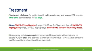
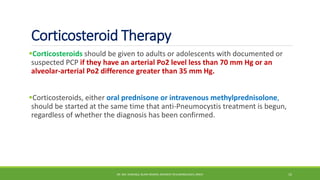
Pneumocystis Pneumonia
D R . M D . S H A F I Q U L I S L A M D E WA N
R E S I D E N T ( P U L M O N O L O G Y )
D E PA RT M E N T O F R E S P I R ATO RY M E D I C I N E
D H A K A M E D I C A L C O L L E G E H O S P I TA L
Pneumocystis pneumonia (PCP)
Pneumocystis pneumonia (PCP) is a serious infection caused by the fungus
Pneumocystis jirovecii.
Most people who get PCP have a medical condition that weakens their immune
system, like HIV/AIDS, or take medicines (such as corticosteroids).
 PCP remains the most common AIDS-defining opportunistic infection in the
United States.
DR. MD. SHAFIQUL ISLAM DEWAN, RESIDENT (PULMONOLOGY), DMCH 2
Risk Factors
About 30-40% of people who get PCP have HIV/AIDS. (CD4+ lymphocyte count
less than 200 cells//µL)
Other people who get PCP are usually taking medicine (such as corticosteroids)
that lowers the body’s ability to fight germs or sickness or have other medical
conditions, such as:
 Chronic lung diseases
 Cancer
 Inflammatory diseases or autoimmune diseases (for example, lupus or rheumatoid
arthritis)
 Solid organ or stem cell transplant
DR. MD. SHAFIQUL ISLAM DEWAN, RESIDENT (PULMONOLOGY), DMCH 3
Transmission
PCP spreads from person to person through the air.
Some healthy adults can carry the Pneumocystis fungus in their lungs without
having symptoms, and it can spread to other people, including those with
weakened immune systems.
DR. MD. SHAFIQUL ISLAM DEWAN, RESIDENT (PULMONOLOGY), DMCH 4
Clinical Features
Classically, PCP presents with_
 Fever
 Non-productive cough
 dyspnea on exertion.
High temperature, rigors, purulent sputum, and pleuritic chest pain are
uncommon and can be used to distinguish PCP from pyogenic pneumonia.
DR. MD. SHAFIQUL ISLAM DEWAN, RESIDENT (PULMONOLOGY), DMCH 5
Clinical Features
HIV-infected adults, unlike other immunocompromised persons, usually have a
prolonged prodromal illness associated with PCP, often several weeks in
duration.
This duration of symptoms can often be used to distinguish PCP from pyogenic
pneumonia, which typically presents with 3 to 5 days of symptoms.
DR. MD. SHAFIQUL ISLAM DEWAN, RESIDENT (PULMONOLOGY), DMCH 6
Physical examination
Physical examination of the chest may be normal.
When abnormal, the most frequent findings on lung auscultation are
inspiratory crackles, which have been reported to be associated with a greater
disease severity and an increased mortality.
Oxygen desaturation with exertion is a sensitive but nonspecific indicator of
PCP.
DR. MD. SHAFIQUL ISLAM DEWAN, RESIDENT (PULMONOLOGY), DMCH 7
Investigation
Imaging (CXR, CT Chest)
Microscopic Examination ( Sputum, BAL, Lung tissue)
Biopsy
PCR
DR. MD. SHAFIQUL ISLAM DEWAN, RESIDENT (PULMONOLOGY), DMCH 8
Microscopic Examination
Microscopic visualization of P. jirovecii cysts or trophic forms (or both) on
stained respiratory specimens.
Standard methods for detection of P. jirovecii have been with cyst wall stains,
such as methenamine silver and toluidine blue-O or with Giemsa and Diff-Quik,
which stain both the cystic and trophic forms.
DR. MD. SHAFIQUL ISLAM DEWAN, RESIDENT (PULMONOLOGY), DMCH 9
Sputum and BAL
Sputum induction is an appropriate initial diagnostic procedure for PCP, but the
sensitivity of induced sputum testing is less than 100%, and thus a negative
result should be followed by bronchoscopy with BAL performed in the most
affected lobe visualized on chest radiograph.
For patients with diffuse radiographic disease, BAL in the right middle lobe is
usually performed.
For patients with an upper lung zone predominance on radiograph, lavage in
both an upper lobe and the right middle lobe should be considered.
DR. MD. SHAFIQUL ISLAM DEWAN, RESIDENT (PULMONOLOGY), DMCH 10
Imaging
Classically, PCP presents with bilateral, symmetrical reticular opacities or as
granular opacities.
These opacities typically begin in the perihilar region and extend outward as
the disease severity increases.
On occasion, the opacities are unilateral or asymmetrical But a lobar or focal
radiographic pattern is uncommon.
Thin-walled cysts, or pneumatoceles, are seen in 10–20% of cases.
Pneumatoceles may be single or multiple, and small or large, and predispose
patients to pneumothorax.
Usually pneumatoceles resolve, but occasionally they persist despite
successful therapy.
DR. MD. SHAFIQUL ISLAM DEWAN, RESIDENT (PULMONOLOGY), DMCH 11
Imaging
Nearly any radiographic finding, including focal, lobar, or segmental
consolidation, nodules with or without cavitation, and a miliary pattern, can
be seen occasionally.
Intrathoracic adenopathy and pleural effusions are rarely due to PCP.
These radiographic findings should prompt a search for an alternate or at least a
coexisting process, such as bacterial pneumonia, TB, fungal pneumonia, or
pulmonary.
In persons with a high clinical suspicion for PCP but a normal chest radiograph,
high-resolution chest CT can be useful.
In CT scan ground-glass opacity appearance is particularly associated PCP.
DR. MD. SHAFIQUL ISLAM DEWAN, RESIDENT (PULMONOLOGY), DMCH 12
DR. MD. SHAFIQUL ISLAM DEWAN, RESIDENT (PULMONOLOGY), DMCH 13
Treatment
Treatment of choice for patients with mild, moderate, and severe PCP remains
TMP-SMX administered for 21 days.
Dose: TMP is 15 mg/kg/day (range, 15–20 mg/kg/day), and that of SMX is 75
mg/kg/day (range, 75–100 mg/kg/day), divided into three or four daily doses.
Dosing may be intravenous (recommended for patients with moderate or
severe PCP) or oral, and patients started on intravenous TMP-SMX can switch to
oral formulations after clinical improvement.
DR. MD. SHAFIQUL ISLAM DEWAN, RESIDENT (PULMONOLOGY), DMCH 14
Corticosteroid Therapy
Corticosteroids should be given to adults or adolescents with documented or
suspected PCP if they have an arterial Po2 level less than 70 mm Hg or an
alveolar-arterial Po2 difference greater than 35 mm Hg.
Corticosteroids, either oral prednisone or intravenous methylprednisolone,
should be started at the same time that anti-Pneumocystis treatment is begun,
regardless of whether the diagnosis has been confirmed.
DR. MD. SHAFIQUL ISLAM DEWAN, RESIDENT (PULMONOLOGY), DMCH 15
DR. MD. SHAFIQUL ISLAM DEWAN, RESIDENT (PULMONOLOGY), DMCH 16
Thank You

Pneumocystis Pneumonia.pptx

  • 1.
    Pneumocystis Pneumonia D R. M D . S H A F I Q U L I S L A M D E WA N R E S I D E N T ( P U L M O N O L O G Y ) D E PA RT M E N T O F R E S P I R ATO RY M E D I C I N E D H A K A M E D I C A L C O L L E G E H O S P I TA L
  • 2.
    Pneumocystis pneumonia (PCP) Pneumocystispneumonia (PCP) is a serious infection caused by the fungus Pneumocystis jirovecii. Most people who get PCP have a medical condition that weakens their immune system, like HIV/AIDS, or take medicines (such as corticosteroids).  PCP remains the most common AIDS-defining opportunistic infection in the United States. DR. MD. SHAFIQUL ISLAM DEWAN, RESIDENT (PULMONOLOGY), DMCH 2
  • 3.
    Risk Factors About 30-40%of people who get PCP have HIV/AIDS. (CD4+ lymphocyte count less than 200 cells//µL) Other people who get PCP are usually taking medicine (such as corticosteroids) that lowers the body’s ability to fight germs or sickness or have other medical conditions, such as:  Chronic lung diseases  Cancer  Inflammatory diseases or autoimmune diseases (for example, lupus or rheumatoid arthritis)  Solid organ or stem cell transplant DR. MD. SHAFIQUL ISLAM DEWAN, RESIDENT (PULMONOLOGY), DMCH 3
  • 4.
    Transmission PCP spreads fromperson to person through the air. Some healthy adults can carry the Pneumocystis fungus in their lungs without having symptoms, and it can spread to other people, including those with weakened immune systems. DR. MD. SHAFIQUL ISLAM DEWAN, RESIDENT (PULMONOLOGY), DMCH 4
  • 5.
    Clinical Features Classically, PCPpresents with_  Fever  Non-productive cough  dyspnea on exertion. High temperature, rigors, purulent sputum, and pleuritic chest pain are uncommon and can be used to distinguish PCP from pyogenic pneumonia. DR. MD. SHAFIQUL ISLAM DEWAN, RESIDENT (PULMONOLOGY), DMCH 5
  • 6.
    Clinical Features HIV-infected adults,unlike other immunocompromised persons, usually have a prolonged prodromal illness associated with PCP, often several weeks in duration. This duration of symptoms can often be used to distinguish PCP from pyogenic pneumonia, which typically presents with 3 to 5 days of symptoms. DR. MD. SHAFIQUL ISLAM DEWAN, RESIDENT (PULMONOLOGY), DMCH 6
  • 7.
    Physical examination Physical examinationof the chest may be normal. When abnormal, the most frequent findings on lung auscultation are inspiratory crackles, which have been reported to be associated with a greater disease severity and an increased mortality. Oxygen desaturation with exertion is a sensitive but nonspecific indicator of PCP. DR. MD. SHAFIQUL ISLAM DEWAN, RESIDENT (PULMONOLOGY), DMCH 7
  • 8.
    Investigation Imaging (CXR, CTChest) Microscopic Examination ( Sputum, BAL, Lung tissue) Biopsy PCR DR. MD. SHAFIQUL ISLAM DEWAN, RESIDENT (PULMONOLOGY), DMCH 8
  • 9.
    Microscopic Examination Microscopic visualizationof P. jirovecii cysts or trophic forms (or both) on stained respiratory specimens. Standard methods for detection of P. jirovecii have been with cyst wall stains, such as methenamine silver and toluidine blue-O or with Giemsa and Diff-Quik, which stain both the cystic and trophic forms. DR. MD. SHAFIQUL ISLAM DEWAN, RESIDENT (PULMONOLOGY), DMCH 9
  • 10.
    Sputum and BAL Sputuminduction is an appropriate initial diagnostic procedure for PCP, but the sensitivity of induced sputum testing is less than 100%, and thus a negative result should be followed by bronchoscopy with BAL performed in the most affected lobe visualized on chest radiograph. For patients with diffuse radiographic disease, BAL in the right middle lobe is usually performed. For patients with an upper lung zone predominance on radiograph, lavage in both an upper lobe and the right middle lobe should be considered. DR. MD. SHAFIQUL ISLAM DEWAN, RESIDENT (PULMONOLOGY), DMCH 10
  • 11.
    Imaging Classically, PCP presentswith bilateral, symmetrical reticular opacities or as granular opacities. These opacities typically begin in the perihilar region and extend outward as the disease severity increases. On occasion, the opacities are unilateral or asymmetrical But a lobar or focal radiographic pattern is uncommon. Thin-walled cysts, or pneumatoceles, are seen in 10–20% of cases. Pneumatoceles may be single or multiple, and small or large, and predispose patients to pneumothorax. Usually pneumatoceles resolve, but occasionally they persist despite successful therapy. DR. MD. SHAFIQUL ISLAM DEWAN, RESIDENT (PULMONOLOGY), DMCH 11
  • 12.
    Imaging Nearly any radiographicfinding, including focal, lobar, or segmental consolidation, nodules with or without cavitation, and a miliary pattern, can be seen occasionally. Intrathoracic adenopathy and pleural effusions are rarely due to PCP. These radiographic findings should prompt a search for an alternate or at least a coexisting process, such as bacterial pneumonia, TB, fungal pneumonia, or pulmonary. In persons with a high clinical suspicion for PCP but a normal chest radiograph, high-resolution chest CT can be useful. In CT scan ground-glass opacity appearance is particularly associated PCP. DR. MD. SHAFIQUL ISLAM DEWAN, RESIDENT (PULMONOLOGY), DMCH 12
  • 13.
    DR. MD. SHAFIQULISLAM DEWAN, RESIDENT (PULMONOLOGY), DMCH 13
  • 14.
    Treatment Treatment of choicefor patients with mild, moderate, and severe PCP remains TMP-SMX administered for 21 days. Dose: TMP is 15 mg/kg/day (range, 15–20 mg/kg/day), and that of SMX is 75 mg/kg/day (range, 75–100 mg/kg/day), divided into three or four daily doses. Dosing may be intravenous (recommended for patients with moderate or severe PCP) or oral, and patients started on intravenous TMP-SMX can switch to oral formulations after clinical improvement. DR. MD. SHAFIQUL ISLAM DEWAN, RESIDENT (PULMONOLOGY), DMCH 14
  • 15.
    Corticosteroid Therapy Corticosteroids shouldbe given to adults or adolescents with documented or suspected PCP if they have an arterial Po2 level less than 70 mm Hg or an alveolar-arterial Po2 difference greater than 35 mm Hg. Corticosteroids, either oral prednisone or intravenous methylprednisolone, should be started at the same time that anti-Pneumocystis treatment is begun, regardless of whether the diagnosis has been confirmed. DR. MD. SHAFIQUL ISLAM DEWAN, RESIDENT (PULMONOLOGY), DMCH 15
  • 16.
    DR. MD. SHAFIQULISLAM DEWAN, RESIDENT (PULMONOLOGY), DMCH 16 Thank You

Editor's Notes