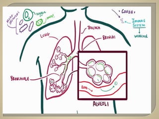
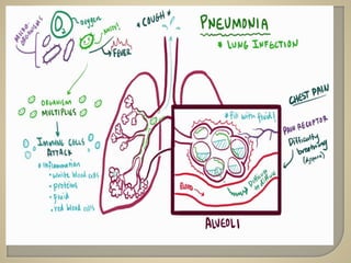
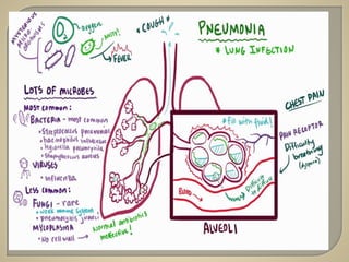
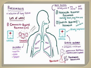
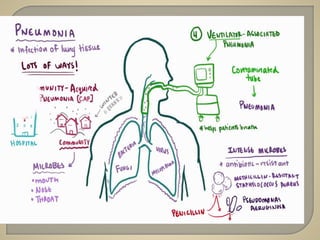
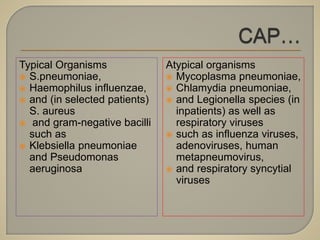
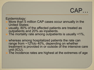
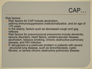
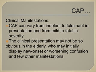
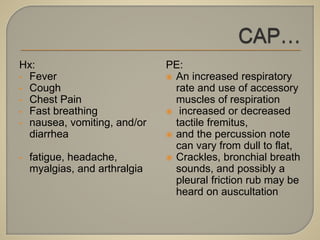
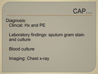
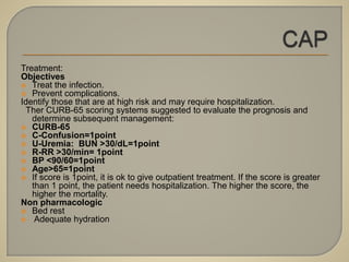
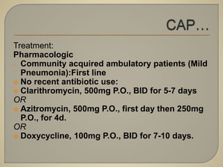
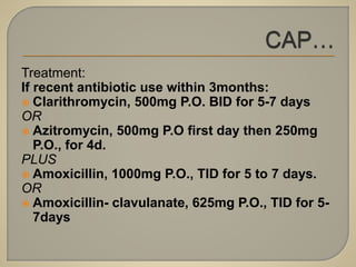
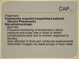
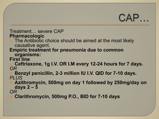
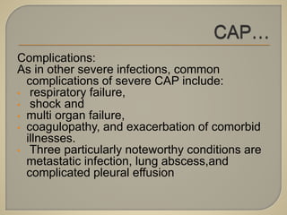
Melaku Yitbarek(M.D)
Internal Medicine Unit,
March 2018
• Definition
• Pathophysiology
• Classification
• Clinical features
• Treatment
• Complications
• Prevention
Pneumonia is an infection of the
pulmonary parenchyma.
Despite being the cause of significant
morbidity and mortality, pneumonia is often
misdiagnosed, mistreated, and
underestimated
 Pneumonia results from the proliferation of microbial
pathogens at the alveolar level and the host’s response to
those pathogens.
 Microorganisms gain access to the lower respiratory tract in
several ways, the most common is by aspiration from the
oropharynx.
 Small-volume aspiration occurs frequently during sleep
(especially in the elderly) and in patients with decreased levels
of consciousness.
 Many pathogens are inhaled as contaminated droplets.
 Rarely,pneumonia occurs via hematogenous spread (e.g.,
from tricuspid endocarditis)or by contiguous extension from an
infected pleural or Mediastinal space.
 Mechanical factors are critically important in host
defense.
 The hairs and turbinates of the nares capture
larger inhaled particles before they reach the lower
respiratory tract.
 The branching architecture of the tracheobronchial
tree traps microbes on the airway lining,where
mucociliary clearance and local antibacterial
factors either clear or kill the potential pathogen.
 The gag reflex and the cough mechanism offer
critical protection from aspiration.
 When these barriers are overcome or when microorganisms are
small enough to be inhaled to the alveolar level, resident alveolar
macrophages are extremely efficient at clearing and killing
pathogens.
 Macrophages are assisted by proteins that are produced by the
alveolar epithelial cells (e.g., surfactant proteins A and D) and that
have intrinsic opsonizing properties or antibacterial or antiviral
activity.
 Once engulfed by the macrophage, the pathogens—even if they are
not killed—are eliminated via either the mucociliary elevator or the
lymphatics and no longer represent an infectious challenge.
 Only when The capacity of the alveolar macrophages to ingest or kill
the Microorganisms is exceedes does clinical pneumonia become
manifest
 In that situation, the alveolar macrophages initiate the
inflammatory response to bolster lower respiratory tract
defenses
 The release of inflammatory mediators, such as interleukin 1
and tumor necrosis factor, results in fever.
 Chemokines,such as interleukin 8 and granulocyte colony-
stimulating factor,stimulate the release of neutrophils and their
attraction to the lung,producing both peripheral leukocytosis
and increased purulent secretions
 Inflammatory mediators released by macrophages and the
Newly recruited neutrophils create an alveolarcapillary leak
The capillary leak results in a radiographic
infiltrate and rales detectable on
auscultation, and hypoxemia results from
alveolar filling.
Moreover, some bacterial pathogens
appear to interfere with the hypoxemic
vasoconstriction that would normally occur
with fluid filled alveoli, and this interference
can result in severe hypoxemia.
Etiology:
 The extensive list of potential etiologic agents
in CAP includes bacteria, fungi, viruses, and
protozoa
 Most cases of CAP, however, are caused by
relatively few pathogens
 Although Streptococcus pneumoniae is most
common, other organisms also must be
considered in light of the patient’s risk factors
and severity of illness
Typical Organisms
 S.pneumoniae,
 Haemophilus influenzae,
 and (in selected patients)
S. aureus
 and gram-negative bacilli
such as
 Klebsiella pneumoniae
and Pseudomonas
aeruginosa
Atypical organisms
 Mycoplasma pneumoniae,
 Chlamydia pneumoniae,
 and Legionella species (in
inpatients) as well as
respiratory viruses
 such as influenza viruses,
adenoviruses, human
metapneumovirus,
 and respiratory syncytial
viruses
Epidemiology:
 More than 5 million CAP cases occur annually in the
United States;
 usually, 80% of the affected patients are treated as
outpatients and 20% as inpatients.
 The mortality rate among outpatients is usually <1%,
 whereas among hospitalized patients the rate can
range from ~12%to 40%, depending on whether
treatment is provided in or outside of the intensive care
unit (ICU).
 The incidence rates are highest at the extremes of age
Risk factors:
 Risk factors for CAP include alcoholism,
asthma,immunosuppression,institutionalization, and an age of
>70 years
 In the elderly, factors such as decreased cough and gag
reflexes
 Risk factors for pneumococcal pneumonia include dementia,
seizure disorders, heart failure, cerebrovascular disease,
alcoholism, tobacco smoking, chronic obstructive pulmonary
disease, and HIV infection
 P. aeruginosa is a particular problem in patients with severe
structural lung disease, such as bronchiectasis, cystic
fibrosis, or severe chronic obstructive pulmonary disease
Clinical Manifestations:
CAP can vary from indolent to fulminant in
presentation and from mild to fatal in
severity.
The clinical presentation may not be so
obvious in the elderly, who may initially
display new-onset or worsening confusion
and few other manifestations
Hx:
• Fever
• Cough
• Chest Pain
• Fast breathing
• nausea, vomiting, and/or
diarrhea
• fatigue, headache,
myalgias, and arthralgia
PE:
 An increased respiratory
rate and use of accessory
muscles of respiration
 increased or decreased
tactile fremitus,
 and the percussion note
can vary from dull to flat,
 Crackles, bronchial breath
sounds, and possibly a
pleural friction rub may be
heard on auscultation
Diagnosis:
• Clincal: Hx and PE
• Laboratory findings: sputum gram stain
and culture
• Blood culture
• Imaging: Chest x-ray
Treatment:
Objectives
 Treat the infection.
 Prevent complications.
Identify those that are at high risk and may require hospitalization.
Ther CURB-65 scoring systems suggested to evaluate the prognosis and
determine subsequent management:
 CURB-65
 C-Confusion=1point
 U-Uremia: BUN >30/dL=1point
 R-RR >30/min= 1point
 BP <90/60=1point
 Age>65=1point
 If score is 1point, it is ok to give outpatient treatment. If the score is greater
than 1 point, the patient needs hospitalization. The higher the score, the
higher the mortality.
Non pharmacologic
 Bed rest
 Adequate hydration
Treatment:
Pharmacologic
• Community acquired ambulatory patients (Mild
Pneumonia):First line
 No recent antibiotic use:
 Clarithromycin, 500mg P.O., BID for 5-7 days
OR
 Azitromycin, 500mg P.O., first day then 250mg
P.O., for 4d.
OR
 Doxycycline, 100mg P.O., BID for 7-10 days.
Treatment:
If recent antibiotic use within 3months:
 Clarithromycin, 500mg P.O. BID for 5-7 days
OR
 Azitromycin, 500mg P.O first day then 250mg
P.O., for 4d.
PLUS
 Amoxicillin, 1000mg P.O., TID for 5 to 7 days.
OR
 Amoxicillin- clavulanate, 625mg P.O., TID for 5-
7days
Treatment:
Community acquired hospitalized patients
(Severe Pneumonia)
Non-pharmacologic
 Bed rest
 Frequent monitoring of temperature, blood
pressure and pulse rate in order to detect
complications early and to monitor response to
therapy.
 Give attention to fluid and nutritional replacements.
 Administer Oxygen via nasal prongs or face mask
Treatment… severe CAP
Pharmacologic
 The Antibiotic choice should be aimed at the most likely
causative agent.
Empiric treatment for pneumonia due to common
organisms:
First line
 Ceftriaxone, 1g I.V. OR I.M every 12-24 hours for 7 days.
OR
 Benzyl penicillin, 2-3 million IU I.V. QID for 7-10 days.
PLUS
 Azithromycin, 500mg on day 1 followed by 250mg/day on
days 2 – 5
OR
 Clarithromycin, 500mg P.O., BID for 7-10 days
Complications:
As in other severe infections, common
complications of severe CAP include:
• respiratory failure,
• shock and
• multi organ failure,
• coagulopathy, and exacerbation of comorbid
illnesses.
• Three particularly noteworthy conditions are
metastatic infection, lung abscess,and
complicated pleural effusion
Prevention:
• The main preventive measure is
vaccination :influenza and pneumococcal
vaccines
• smokers should be strongly encouraged
to stop smoking
 While significantly less well studied than VAP,
HAP in nonintubated patients—both inside
and outside the ICU—is similar to VAP.
 The main differences are the higher
frequency of non-MDR pathogens and the
better underlying host immunity in non
intubated patients.
 The lower frequency of MDR pathogens
allows monotherapy in a larger proportion of
cases of HAP than of VAP
Treatment:
Empiric treatment for commonly suspected etiologies of HAP
First line
 Ceftazidime, 1gm I.V. TID for 10-14days
PLUS
 Vancomycin 1g I.V. BID for 10-14 days
OR (particularly in the ICU setup and in ventiltor associated pneumonia)
 Imipenem-cilastatin, 500mg IV (infused slowly over 1hour) Q6h
OR
 Meropenem, 1gm IV (infused slowly over 30min) Q8h
Alternatives
 Ceftriaxone, 1-2g I.V. OR I.M. BID for 7 days.
PLUS
 Gentamicin, 3-5mg/kg I.V. QDdaily in divided doses for 7 days.
OR
 Ciprofloxacin, 500mg P.O./I.V. BID every 12 hours for 7 days.
If methicillin-resistant (MRSA) suspected
 Vancomycin, 1 g I.V. BID should be added to the existing empric
regimen
 HCAP represents a transition between classic
CAP and typical HAP
 Several early studies were limited topatients
with culture-positive pneumonia.
 In these studies, the incidence of MDR
pathogens in HCAP was as high as or higher
than in HAP/VAP
 Patients in nursing homes and those who
have recent hospitalization(i.e with in 90
days) are at higher risk
Potential etiologic agents of VAP include
both MDR and non-MDR bacterial
pathogens
Most hospitals have problems with
P.aeruginosa and MRSA
Less commonly, fungal and viral
pathogens cause VAP, usually affecting
severely immunocompromised patients.
• Harrison’s Principles of Internal
Medicine,19th Edition,
• Standard Treatment Guideline for general
Hospital,2014
• Uptodate 21.6
Thank You…

Pneumonia

  • 1.
  • 2.
    • Definition • Pathophysiology •Classification • Clinical features • Treatment • Complications • Prevention
  • 3.
    Pneumonia is aninfection of the pulmonary parenchyma. Despite being the cause of significant morbidity and mortality, pneumonia is often misdiagnosed, mistreated, and underestimated
  • 4.
     Pneumonia resultsfrom the proliferation of microbial pathogens at the alveolar level and the host’s response to those pathogens.  Microorganisms gain access to the lower respiratory tract in several ways, the most common is by aspiration from the oropharynx.  Small-volume aspiration occurs frequently during sleep (especially in the elderly) and in patients with decreased levels of consciousness.  Many pathogens are inhaled as contaminated droplets.  Rarely,pneumonia occurs via hematogenous spread (e.g., from tricuspid endocarditis)or by contiguous extension from an infected pleural or Mediastinal space.
  • 5.
     Mechanical factorsare critically important in host defense.  The hairs and turbinates of the nares capture larger inhaled particles before they reach the lower respiratory tract.  The branching architecture of the tracheobronchial tree traps microbes on the airway lining,where mucociliary clearance and local antibacterial factors either clear or kill the potential pathogen.  The gag reflex and the cough mechanism offer critical protection from aspiration.
  • 6.
     When thesebarriers are overcome or when microorganisms are small enough to be inhaled to the alveolar level, resident alveolar macrophages are extremely efficient at clearing and killing pathogens.  Macrophages are assisted by proteins that are produced by the alveolar epithelial cells (e.g., surfactant proteins A and D) and that have intrinsic opsonizing properties or antibacterial or antiviral activity.  Once engulfed by the macrophage, the pathogens—even if they are not killed—are eliminated via either the mucociliary elevator or the lymphatics and no longer represent an infectious challenge.  Only when The capacity of the alveolar macrophages to ingest or kill the Microorganisms is exceedes does clinical pneumonia become manifest
  • 7.
     In thatsituation, the alveolar macrophages initiate the inflammatory response to bolster lower respiratory tract defenses  The release of inflammatory mediators, such as interleukin 1 and tumor necrosis factor, results in fever.  Chemokines,such as interleukin 8 and granulocyte colony- stimulating factor,stimulate the release of neutrophils and their attraction to the lung,producing both peripheral leukocytosis and increased purulent secretions  Inflammatory mediators released by macrophages and the Newly recruited neutrophils create an alveolarcapillary leak
  • 8.
    The capillary leakresults in a radiographic infiltrate and rales detectable on auscultation, and hypoxemia results from alveolar filling. Moreover, some bacterial pathogens appear to interfere with the hypoxemic vasoconstriction that would normally occur with fluid filled alveoli, and this interference can result in severe hypoxemia.
  • 16.
    Etiology:  The extensivelist of potential etiologic agents in CAP includes bacteria, fungi, viruses, and protozoa  Most cases of CAP, however, are caused by relatively few pathogens  Although Streptococcus pneumoniae is most common, other organisms also must be considered in light of the patient’s risk factors and severity of illness
  • 17.
    Typical Organisms  S.pneumoniae, Haemophilus influenzae,  and (in selected patients) S. aureus  and gram-negative bacilli such as  Klebsiella pneumoniae and Pseudomonas aeruginosa Atypical organisms  Mycoplasma pneumoniae,  Chlamydia pneumoniae,  and Legionella species (in inpatients) as well as respiratory viruses  such as influenza viruses, adenoviruses, human metapneumovirus,  and respiratory syncytial viruses
  • 18.
    Epidemiology:  More than5 million CAP cases occur annually in the United States;  usually, 80% of the affected patients are treated as outpatients and 20% as inpatients.  The mortality rate among outpatients is usually <1%,  whereas among hospitalized patients the rate can range from ~12%to 40%, depending on whether treatment is provided in or outside of the intensive care unit (ICU).  The incidence rates are highest at the extremes of age
  • 19.
    Risk factors:  Riskfactors for CAP include alcoholism, asthma,immunosuppression,institutionalization, and an age of >70 years  In the elderly, factors such as decreased cough and gag reflexes  Risk factors for pneumococcal pneumonia include dementia, seizure disorders, heart failure, cerebrovascular disease, alcoholism, tobacco smoking, chronic obstructive pulmonary disease, and HIV infection  P. aeruginosa is a particular problem in patients with severe structural lung disease, such as bronchiectasis, cystic fibrosis, or severe chronic obstructive pulmonary disease
  • 20.
    Clinical Manifestations: CAP canvary from indolent to fulminant in presentation and from mild to fatal in severity. The clinical presentation may not be so obvious in the elderly, who may initially display new-onset or worsening confusion and few other manifestations
  • 21.
    Hx: • Fever • Cough •Chest Pain • Fast breathing • nausea, vomiting, and/or diarrhea • fatigue, headache, myalgias, and arthralgia PE:  An increased respiratory rate and use of accessory muscles of respiration  increased or decreased tactile fremitus,  and the percussion note can vary from dull to flat,  Crackles, bronchial breath sounds, and possibly a pleural friction rub may be heard on auscultation
  • 22.
    Diagnosis: • Clincal: Hxand PE • Laboratory findings: sputum gram stain and culture • Blood culture • Imaging: Chest x-ray
  • 23.
    Treatment: Objectives  Treat theinfection.  Prevent complications. Identify those that are at high risk and may require hospitalization. Ther CURB-65 scoring systems suggested to evaluate the prognosis and determine subsequent management:  CURB-65  C-Confusion=1point  U-Uremia: BUN >30/dL=1point  R-RR >30/min= 1point  BP <90/60=1point  Age>65=1point  If score is 1point, it is ok to give outpatient treatment. If the score is greater than 1 point, the patient needs hospitalization. The higher the score, the higher the mortality. Non pharmacologic  Bed rest  Adequate hydration
  • 24.
    Treatment: Pharmacologic • Community acquiredambulatory patients (Mild Pneumonia):First line  No recent antibiotic use:  Clarithromycin, 500mg P.O., BID for 5-7 days OR  Azitromycin, 500mg P.O., first day then 250mg P.O., for 4d. OR  Doxycycline, 100mg P.O., BID for 7-10 days.
  • 25.
    Treatment: If recent antibioticuse within 3months:  Clarithromycin, 500mg P.O. BID for 5-7 days OR  Azitromycin, 500mg P.O first day then 250mg P.O., for 4d. PLUS  Amoxicillin, 1000mg P.O., TID for 5 to 7 days. OR  Amoxicillin- clavulanate, 625mg P.O., TID for 5- 7days
  • 26.
    Treatment: Community acquired hospitalizedpatients (Severe Pneumonia) Non-pharmacologic  Bed rest  Frequent monitoring of temperature, blood pressure and pulse rate in order to detect complications early and to monitor response to therapy.  Give attention to fluid and nutritional replacements.  Administer Oxygen via nasal prongs or face mask
  • 27.
    Treatment… severe CAP Pharmacologic The Antibiotic choice should be aimed at the most likely causative agent. Empiric treatment for pneumonia due to common organisms: First line  Ceftriaxone, 1g I.V. OR I.M every 12-24 hours for 7 days. OR  Benzyl penicillin, 2-3 million IU I.V. QID for 7-10 days. PLUS  Azithromycin, 500mg on day 1 followed by 250mg/day on days 2 – 5 OR  Clarithromycin, 500mg P.O., BID for 7-10 days
  • 28.
    Complications: As in othersevere infections, common complications of severe CAP include: • respiratory failure, • shock and • multi organ failure, • coagulopathy, and exacerbation of comorbid illnesses. • Three particularly noteworthy conditions are metastatic infection, lung abscess,and complicated pleural effusion
  • 29.
    Prevention: • The mainpreventive measure is vaccination :influenza and pneumococcal vaccines • smokers should be strongly encouraged to stop smoking
  • 30.
     While significantlyless well studied than VAP, HAP in nonintubated patients—both inside and outside the ICU—is similar to VAP.  The main differences are the higher frequency of non-MDR pathogens and the better underlying host immunity in non intubated patients.  The lower frequency of MDR pathogens allows monotherapy in a larger proportion of cases of HAP than of VAP
  • 31.
    Treatment: Empiric treatment forcommonly suspected etiologies of HAP First line  Ceftazidime, 1gm I.V. TID for 10-14days PLUS  Vancomycin 1g I.V. BID for 10-14 days OR (particularly in the ICU setup and in ventiltor associated pneumonia)  Imipenem-cilastatin, 500mg IV (infused slowly over 1hour) Q6h OR  Meropenem, 1gm IV (infused slowly over 30min) Q8h Alternatives  Ceftriaxone, 1-2g I.V. OR I.M. BID for 7 days. PLUS  Gentamicin, 3-5mg/kg I.V. QDdaily in divided doses for 7 days. OR  Ciprofloxacin, 500mg P.O./I.V. BID every 12 hours for 7 days. If methicillin-resistant (MRSA) suspected  Vancomycin, 1 g I.V. BID should be added to the existing empric regimen
  • 32.
     HCAP representsa transition between classic CAP and typical HAP  Several early studies were limited topatients with culture-positive pneumonia.  In these studies, the incidence of MDR pathogens in HCAP was as high as or higher than in HAP/VAP  Patients in nursing homes and those who have recent hospitalization(i.e with in 90 days) are at higher risk
  • 33.
    Potential etiologic agentsof VAP include both MDR and non-MDR bacterial pathogens Most hospitals have problems with P.aeruginosa and MRSA Less commonly, fungal and viral pathogens cause VAP, usually affecting severely immunocompromised patients.
  • 34.
    • Harrison’s Principlesof Internal Medicine,19th Edition, • Standard Treatment Guideline for general Hospital,2014 • Uptodate 21.6
  • 35.