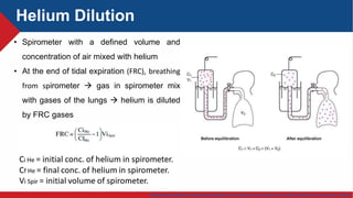
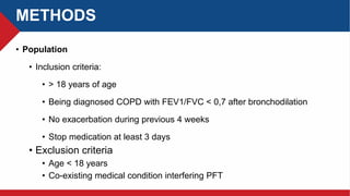
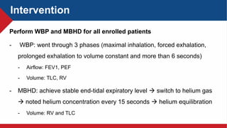
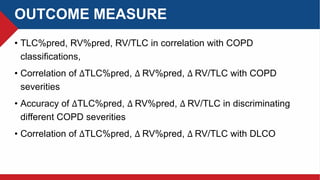
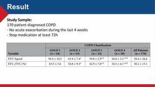
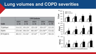
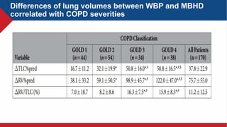
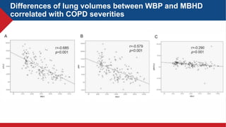
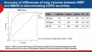
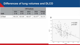
Journal Club
The measurement of lung volumes using body
plethysmography and helium dilution methods in COPD
patients: a correlation and diagnosis analysis
Sci Rep. 2016 Nov 23;6:37550. doi: 10.1038/srep37550
Tien Thanh, MD
BACKGROUND
• “Plethysmo-”: full
• Plethysmography: measures changes in volume in different parts of
the body.
• Check for blood clots in the arms and legs.
• Air in lungs
• Type:
• Limb plethysmography
• Body/lung plethysmography
https://www.uptodate.com/contents/epidemiology-clinical-presentation-and-diagnostic-evaluation-of-parapneumonic-effusion-and-empyema-in-adults
Body/lung Plethysmography
Boyle – Marriote law: at constant temperature, the volume V of a gas mass is inversely proportional to its
pressure P
P.V = const
 At the end of normal expiration:
P box (PB) = P mouth = Equilibrium alveolar pressure
V lung (VL) = FRC
 The shutter closes, the lung is a closed compartment with a fixed amount of
gas and object starts inspiration:
PB.VL= (PB- ∆P) . (VL+ ∆V)
PB.VL= PB.VL + PB. ∆V - ∆P. VL - ∆P.∆V
∆P. VL= PB . ∆V
Body/lung Plethysmography
∆V lung = - ∆V pleth (shift volume)
Boyle – Mariotte law:
∆ 𝑽 𝒍𝒖𝒏𝒈
𝑭𝑹𝑪
=
𝑨𝒗𝒆𝒐𝒍𝒂𝒓 𝒑𝒓𝒆𝒔𝒔𝒖𝒓𝒆 𝒄𝒉𝒂𝒏𝒈𝒆
𝑬𝒒𝒖𝒊𝒍𝒊𝒃𝒊𝒓𝒊𝒖𝒎 𝒂𝒗𝒆𝒐𝒍𝒂𝒓 𝒑𝒓𝒆𝒔𝒔𝒖𝒓𝒆
RV = FRC - ERV
TLC = FRC + IC = RV +IVC
Body/lung Plethysmography
https://www.uptodate.com/contents/management-and-prognosis-of-parapneumonic-pleural-effusion-and-empyema-in-adults
Helium Dilution
• Spirometer with a defined volume and
concentration of air mixed with helium
• At the end of tidal expiration (FRC), breathing
from spirometer  gas in spirometer mix
with gases of the lungs  helium is diluted
by FRC gases
The measurement of lung volumes using body
plethysmography and helium dilution methods in COPD
patients: a correlation and diagnosis analysis
Tang Y, Zhang M, Feng Y, Liang B
Sci Rep. 2016 Nov 23;6:37550. doi: 10.1038/srep37550. PMID: 27876834; PMCID: PMC5120321.
PICO
• Patient: COPD patients
• Intervention: Whole Body Plethysmography(WBP) and Multi Breath
Helium Dilution method (MBHD)
• Comparison: lung volumes with COPD severities
• Outcome: correlation between lung volumes and differences
between WBP and MBHD with level of airway limitation, DLCO
METHODS
• Study design:
• Prospective, descriptive study
• Site: West China Hospital of Sichuan University
• Time: January 2014 to March 2015
METHODS
• Population
• Inclusion criteria:
• > 18 years of age
• Being diagnosed COPD with FEV1/FVC < 0,7 after bronchodilation
• No exacerbation during previous 4 weeks
• Stop medication at least 3 days
• Exclusion criteria
• Age < 18 years
• Co-existing medical condition interfering PFT
Intervention
Perform WBP and MBHD for all enrolled patients
- WBP: went through 3 phases (maximal inhalation, forced exhalation,
prolonged exhalation to volume constant and more than 6 seconds)
- Airflow: FEV1, PEF
- Volume: TLC, RV
- MBHD: achieve stable end-tidal expiratory level  switch to helium gas
 noted helium concentration every 15 seconds  helium equilibration
- Volume: RV and TLC
OUTCOME MEASURE
• TLC%pred, RV%pred, RV/TLC in correlation with COPD
classifications,
• Correlation of ∆TLC%pred, ∆ RV%pred, ∆ RV/TLC with COPD
severities
• Accuracy of ∆TLC%pred, ∆ RV%pred, ∆ RV/TLC in discriminating
different COPD severities
• Correlation of ∆TLC%pred, ∆ RV%pred, ∆ RV/TLC with DLCO
STUDY RESULT
Result
Study Sample:
170 patient diagnosed COPD
- No acute exacerbation during the last 4 weeks
- Stop medication at least 72h
Lung volumes and COPD severities
Differences of lung volumes between WBP and MBHD
correlated with COPD severities
Differences of lung volumes between WBP and MBHD
correlated with COPD severities
Accuracy of differences of lung volumes between WBP
and MBHD in discriminating COPD severities
AUC of ∆TLC%pred: 0.886 (95%CI 0.834 – 0.939)
Differences of lung volumes and DLCO
Conclusion
• TLC%pred, RV%pred and RV/TLC as measured by WBP were significantly increased as
COPD exacerbated
• TLC%pred, RV%pred and RV/TLC measured by WBP were significantly higher than those
measured by MBHD method
• ∆TLC%pred, ∆ RV%pred, ∆ RV/TLC by these two methods were negatively correlated
with FEV1%pred and effectively differentiated COPD severity
.
REVIEW
• Limitation:
• The sample size is small (32 participants)
• Lack of assessment of related factors: history of exacerbation, comorbidities,
demographic (age, educational level),
• Any connection with clinical symptom control
• Application
• Lung volume measurement of the differences between WBP and MBHD may be an
alternative marker to reflect the degree of airflow obstruction and gas trapping
THANK YOU FOR YOUR ATTENTION!

Plethysmography vs Helium dilution in COPD.pptx

  • 1.
    Journal Club The measurementof lung volumes using body plethysmography and helium dilution methods in COPD patients: a correlation and diagnosis analysis Sci Rep. 2016 Nov 23;6:37550. doi: 10.1038/srep37550 Tien Thanh, MD
  • 2.
    BACKGROUND • “Plethysmo-”: full •Plethysmography: measures changes in volume in different parts of the body. • Check for blood clots in the arms and legs. • Air in lungs • Type: • Limb plethysmography • Body/lung plethysmography https://www.uptodate.com/contents/epidemiology-clinical-presentation-and-diagnostic-evaluation-of-parapneumonic-effusion-and-empyema-in-adults
  • 3.
  • 4.
    Body/lung Plethysmography Boyle –Marriote law: at constant temperature, the volume V of a gas mass is inversely proportional to its pressure P P.V = const  At the end of normal expiration: P box (PB) = P mouth = Equilibrium alveolar pressure V lung (VL) = FRC  The shutter closes, the lung is a closed compartment with a fixed amount of gas and object starts inspiration: PB.VL= (PB- ∆P) . (VL+ ∆V) PB.VL= PB.VL + PB. ∆V - ∆P. VL - ∆P.∆V ∆P. VL= PB . ∆V
  • 5.
    Body/lung Plethysmography ∆V lung= - ∆V pleth (shift volume) Boyle – Mariotte law: ∆ 𝑽 𝒍𝒖𝒏𝒈 𝑭𝑹𝑪 = 𝑨𝒗𝒆𝒐𝒍𝒂𝒓 𝒑𝒓𝒆𝒔𝒔𝒖𝒓𝒆 𝒄𝒉𝒂𝒏𝒈𝒆 𝑬𝒒𝒖𝒊𝒍𝒊𝒃𝒊𝒓𝒊𝒖𝒎 𝒂𝒗𝒆𝒐𝒍𝒂𝒓 𝒑𝒓𝒆𝒔𝒔𝒖𝒓𝒆 RV = FRC - ERV TLC = FRC + IC = RV +IVC Body/lung Plethysmography
  • 6.
    https://www.uptodate.com/contents/management-and-prognosis-of-parapneumonic-pleural-effusion-and-empyema-in-adults Helium Dilution • Spirometerwith a defined volume and concentration of air mixed with helium • At the end of tidal expiration (FRC), breathing from spirometer  gas in spirometer mix with gases of the lungs  helium is diluted by FRC gases
  • 7.
    The measurement oflung volumes using body plethysmography and helium dilution methods in COPD patients: a correlation and diagnosis analysis Tang Y, Zhang M, Feng Y, Liang B Sci Rep. 2016 Nov 23;6:37550. doi: 10.1038/srep37550. PMID: 27876834; PMCID: PMC5120321.
  • 8.
    PICO • Patient: COPDpatients • Intervention: Whole Body Plethysmography(WBP) and Multi Breath Helium Dilution method (MBHD) • Comparison: lung volumes with COPD severities • Outcome: correlation between lung volumes and differences between WBP and MBHD with level of airway limitation, DLCO
  • 9.
    METHODS • Study design: •Prospective, descriptive study • Site: West China Hospital of Sichuan University • Time: January 2014 to March 2015
  • 10.
    METHODS • Population • Inclusioncriteria: • > 18 years of age • Being diagnosed COPD with FEV1/FVC < 0,7 after bronchodilation • No exacerbation during previous 4 weeks • Stop medication at least 3 days • Exclusion criteria • Age < 18 years • Co-existing medical condition interfering PFT
  • 11.
    Intervention Perform WBP andMBHD for all enrolled patients - WBP: went through 3 phases (maximal inhalation, forced exhalation, prolonged exhalation to volume constant and more than 6 seconds) - Airflow: FEV1, PEF - Volume: TLC, RV - MBHD: achieve stable end-tidal expiratory level  switch to helium gas  noted helium concentration every 15 seconds  helium equilibration - Volume: RV and TLC
  • 12.
    OUTCOME MEASURE • TLC%pred,RV%pred, RV/TLC in correlation with COPD classifications, • Correlation of ∆TLC%pred, ∆ RV%pred, ∆ RV/TLC with COPD severities • Accuracy of ∆TLC%pred, ∆ RV%pred, ∆ RV/TLC in discriminating different COPD severities • Correlation of ∆TLC%pred, ∆ RV%pred, ∆ RV/TLC with DLCO
  • 13.
  • 14.
    Result Study Sample: 170 patientdiagnosed COPD - No acute exacerbation during the last 4 weeks - Stop medication at least 72h
  • 15.
    Lung volumes andCOPD severities
  • 16.
    Differences of lungvolumes between WBP and MBHD correlated with COPD severities
  • 17.
    Differences of lungvolumes between WBP and MBHD correlated with COPD severities
  • 18.
    Accuracy of differencesof lung volumes between WBP and MBHD in discriminating COPD severities AUC of ∆TLC%pred: 0.886 (95%CI 0.834 – 0.939)
  • 19.
    Differences of lungvolumes and DLCO
  • 20.
    Conclusion • TLC%pred, RV%predand RV/TLC as measured by WBP were significantly increased as COPD exacerbated • TLC%pred, RV%pred and RV/TLC measured by WBP were significantly higher than those measured by MBHD method • ∆TLC%pred, ∆ RV%pred, ∆ RV/TLC by these two methods were negatively correlated with FEV1%pred and effectively differentiated COPD severity .
  • 21.
    REVIEW • Limitation: • Thesample size is small (32 participants) • Lack of assessment of related factors: history of exacerbation, comorbidities, demographic (age, educational level), • Any connection with clinical symptom control • Application • Lung volume measurement of the differences between WBP and MBHD may be an alternative marker to reflect the degree of airflow obstruction and gas trapping
  • 22.
    THANK YOU FORYOUR ATTENTION!

Editor's Notes

  • #4 You will sit in a large airtight cabin known as a body box. The walls of the cabin are clear so that you and the health care provider can see each other. You will breathe or pant against a mouthpiece. Clips will be put on your nose to close off your nostrils. Depending on the information your doctor is looking for, the mouthpiece may be open at first, and then closed. You will breathe against the mouthpiece in both the open and closed positions. The positions give different information to the doctor. As your chest moves while you breathe or pant, it changes the pressure and amount of air in the room and against the mouthpiece. From these changes, the doctor can get an accurate measure of the amount of air in your lungs. Depending on the purpose of the test, you may be given medicine before the test to most accurately measure the volume.
  • #5 You will sit in a large airtight cabin known as a body box. The walls of the cabin are clear so that you and the health care provider can see each other. You will breathe or pant against a mouthpiece. Clips will be put on your nose to close off your nostrils. Depending on the information your doctor is looking for, the mouthpiece may be open at first, and then closed. You will breathe against the mouthpiece in both the open and closed positions. The positions give different information to the doctor. As your chest moves while you breathe or pant, it changes the pressure and amount of air in the room and against the mouthpiece. From these changes, the doctor can get an accurate measure of the amount of air in your lungs. Depending on the purpose of the test, you may be given medicine before the test to most accurately measure the volume.
  • #6 You will sit in a large airtight cabin known as a body box. The walls of the cabin are clear so that you and the health care provider can see each other. You will breathe or pant against a mouthpiece. Clips will be put on your nose to close off your nostrils. Depending on the information your doctor is looking for, the mouthpiece may be open at first, and then closed. You will breathe against the mouthpiece in both the open and closed positions. The positions give different information to the doctor. As your chest moves while you breathe or pant, it changes the pressure and amount of air in the room and against the mouthpiece. From these changes, the doctor can get an accurate measure of the amount of air in your lungs. Depending on the purpose of the test, you may be given medicine before the test to most accurately measure the volume.