- Plasma cell neoplasms originate from terminally differentiated B cells and produce monoclonal immunoglobulins. They commonly affect people around age 70 and have a male predominance.
- Risk factors include prior radiation exposure and certain chemical exposures. They are caused by genetic mutations that allow a clonal plasma cell population to proliferate in the bone marrow.
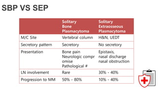
- Presentations depend on the type but commonly include anemia, bone lesions, kidney dysfunction, and infections. Workup involves blood and urine tests and imaging like skeletal surveys and PET scans.
- Treatment involves chemotherapy, radiation, stem cell transplants, surgery, and palliative care. Prognosis depends on the stage and type, ranging from potentially cur


![Standard Laboratory tests
Unilateral bone marrow aspirate and biopsy.
Bone marrow immunohistochemistry and flow cytometry
Gene expression profiling is increasingly used for prognostic classification and to check for minimal
residual disease.
Cytogenetic/karyotype for hyper/hypodiploidy. Hyperdiploidy has better prognosis.
FISH [del 13, del 17, t(4;14), t(11;14), t(14;16)].
Work Up](https://image.slidesharecdn.com/plasmacellneoplasms-181003152550/85/Plasma-cell-neoplasms-17-320.jpg)


![IFM [Intergroupe Francophone du Mye’lome]
trial 9502
Melphalan, 200 mg/m2
alone
Toxic death rate 0%
The event-free survival: No
Difference
45m OS - 65.8%, P = .05
M200
Melphalan 140 mg/m2 +
TBI (8 Gy in 4 #)
Toxic death rate in the 3.6%
The event-free survival: No
Difference
45m OS - 45.5%; P = .05
grade 3/4 mucosal toxicity,
heavier transfusion
longer hospitalization stay
M140/TBI
EFS: The length of time after primary tretment the patient remains free of certain complication or
events that the treament was intended to prevent or delay](https://image.slidesharecdn.com/plasmacellneoplasms-181003152550/85/Plasma-cell-neoplasms-34-320.jpg)


![Management
Local External Beam for Palliation
Palliative RT for cord compression
Motor improvement is expected in approximately 50% of irradiated patients
30 Gy in 10 fractions or higher was associated with better neurologic recovery[1] than 20 Gy in 5 fractio
ns or a single 8 Gy.
Rades D, Stalpers LJ, Veninga T, et al. Evaluation of five radiation schedules and prognostic factors for metastatic spina
l cord compression. J Clin Oncol 2005; 23(15):3366–3375.](https://image.slidesharecdn.com/plasmacellneoplasms-181003152550/85/Plasma-cell-neoplasms-38-320.jpg)


![Reesident talk 4 12 17 anderson pcd final[2]](https://cdn.slidesharecdn.com/ss_thumbnails/reesidenttalk41217andersonpcdfinal2-170412172459-thumbnail.jpg?width=640&height=640&fit=bounds)