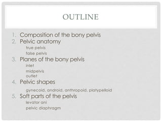
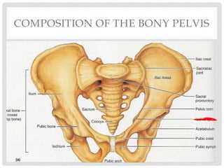
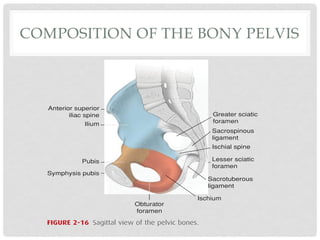
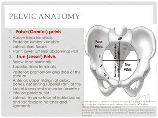
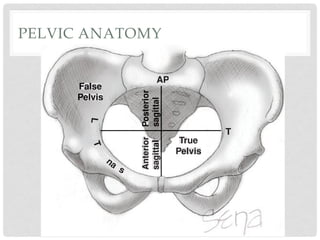
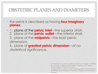
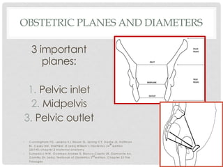
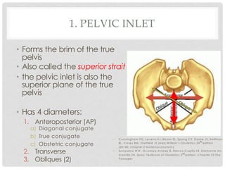
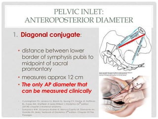
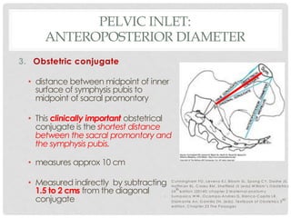
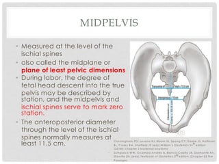
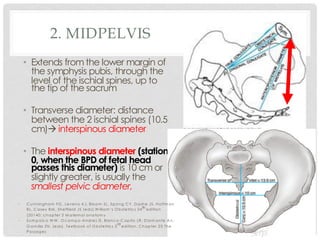
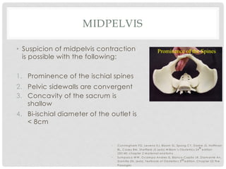
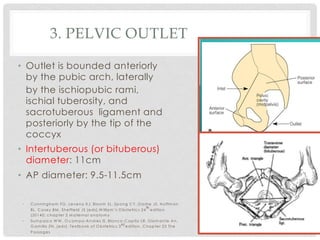
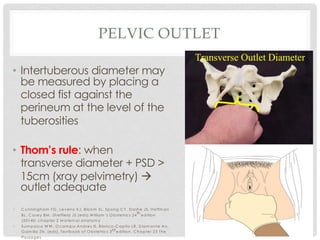
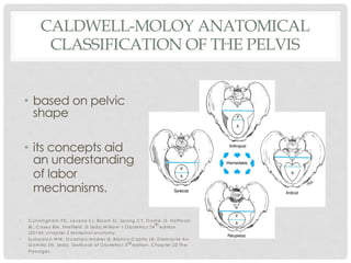
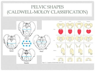
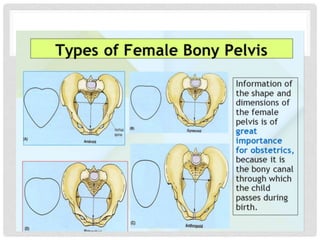
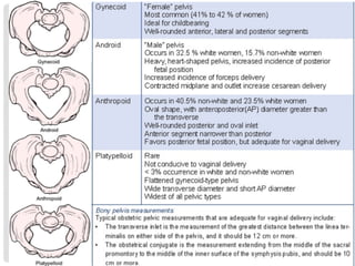
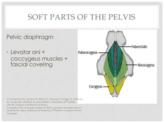
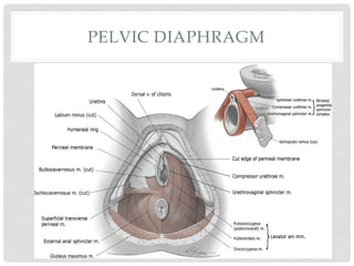
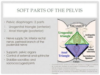
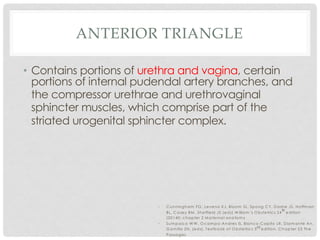
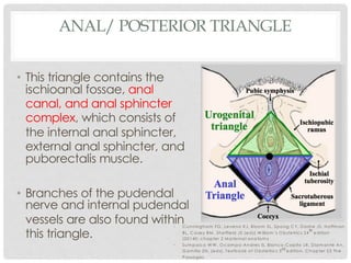
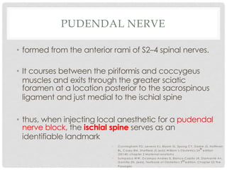
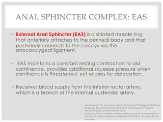
EVALUATION OF THE
PELVIS
Ina S. Irabon, MD, FPOGS, FPSRM, FPSGE
Obstetrics and Gynecology
Reproductive Endocrinology and Infertility
Laparoscopy and Hysteroscopy
TO DOWNLOAD THIS LECTURE DECK:
• www.slideshare.net à type “Ina Irabon” in
SEARCH bar
• www.wordpress.com
REFERENCE
• Cunningham FG, Leveno KJ, Bloom SL, Spong CY,
Dashe JS, Hoffman BL, Casey BM, Sheffield JS
(eds).William’s Obstetrics 24th edition (20140;
chapter 2 Maternal anatomy
• Sumpaico WW, Ocampo-Andres IS, Blanco-Capito
LR, Diamante An, Gamilla ZN. (eds). Textbook of
Obstetrics 3rd edition. Chapter 23 The Passages
OUTLINE
1. Composition of the bony pelvis
2. Pelvic anatomy
true pelvis
false pelvis
3. Planes of the bony pelvis
inlet
midpelvis
outlet
4. Pelvic shapes
gynecoid, android, anthropoid, platypelloid
5. Soft parts of the pelvis
levator ani
pelvic diaphragm
COMPOSITION OF THE BONY PELVIS
nctioning
muscle is
l layer. In
e replaced
muscular
ntractions
arian hor-
ar epithe-
n a sparse
ying myo-
fimbriated
e patches.
e two cell
mucosa is
vely more
n is occu-
The current
of flow is
y cilia and
ant factor
blood ves-
e tubes is
tion. This
Anterior superior
iliac spine
Ilium
Pubis
Symphysis pubis
Greater sciatic
foramen
Sacrospinous
ligament
Ischial spine
Lesser sciatic
foramen
SacrotuberousS
ligament
Ischium
Obturator
foramen
FIGURE 2-16 Sagittal view of the pelvic bones.
linea terminalis serves as the superior
border, whereas the pelvic outlet is the inferior margin. The
posterior boundary is the anterior surface of the sacrum, and
the lateral limits are formed by the inner surface of the ischial
bones and the sacrosciatic notches and ligaments. In front,
PELVIC ANATOMY
1. False (Greater) pelvis
• Above linea terminalis
• Posterior: lumbar vertebra
• Lateral: iliac fossae
• Front: lower anterior abdominal wall
2. True (Lesser) Pelvis
• Below linea terminalis
• Superior: linea terminalis
• Posterior: promontory and alae of the
sacrum
• Anterior: upper margin of pubic
bones, ascending superior rami of the
ischial bones and obturator foramina
• Inferior: pelvic outlet
• Lateral: inner surface of ischial bones
and sacrosciatic notches and
ligaments
32 Maternal Anatomy and Physiology
SECTION2
TTTT
APAPAPAP
True
Pelvis
FFFaFaFaFallslslsseee
PPPPellele viiv sss
PPPPPPPPPososssososossososotetetetetettriiiririiirirrrorrororrorooo
sasaasasaasaaasasgigigigiigggttttttttttttalalalaa
Anterior
sagittttttttttttttlalalalalalaaaaa
FIGURE 2-17 Anteroposterior view of a normal female pelvis.
Anteroposterior (AP) and transverse (T) diameters of the pelvic
• C unningham FG , Leveno KJ, Bloom SL, Spong C Y, D ashe JS, Hoffm an
BL, C asey BM , Sheffield JS (eds).W illiam ’s O bstetrics 24
th
edition
(20140; chapter 2 M aternal anatom y
• Sum paico W W , O cam po-Andres IS, Blanco-C apito LR, D iam ante An,
G am illa ZN. (eds). Textbook of O bstetrics 3rd edition. C hapter 23 The
Passages
PELVIC ANATOMY
32 Maternal Anatomy and Physiology
outle
to ro
force
the d
mob
ver o
a cas
have
ting
squa
the p
obser
birth
■ P
TTTT
APAPAPAP
True
Pelvis
FFFaFaFaFallslslsseee
PPPPellele viiv sss
PPPPPPPPPososssososossososotetetetetettriiiririiirirrrorrororrorooo
sasaasasaasaaasasgigigigiigggttttttttttttalalalaa
Anterior
sagittttttttttttttlalalalalalaaaaa
OBSTETRIC PLANES AND DIAMETERS
• the pelvis is described as having four imaginary
planes:
• 1. plane of the pelvic inlet—the superior strait.
2. plane of the pelvic outlet—the inferior strait.
3. plane of the midpelvis—the least pelvic
dimensions.
4. plane of greatest pelvic dimension—of no
obstetrical significance.
• C unningham FG , Leveno KJ, Bloom SL, Spong C Y, D ashe JS, Hoffm an
BL, C asey BM , Sheffield JS (eds).W illiam ’s O bstetrics 24
th
edition
(20140; chapter 2 M aternal anatom y
• Sum paico W W , O cam po-Andres IS, Blanco-C apito LR, D iam ante An,
G am illa ZN. (eds). Textbook of O bstetrics 3rd edition. C hapter 23 The
Passages
OBSTETRIC PLANES AND DIAMETERS
3 important
planes:
1. Pelvic inlet
2. Midpelvis
3. Pelvic outlet
• C unningham FG , Leveno KJ, Bloom SL, Spong C Y, D ashe JS, Hoffm an
BL, C asey BM , Sheffield JS (eds).W illiam ’s O bstetrics 24
th
edition
(20140; chapter 2 M aternal anatom y
• Sum paico W W , O cam po-Andres IS, Blanco-C apito LR, D iam ante An,
G am illa ZN. (eds). Textbook of O bstetrics 3rd edition. C hapter 23 The
Passages
1. PELVIC INLET
• Forms the brim of the true
pelvis
• Also called the superior strait
• the pelvic inlet is also the
superior plane of the true
pelvis
• Has 4 diameters:
1. Anteroposterior (AP)
a) Diagonal conjugate
b) True conjugate
c) Obstetric conjugate
2. Transverse
3. Obliques (2)
• C unningham FG , Leveno KJ, Bloom SL, Spong C Y, D ashe JS, Hoffm an
BL, C asey BM , Sheffield JS (eds).W illiam ’s O bstetrics 24th edition
(20140; chapter 2 M aternal anatom y
• Sum paico W W , O cam po-Andres IS, Blanco-C apito LR, D iam ante An,
G am illa ZN. (eds). Textbook of O bstetrics 3rd edition. C hapter 23 The
Passages
PELVIC INLET:
ANTEROPOSTERIOR DIAMETER
1. Diagonal conjugate:
• distance between lower
border of symphysis pubis to
midpoint of sacral
promontory
• measures approx 12 cm
• The only AP diameter that
can be measured clinically
• C unningham FG , Leveno KJ, Bloom SL, Spong C Y, D ashe JS, Hoffm an
BL, C asey BM , Sheffield JS (eds).W illiam ’s O bstetrics 24
th
edition
(20140; chapter 2 M aternal anatom y
• Sum paico W W , O cam po-Andres IS, Blanco-C apito LR, D iam ante An,
G am illa ZN. (eds). Textbook of O bstetrics 3rd edition. C hapter 23 The
Passages
VAGINAL EXAMINATION TO DETERMINE
THE DIAGONAL CONJUGATE
FIG
det
P ==
PELVIC INLET:
ANTEROPOSTERIOR DIAMETER
2. True or anatomic
conjugate
• distance between upper
margin of symphysis pubis to
midpoint of sacral promontory
• measures approx 11 cm
• Measured indirectly by
subtracting 1.2 cms from the
diagonal conjugate
• C unningham FG , Leveno KJ, Bloom SL, Spong C Y, D ashe JS, Hoffm an
BL, C asey BM , Sheffield JS (eds).W illiam ’s O bstetrics 24th edition
(20140; chapter 2 M aternal anatom y
• Sum paico W W , O cam po-Andres IS, Blanco-C apito LR, D iam ante An,
G am illa ZN. (eds). Textbook of O bstetrics 3
rd
edition. C hapter 23 The
Passages
PELVIC INLET:
ANTEROPOSTERIOR DIAMETER
3. Obstetric conjugate
• distance between midpoint of inner
surface of symphysis pubis to
midpoint of sacral promontory
• This clinically important obstetrical
conjugate is the shortest distance
between the sacral promontory and
the symphysis pubis.
• measures approx 10 cm
• Measured indirectly by subtracting
1.5 to 2 cms from the diagonal
conjugate
• C unningham FG , Leveno KJ, Bloom SL, Spong C Y, D ashe JS,
Hoffm an BL, C asey BM , Sheffield JS (eds).W illiam ’s O bstetrics
24
th
edition (20140; chapter 2 M aternal anatom y
• Sum paico W W , O cam po-Andres IS, Blanco-C apito LR,
D iam ante An, G am illa ZN. (eds). Textbook of O bstetrics 3
rd
edition. C hapter 23 The Passages
PELVIC INLET:
TRANSVERSE DIAMETER
• constructed at right
angles to the obstetrical
conjugate and represents
the greatest distance
between the linea
terminalis on either side.
• It usually intersects the
obstetrical conjugate at
a point approximately 5
cm in front of the
promontory
• Average: 13 cm
• Divides the inlet into
anterior and posterior
segments
Posterior sagittal
diameter
• C unningham FG , Leveno KJ, Bloom SL, Spong C Y, D ashe JS, Hoffm an
BL, C asey BM , Sheffield JS (eds).W illiam ’s O bstetrics 24th edition
(20140; chapter 2 M aternal anatom y
• Sum paico W W , O cam po-Andres IS, Blanco-C apito LR, D iam ante An,
G am illa ZN. (eds). Textbook of O bstetrics 3rd edition. C hapter 23 The
Passages
PELVIC INLET:
OBLIQUE DIAMETERS
• Extend from the
sacroiliac joints to
the opposite
iliopubic eminence
• Measures <13 cm
• C unningham FG , Leveno KJ, Bloom SL, Spong C Y, D ashe JS, Hoffm an
BL, C asey BM , Sheffield JS (eds).W illiam ’s O bstetrics 24
th
edition
(20140; chapter 2 M aternal anatom y
• Sum paico W W , O cam po-Andres IS, Blanco-C apito LR, D iam ante An,
G am illa ZN. (eds). Textbook of O bstetrics 3rd edition. C hapter 23 The
Passages
PLANE OF THE GREATEST PELVIC
DIAMETER
• Corresponds to the
roomiest plane of the
pelvis
• Posterior: 3rd to 4th sacral
vertebrae
• Lateral: ischial bones
• Anterior: middle surface of
symphysis pubis
• Its AP diameter and
transverse diameters
average 12.5cm
• C unningham FG , Leveno KJ, Bloom SL, Spong C Y, D ashe JS, Hoffm an
BL, C asey BM , Sheffield JS (eds).W illiam ’s O bstetrics 24
th
edition
(20140; chapter 2 M aternal anatom y
• Sum paico W W , O cam po-Andres IS, Blanco-C apito LR, D iam ante An,
G am illa ZN. (eds). Textbook of O bstetrics 3rd edition. C hapter 23 The
Passages
MIDPELVIS
• Measured at the level of the
ischial spines
• also called the midplane or
plane of least pelvic dimensions
• During labor, the degree of
fetal head descent into the true
pelvis may be described by
station, and the midpelvis and
ischial spines serve to mark zero
station.
• The anteroposterior diameter
through the level of the ischial
spines normally measures at
least 11.5 cm.
the true pelvis may be described by station, and the midpelvis
and ischial spines serve to mark zero station. The interspinous
diameter is 10 cm or slightly greater, is usually the smallest
pelvicdiameter,and,incasesofobstructedlabor,isparticularly
b
tu
in
an
w
th
tr
p
n
■
Th
th
in
di
se
an
ty
de
pu
an
an
co
Interspinous = 10 cmInterspinous = 10 cm
Transverse of inlet = 13.5 cm
Obstetrical
Conj=10.5cm
FIGURE 2-19 Adult female pelvis demonstrating the interspinous
diameter of the midpelvis. The anteroposterior and transverse
diameters of the pelvic inlet are also shown.
• C unningham FG , Leveno KJ, Bloom SL, Spong C Y, D ashe JS, Hoffm an
BL, C asey BM , Sheffield JS (eds).W illiam ’s O bstetrics 24
th
edition
(20140; chapter 2 M aternal anatom y
• Sum paico W W , O cam po-Andres IS, Blanco-C apito LR, D iam ante An,
G am illa ZN. (eds). Textbook of O bstetrics 3rd edition. C hapter 23 The
Passages
2. MIDPELVIS
• Extends from the lower margin of
the symphysis pubis, through the
level of the ischial spines, up to
the tip of the sacrum
• Transverse diameter: distance
between the 2 ischial spines (10.5
cm)à interspinous diameter
• The interspinous diameter (station
0, when the BPD of fetal head
passes this diameter) is 10 cm or
slightly greater, is usually the
smallest pelvic diameter,
is a minor elevation that marks the union site of the ilium and
pubis. These oblique diameters average less than 13 cm.
Midpelvis and Pelvic Outlet
The midpelvis is measured at the level of the ischial spines,
also called the midplane or plane of least pelvic dimensions
(Fig. 2-19). During labor, the degree of fetal head descent into
the true pelvis may be described by station, and the midpelvis
and ischial spines serve to mark zero station. The interspinous
diameter is 10 cm or slightly greater, is usually the smallest
pelvic diameter, and, in cases of obstructed labor, is particularly
Interspinous = 10 cmInterspinous = 10 cm
Transverse of inlet = 13.5 cm
Obstetrical
Conj=10.5cm
• C unningham FG , Leveno KJ, Bloom SL, Spong C Y, D ashe JS, Hoffm an
BL, C asey BM , Sheffield JS (eds).W illiam ’s O bstetrics 24th edition
(20140; chapter 2 M aternal anatom y
• Sum paico W W , O cam po-Andres IS, Blanco-C apito LR, D iam ante An,
G am illa ZN. (eds). Textbook of O bstetrics 3
rd
edition. C hapter 23 The
Passages
MIDPELVIS
• Suspicion of midpelvis contraction
is possible with the following:
1. Prominence of the ischial spines
2. Pelvic sidewalls are convergent
3. Concavity of the sacrum is
shallow
4. Bi-ischial diameter of the outlet is
< 8cm
• C unningham FG , Leveno KJ, Bloom SL, Spong C Y, D ashe JS, Hoffm an
BL, C asey BM , Sheffield JS (eds).W illiam ’s O bstetrics 24
th
edition
(20140; chapter 2 M aternal anatom y
• Sum paico W W , O cam po-Andres IS, Blanco-C apito LR, D iam ante An,
G am illa ZN. (eds). Textbook of O bstetrics 3rd edition. C hapter 23 The
Passages
3. PELVIC OUTLET
• Outlet is bounded anteriorly
by the pubic arch, laterally
by the ischiopubic rami,
ischial tuberosity, and
sacrotuberous ligament and
posteriorly by the tip of the
coccyx
• Intertuberous (or bituberous)
diameter: 11cm
• AP diameter: 9.5-11.5cm
• C unningham FG , Leveno KJ, Bloom SL, Spong C Y, D ashe JS, Hoffm an
BL, C asey BM , Sheffield JS (eds).W illiam ’s O bstetrics 24
th
edition
(20140; chapter 2 M aternal anatom y
• Sum paico W W , O cam po-Andres IS, Blanco-C apito LR, D iam ante An,
G am illa ZN. (eds). Textbook of O bstetrics 3rd edition. C hapter 23 The
Passages
PELVIC OUTLET
• The pelvic outlet consists of two
approximately triangular areas.
• Posterior triangle
• Anterior triangle
• They have a common base, which
is a line drawn between the two
ischial tuberosities.
• Clinically, three diameters of the
pelvic outlet usually are
described—the anteroposterior,
transverse, and posterior sagittal.
• C unningham FG , Leveno KJ, Bloom SL, Spong C Y, D ashe JS,
Hoffm an BL, C asey BM , Sheffield JS (eds).W illiam ’s O bstetrics
24th edition (20140; chapter 2 M aternal anatom y
• Sum paico W W , O cam po-Andres IS, Blanco-C apito LR,
D iam ante An, G am illa ZN. (eds). Textbook of O bstetrics 3rd
edition. C hapter 23 The Passages
PELVIC OUTLET
• Intertuberous diameter may
be measured by placing a
closed fist against the
perineum at the level of the
tuberosities
• Thom’s rule: when
transverse diameter + PSD >
15cm (xray pelvimetry) à
outlet adequate
• C unningham FG , Leveno KJ, Bloom SL, Spong C Y, D ashe JS, Hoffm an
BL, C asey BM , Sheffield JS (eds).W illiam ’s O bstetrics 24
th
edition
(20140; chapter 2 M aternal anatom y
• Sum paico W W , O cam po-Andres IS, Blanco-C apito LR, D iam ante An,
G am illa ZN. (eds). Textbook of O bstetrics 3rd edition. C hapter 23 The
Passages
CALDWELL-MOLOY ANATOMICAL
CLASSIFICATION OF THE PELVIS
• based on pelvic
shape
• its concepts aid
an understanding
of labor
mechanisms.
34 Maternal Anatomy and Physiology
SECTION2
FIGURE 2-20 The four parent pelvic types of the Caldwell–Moloy classification. A line passing through the widest transverse diameter
divides the inlets into posterior (P) and anterior (A) segments.
• C unningham FG , Leveno KJ, Bloom SL, Spong C Y, D ashe JS, Hoffm an
BL, C asey BM , Sheffield JS (eds).W illiam ’s O bstetrics 24
th
edition
(20140; chapter 2 M aternal anatom y
• Sum paico W W , O cam po-Andres IS, Blanco-C apito LR, D iam ante An,
G am illa ZN. (eds). Textbook of O bstetrics 3rd edition. C hapter 23 The
Passages
• the greatest transverse diameter of the inlet and its
division into anterior and posterior segments are used to
classify the pelvis as gynecoid, anthropoid, android, or
platypelloid.
• The posterior segment determines the type of pelvis,
whereas the anterior segment determines the tendency.
• For example: a gynecoid pelvis with an android
tendency à posterior pelvis is gynecoid and the anterior
pelvis is android shaped.
PELVIC SHAPES
(CALDWELL-MOLOY CLASSIFICATION)
• C unningham FG , Leveno KJ, Bloom SL, Spong C Y,
D ashe JS, Hoffm an BL, C asey BM , Sheffield JS
(eds).W illiam ’s O bstetrics 24th edition (20140; chapter 2
M aternal anatom y
• Sum paico W W , O cam po-Andres IS, Blanco-C apito LR,
D iam ante An, G am illa ZN. (eds). Textbook of
O bstetrics 3rd edition. C hapter 23 The Passages
PELVIC SHAPES
(CALDWELL-MOLOY CLASSIFICATION)
Maternal Anatomy and Physiology
Corton MM: Anatomy of the pelvis: how the pelvis is built for support. Clin
FIGURE 2-20 The four parent pelvic types of the Caldwell–Moloy classification. A line passing through the widest transverse diameter
divides the inlets into posterior (P) and anterior (A) segments.
CALDWELL MOLOY CLASSIFICATION
• many pelves are not pure but are of mixed types.
• For example, a gynecoid pelvis with an android
tendency means that the posterior pelvis is
gynecoid and the anterior pelvis is android shaped.
• C unningham FG , Leveno KJ, Bloom SL, Spong C Y, D ashe JS, Hoffm an
BL, C asey BM , Sheffield JS (eds).W illiam ’s O bstetrics 24
th
edition
(20140; chapter 2 M aternal anatom y
• Sum paico W W , O cam po-Andres IS, Blanco-C apito LR, D iam ante An,
G am illa ZN. (eds). Textbook of O bstetrics 3rd edition. C hapter 23 The
Passages
SOFT PARTS OF THE PELVIS
• Pelvic floor: muscular
partition that separates
pelvic cavity from
perineum
• 3 sets of muscles
1. Pubococcygeus
(pubovisceral)
2. Iliococcygeus
3. puborectalis
àCollectively known as
LEVATOR ANI
• C unningham FG , Leveno KJ, Bloom SL, Spong C Y, D ashe JS, Hoffm an
BL, C asey BM , Sheffield JS (eds).W illiam ’s O bstetrics 24
th
edition
(20140; chapter 2 M aternal anatom y
• Sum paico W W , O cam po-Andres IS, Blanco-C apito LR, D iam ante An,
G am illa ZN. (eds). Textbook of O bstetrics 3rd edition. C hapter 23 The
Passages
SOFT PARTS OF THE PELVIS
• Vaginal birth conveys significant risk for damage to
the levator ani or to its innervation
• Of these muscles, the pubococcygeus muscle is
more commonly damaged
• Evidence supports that these injuries may
predispose women to greater risk of pelvic organ
prolapse or urinary incontinence
• C unningham FG , Leveno KJ, Bloom SL, Spong C Y, D ashe JS, Hoffm an
BL, C asey BM , Sheffield JS (eds).W illiam ’s O bstetrics 24
th
edition
(20140; chapter 2 M aternal anatom y
• Sum paico W W , O cam po-Andres IS, Blanco-C apito LR, D iam ante An,
G am illa ZN. (eds). Textbook of O bstetrics 3rd edition. C hapter 23 The
Passages
SOFT PARTS OF THE PELVIS
Pelvic diaphragm
• Levator ani +
coccygeus muscles +
fascial covering
• C unningham FG , Leveno KJ, Bloom SL, Spong C Y, D ashe JS, Hoffm an
BL, C asey BM , Sheffield JS (eds).W illiam ’s O bstetrics 24
th
edition
(20140; chapter 2 M aternal anatom y
• Sum paico W W , O cam po-Andres IS, Blanco-C apito LR, D iam ante An,
G am illa ZN. (eds). Textbook of O bstetrics 3rd edition. C hapter 23 The
Passages
PELVIC DIAPHRAGM 23Maternal Anatomy
CHAPTER2
SOFT PARTS OF THE PELVIS
• Pelvic diaphragm: 2 parts
1. Urogenital triangle (anterior)
2. Anal triangle (posterior)
• Nerve supply: S4, inferior rectal
nerve, perineal branch of the
pudendal nerve
• Supports pelvic organs
• Control of external anal sphincter
• Stabilize sacroiliac and
sacrococcygeal joints • C unningham FG , Leveno KJ, Bloom SL, Spong C Y, D ashe JS, Hoffm an
BL, C asey BM , Sheffield JS (eds).W illiam ’s O bstetrics 24
th
edition
(20140; chapter 2 M aternal anatom y
• Sum paico W W , O cam po-Andres IS, Blanco-C apito LR, D iam ante An,
G am illa ZN. (eds). Textbook of O bstetrics 3rd edition. C hapter 23 The
Passages
ANTERIOR TRIANGLE
• Contains portions of urethra and vagina, certain
portions of internal pudendal artery branches, and
the compressor urethrae and urethrovaginal
sphincter muscles, which comprise part of the
striated urogenital sphincter complex.
• C unningham FG , Leveno KJ, Bloom SL, Spong C Y, D ashe JS, Hoffm an
BL, C asey BM , Sheffield JS (eds).W illiam ’s O bstetrics 24
th
edition
(20140; chapter 2 M aternal anatom y
• Sum paico W W , O cam po-Andres IS, Blanco-C apito LR, D iam ante An,
G am illa ZN. (eds). Textbook of O bstetrics 3rd edition. C hapter 23 The
Passages
ANAL/ POSTERIOR TRIANGLE
• This triangle contains the
ischioanal fossae, anal
canal, and anal sphincter
complex, which consists of
the internal anal sphincter,
external anal sphincter, and
puborectalis muscle.
• Branches of the pudendal
nerve and internal pudendal
vessels are also found within
this triangle.
• C unningham FG , Leveno KJ, Bloom SL, Spong C Y, D ashe JS, Hoffm an
BL, C asey BM , Sheffield JS (eds).W illiam ’s O bstetrics 24
th
edition
(20140; chapter 2 M aternal anatom y
• Sum paico W W , O cam po-Andres IS, Blanco-C apito LR, D iam ante An,
G am illa ZN. (eds). Textbook of O bstetrics 3rd edition. C hapter 23 The
Passages
PUDENDAL NERVE
• formed from the anterior rami of S2–4 spinal nerves.
• It courses between the piriformis and coccygeus
muscles and exits through the greater sciatic
foramen at a location posterior to the sacrospinous
ligament and just medial to the ischial spine
• thus, when injecting local anesthetic for a pudendal
nerve block, the ischial spine serves as an
identifiable landmark
• C unningham FG , Leveno KJ, Bloom SL, Spong C Y, D ashe JS, Hoffm an
BL, C asey BM , Sheffield JS (eds).W illiam ’s O bstetrics 24
th
edition
(20140; chapter 2 M aternal anatom y
• Sum paico W W , O cam po-Andres IS, Blanco-C apito LR, D iam ante An,
G am illa ZN. (eds). Textbook of O bstetrics 3rd edition. C hapter 23 The
Passages
ANAL SPHINCTER COMPLEX: IAS
• Two sphincters surround the anal canal to provide fecal
continence—the external and internal anal sphincters.
• Both lie proximate to the vagina, and one or both may
be torn during vaginal delivery.
• Internal Anal Sphincter (IAS) is a distal continuation of
the rectal circular smooth muscle layer. It receives
predominantly parasympathetic fibers, which pass
through the pelvic splanchnic nerves.
• this sphincter is supplied by the superior, middle, and inferior
rectal arteries.
• IAS contributes the bulk of anal canal resting pressure for fecal
continence and relaxes prior to defecation.
• C unningham FG , Leveno KJ, Bloom SL, Spong C Y, D ashe JS, Hoffm an
BL, C asey BM , Sheffield JS (eds).W illiam ’s O bstetrics 24
th
edition
(20140; chapter 2 M aternal anatom y
• Sum paico W W , O cam po-Andres IS, Blanco-C apito LR, D iam ante An,
G am illa ZN. (eds). Textbook of O bstetrics 3rd edition. C hapter 23 The
ANAL SPHINCTER COMPLEX: EAS
• External Anal Sphincter (EAS) is a striated muscle ring
that anteriorly attaches to the perineal body and that
posteriorly connects to the coccyx via the
anococcygeal ligament.
• EAS maintains a constant resting contraction to aid
continence, provides additional squeeze pressure when
continence is threatened, yet relaxes for defecation.
• Receives blood supply from the inferior rectal artery,
which is a branch of the internal pudendal artery.
• C unningham FG , Leveno KJ, Bloom SL, Spong C Y, D ashe JS, Hoffm an
BL, C asey BM , Sheffield JS (eds).W illiam ’s O bstetrics 24
th
edition
(20140; chapter 2 M aternal anatom y
• Sum paico W W , O cam po-Andres IS, Blanco-C apito LR, D iam ante An,
G am illa ZN. (eds). Textbook of O bstetrics 3rd edition. C hapter 23 The
Passages
SUMMARY/REVIEW
1. Composition of the bony pelvis
2. Pelvic anatomy
true pelvis
false pelvis
3. Planes of the bony pelvis
inlet
midpelvis
outlet
4. Pelvic shapes
gynecoid, android, anthropoid, platypelloid
5. Soft parts of the pelvis
levator ani
pelvic diaphragm

Evaluation of the pelvis

  • 1.
    EVALUATION OF THE PELVIS InaS. Irabon, MD, FPOGS, FPSRM, FPSGE Obstetrics and Gynecology Reproductive Endocrinology and Infertility Laparoscopy and Hysteroscopy
  • 2.
    TO DOWNLOAD THISLECTURE DECK: • www.slideshare.net à type “Ina Irabon” in SEARCH bar • www.wordpress.com
  • 3.
    REFERENCE • Cunningham FG,Leveno KJ, Bloom SL, Spong CY, Dashe JS, Hoffman BL, Casey BM, Sheffield JS (eds).William’s Obstetrics 24th edition (20140; chapter 2 Maternal anatomy • Sumpaico WW, Ocampo-Andres IS, Blanco-Capito LR, Diamante An, Gamilla ZN. (eds). Textbook of Obstetrics 3rd edition. Chapter 23 The Passages
  • 4.
    OUTLINE 1. Composition ofthe bony pelvis 2. Pelvic anatomy true pelvis false pelvis 3. Planes of the bony pelvis inlet midpelvis outlet 4. Pelvic shapes gynecoid, android, anthropoid, platypelloid 5. Soft parts of the pelvis levator ani pelvic diaphragm
  • 5.
    COMPOSITION OF THEBONY PELVIS
  • 6.
    COMPOSITION OF THEBONY PELVIS nctioning muscle is l layer. In e replaced muscular ntractions arian hor- ar epithe- n a sparse ying myo- fimbriated e patches. e two cell mucosa is vely more n is occu- The current of flow is y cilia and ant factor blood ves- e tubes is tion. This Anterior superior iliac spine Ilium Pubis Symphysis pubis Greater sciatic foramen Sacrospinous ligament Ischial spine Lesser sciatic foramen SacrotuberousS ligament Ischium Obturator foramen FIGURE 2-16 Sagittal view of the pelvic bones. linea terminalis serves as the superior border, whereas the pelvic outlet is the inferior margin. The posterior boundary is the anterior surface of the sacrum, and the lateral limits are formed by the inner surface of the ischial bones and the sacrosciatic notches and ligaments. In front,
  • 7.
    PELVIC ANATOMY 1. False(Greater) pelvis • Above linea terminalis • Posterior: lumbar vertebra • Lateral: iliac fossae • Front: lower anterior abdominal wall 2. True (Lesser) Pelvis • Below linea terminalis • Superior: linea terminalis • Posterior: promontory and alae of the sacrum • Anterior: upper margin of pubic bones, ascending superior rami of the ischial bones and obturator foramina • Inferior: pelvic outlet • Lateral: inner surface of ischial bones and sacrosciatic notches and ligaments 32 Maternal Anatomy and Physiology SECTION2 TTTT APAPAPAP True Pelvis FFFaFaFaFallslslsseee PPPPellele viiv sss PPPPPPPPPososssososossososotetetetetettriiiririiirirrrorrororrorooo sasaasasaasaaasasgigigigiigggttttttttttttalalalaa Anterior sagittttttttttttttlalalalalalaaaaa FIGURE 2-17 Anteroposterior view of a normal female pelvis. Anteroposterior (AP) and transverse (T) diameters of the pelvic • C unningham FG , Leveno KJ, Bloom SL, Spong C Y, D ashe JS, Hoffm an BL, C asey BM , Sheffield JS (eds).W illiam ’s O bstetrics 24 th edition (20140; chapter 2 M aternal anatom y • Sum paico W W , O cam po-Andres IS, Blanco-C apito LR, D iam ante An, G am illa ZN. (eds). Textbook of O bstetrics 3rd edition. C hapter 23 The Passages
  • 8.
    PELVIC ANATOMY 32 MaternalAnatomy and Physiology outle to ro force the d mob ver o a cas have ting squa the p obser birth ■ P TTTT APAPAPAP True Pelvis FFFaFaFaFallslslsseee PPPPellele viiv sss PPPPPPPPPososssososossososotetetetetettriiiririiirirrrorrororrorooo sasaasasaasaaasasgigigigiigggttttttttttttalalalaa Anterior sagittttttttttttttlalalalalalaaaaa
  • 9.
    OBSTETRIC PLANES ANDDIAMETERS • the pelvis is described as having four imaginary planes: • 1. plane of the pelvic inlet—the superior strait. 2. plane of the pelvic outlet—the inferior strait. 3. plane of the midpelvis—the least pelvic dimensions. 4. plane of greatest pelvic dimension—of no obstetrical significance. • C unningham FG , Leveno KJ, Bloom SL, Spong C Y, D ashe JS, Hoffm an BL, C asey BM , Sheffield JS (eds).W illiam ’s O bstetrics 24 th edition (20140; chapter 2 M aternal anatom y • Sum paico W W , O cam po-Andres IS, Blanco-C apito LR, D iam ante An, G am illa ZN. (eds). Textbook of O bstetrics 3rd edition. C hapter 23 The Passages
  • 10.
    OBSTETRIC PLANES ANDDIAMETERS 3 important planes: 1. Pelvic inlet 2. Midpelvis 3. Pelvic outlet • C unningham FG , Leveno KJ, Bloom SL, Spong C Y, D ashe JS, Hoffm an BL, C asey BM , Sheffield JS (eds).W illiam ’s O bstetrics 24 th edition (20140; chapter 2 M aternal anatom y • Sum paico W W , O cam po-Andres IS, Blanco-C apito LR, D iam ante An, G am illa ZN. (eds). Textbook of O bstetrics 3rd edition. C hapter 23 The Passages
  • 11.
    1. PELVIC INLET •Forms the brim of the true pelvis • Also called the superior strait • the pelvic inlet is also the superior plane of the true pelvis • Has 4 diameters: 1. Anteroposterior (AP) a) Diagonal conjugate b) True conjugate c) Obstetric conjugate 2. Transverse 3. Obliques (2) • C unningham FG , Leveno KJ, Bloom SL, Spong C Y, D ashe JS, Hoffm an BL, C asey BM , Sheffield JS (eds).W illiam ’s O bstetrics 24th edition (20140; chapter 2 M aternal anatom y • Sum paico W W , O cam po-Andres IS, Blanco-C apito LR, D iam ante An, G am illa ZN. (eds). Textbook of O bstetrics 3rd edition. C hapter 23 The Passages
  • 12.
    PELVIC INLET: ANTEROPOSTERIOR DIAMETER 1.Diagonal conjugate: • distance between lower border of symphysis pubis to midpoint of sacral promontory • measures approx 12 cm • The only AP diameter that can be measured clinically • C unningham FG , Leveno KJ, Bloom SL, Spong C Y, D ashe JS, Hoffm an BL, C asey BM , Sheffield JS (eds).W illiam ’s O bstetrics 24 th edition (20140; chapter 2 M aternal anatom y • Sum paico W W , O cam po-Andres IS, Blanco-C apito LR, D iam ante An, G am illa ZN. (eds). Textbook of O bstetrics 3rd edition. C hapter 23 The Passages
  • 13.
    VAGINAL EXAMINATION TODETERMINE THE DIAGONAL CONJUGATE FIG det P ==
  • 14.
    PELVIC INLET: ANTEROPOSTERIOR DIAMETER 2.True or anatomic conjugate • distance between upper margin of symphysis pubis to midpoint of sacral promontory • measures approx 11 cm • Measured indirectly by subtracting 1.2 cms from the diagonal conjugate • C unningham FG , Leveno KJ, Bloom SL, Spong C Y, D ashe JS, Hoffm an BL, C asey BM , Sheffield JS (eds).W illiam ’s O bstetrics 24th edition (20140; chapter 2 M aternal anatom y • Sum paico W W , O cam po-Andres IS, Blanco-C apito LR, D iam ante An, G am illa ZN. (eds). Textbook of O bstetrics 3 rd edition. C hapter 23 The Passages
  • 15.
    PELVIC INLET: ANTEROPOSTERIOR DIAMETER 3.Obstetric conjugate • distance between midpoint of inner surface of symphysis pubis to midpoint of sacral promontory • This clinically important obstetrical conjugate is the shortest distance between the sacral promontory and the symphysis pubis. • measures approx 10 cm • Measured indirectly by subtracting 1.5 to 2 cms from the diagonal conjugate • C unningham FG , Leveno KJ, Bloom SL, Spong C Y, D ashe JS, Hoffm an BL, C asey BM , Sheffield JS (eds).W illiam ’s O bstetrics 24 th edition (20140; chapter 2 M aternal anatom y • Sum paico W W , O cam po-Andres IS, Blanco-C apito LR, D iam ante An, G am illa ZN. (eds). Textbook of O bstetrics 3 rd edition. C hapter 23 The Passages
  • 16.
    PELVIC INLET: TRANSVERSE DIAMETER •constructed at right angles to the obstetrical conjugate and represents the greatest distance between the linea terminalis on either side. • It usually intersects the obstetrical conjugate at a point approximately 5 cm in front of the promontory • Average: 13 cm • Divides the inlet into anterior and posterior segments Posterior sagittal diameter • C unningham FG , Leveno KJ, Bloom SL, Spong C Y, D ashe JS, Hoffm an BL, C asey BM , Sheffield JS (eds).W illiam ’s O bstetrics 24th edition (20140; chapter 2 M aternal anatom y • Sum paico W W , O cam po-Andres IS, Blanco-C apito LR, D iam ante An, G am illa ZN. (eds). Textbook of O bstetrics 3rd edition. C hapter 23 The Passages
  • 17.
    PELVIC INLET: OBLIQUE DIAMETERS •Extend from the sacroiliac joints to the opposite iliopubic eminence • Measures <13 cm • C unningham FG , Leveno KJ, Bloom SL, Spong C Y, D ashe JS, Hoffm an BL, C asey BM , Sheffield JS (eds).W illiam ’s O bstetrics 24 th edition (20140; chapter 2 M aternal anatom y • Sum paico W W , O cam po-Andres IS, Blanco-C apito LR, D iam ante An, G am illa ZN. (eds). Textbook of O bstetrics 3rd edition. C hapter 23 The Passages
  • 18.
    PLANE OF THEGREATEST PELVIC DIAMETER • Corresponds to the roomiest plane of the pelvis • Posterior: 3rd to 4th sacral vertebrae • Lateral: ischial bones • Anterior: middle surface of symphysis pubis • Its AP diameter and transverse diameters average 12.5cm • C unningham FG , Leveno KJ, Bloom SL, Spong C Y, D ashe JS, Hoffm an BL, C asey BM , Sheffield JS (eds).W illiam ’s O bstetrics 24 th edition (20140; chapter 2 M aternal anatom y • Sum paico W W , O cam po-Andres IS, Blanco-C apito LR, D iam ante An, G am illa ZN. (eds). Textbook of O bstetrics 3rd edition. C hapter 23 The Passages
  • 19.
    MIDPELVIS • Measured atthe level of the ischial spines • also called the midplane or plane of least pelvic dimensions • During labor, the degree of fetal head descent into the true pelvis may be described by station, and the midpelvis and ischial spines serve to mark zero station. • The anteroposterior diameter through the level of the ischial spines normally measures at least 11.5 cm. the true pelvis may be described by station, and the midpelvis and ischial spines serve to mark zero station. The interspinous diameter is 10 cm or slightly greater, is usually the smallest pelvicdiameter,and,incasesofobstructedlabor,isparticularly b tu in an w th tr p n ■ Th th in di se an ty de pu an an co Interspinous = 10 cmInterspinous = 10 cm Transverse of inlet = 13.5 cm Obstetrical Conj=10.5cm FIGURE 2-19 Adult female pelvis demonstrating the interspinous diameter of the midpelvis. The anteroposterior and transverse diameters of the pelvic inlet are also shown. • C unningham FG , Leveno KJ, Bloom SL, Spong C Y, D ashe JS, Hoffm an BL, C asey BM , Sheffield JS (eds).W illiam ’s O bstetrics 24 th edition (20140; chapter 2 M aternal anatom y • Sum paico W W , O cam po-Andres IS, Blanco-C apito LR, D iam ante An, G am illa ZN. (eds). Textbook of O bstetrics 3rd edition. C hapter 23 The Passages
  • 20.
    2. MIDPELVIS • Extendsfrom the lower margin of the symphysis pubis, through the level of the ischial spines, up to the tip of the sacrum • Transverse diameter: distance between the 2 ischial spines (10.5 cm)à interspinous diameter • The interspinous diameter (station 0, when the BPD of fetal head passes this diameter) is 10 cm or slightly greater, is usually the smallest pelvic diameter, is a minor elevation that marks the union site of the ilium and pubis. These oblique diameters average less than 13 cm. Midpelvis and Pelvic Outlet The midpelvis is measured at the level of the ischial spines, also called the midplane or plane of least pelvic dimensions (Fig. 2-19). During labor, the degree of fetal head descent into the true pelvis may be described by station, and the midpelvis and ischial spines serve to mark zero station. The interspinous diameter is 10 cm or slightly greater, is usually the smallest pelvic diameter, and, in cases of obstructed labor, is particularly Interspinous = 10 cmInterspinous = 10 cm Transverse of inlet = 13.5 cm Obstetrical Conj=10.5cm • C unningham FG , Leveno KJ, Bloom SL, Spong C Y, D ashe JS, Hoffm an BL, C asey BM , Sheffield JS (eds).W illiam ’s O bstetrics 24th edition (20140; chapter 2 M aternal anatom y • Sum paico W W , O cam po-Andres IS, Blanco-C apito LR, D iam ante An, G am illa ZN. (eds). Textbook of O bstetrics 3 rd edition. C hapter 23 The Passages
  • 21.
    MIDPELVIS • Suspicion ofmidpelvis contraction is possible with the following: 1. Prominence of the ischial spines 2. Pelvic sidewalls are convergent 3. Concavity of the sacrum is shallow 4. Bi-ischial diameter of the outlet is < 8cm • C unningham FG , Leveno KJ, Bloom SL, Spong C Y, D ashe JS, Hoffm an BL, C asey BM , Sheffield JS (eds).W illiam ’s O bstetrics 24 th edition (20140; chapter 2 M aternal anatom y • Sum paico W W , O cam po-Andres IS, Blanco-C apito LR, D iam ante An, G am illa ZN. (eds). Textbook of O bstetrics 3rd edition. C hapter 23 The Passages
  • 22.
    3. PELVIC OUTLET •Outlet is bounded anteriorly by the pubic arch, laterally by the ischiopubic rami, ischial tuberosity, and sacrotuberous ligament and posteriorly by the tip of the coccyx • Intertuberous (or bituberous) diameter: 11cm • AP diameter: 9.5-11.5cm • C unningham FG , Leveno KJ, Bloom SL, Spong C Y, D ashe JS, Hoffm an BL, C asey BM , Sheffield JS (eds).W illiam ’s O bstetrics 24 th edition (20140; chapter 2 M aternal anatom y • Sum paico W W , O cam po-Andres IS, Blanco-C apito LR, D iam ante An, G am illa ZN. (eds). Textbook of O bstetrics 3rd edition. C hapter 23 The Passages
  • 23.
    PELVIC OUTLET • Thepelvic outlet consists of two approximately triangular areas. • Posterior triangle • Anterior triangle • They have a common base, which is a line drawn between the two ischial tuberosities. • Clinically, three diameters of the pelvic outlet usually are described—the anteroposterior, transverse, and posterior sagittal. • C unningham FG , Leveno KJ, Bloom SL, Spong C Y, D ashe JS, Hoffm an BL, C asey BM , Sheffield JS (eds).W illiam ’s O bstetrics 24th edition (20140; chapter 2 M aternal anatom y • Sum paico W W , O cam po-Andres IS, Blanco-C apito LR, D iam ante An, G am illa ZN. (eds). Textbook of O bstetrics 3rd edition. C hapter 23 The Passages
  • 24.
    PELVIC OUTLET • Intertuberousdiameter may be measured by placing a closed fist against the perineum at the level of the tuberosities • Thom’s rule: when transverse diameter + PSD > 15cm (xray pelvimetry) à outlet adequate • C unningham FG , Leveno KJ, Bloom SL, Spong C Y, D ashe JS, Hoffm an BL, C asey BM , Sheffield JS (eds).W illiam ’s O bstetrics 24 th edition (20140; chapter 2 M aternal anatom y • Sum paico W W , O cam po-Andres IS, Blanco-C apito LR, D iam ante An, G am illa ZN. (eds). Textbook of O bstetrics 3rd edition. C hapter 23 The Passages
  • 25.
    CALDWELL-MOLOY ANATOMICAL CLASSIFICATION OFTHE PELVIS • based on pelvic shape • its concepts aid an understanding of labor mechanisms. 34 Maternal Anatomy and Physiology SECTION2 FIGURE 2-20 The four parent pelvic types of the Caldwell–Moloy classification. A line passing through the widest transverse diameter divides the inlets into posterior (P) and anterior (A) segments. • C unningham FG , Leveno KJ, Bloom SL, Spong C Y, D ashe JS, Hoffm an BL, C asey BM , Sheffield JS (eds).W illiam ’s O bstetrics 24 th edition (20140; chapter 2 M aternal anatom y • Sum paico W W , O cam po-Andres IS, Blanco-C apito LR, D iam ante An, G am illa ZN. (eds). Textbook of O bstetrics 3rd edition. C hapter 23 The Passages
  • 26.
    • the greatesttransverse diameter of the inlet and its division into anterior and posterior segments are used to classify the pelvis as gynecoid, anthropoid, android, or platypelloid. • The posterior segment determines the type of pelvis, whereas the anterior segment determines the tendency. • For example: a gynecoid pelvis with an android tendency à posterior pelvis is gynecoid and the anterior pelvis is android shaped. PELVIC SHAPES (CALDWELL-MOLOY CLASSIFICATION) • C unningham FG , Leveno KJ, Bloom SL, Spong C Y, D ashe JS, Hoffm an BL, C asey BM , Sheffield JS (eds).W illiam ’s O bstetrics 24th edition (20140; chapter 2 M aternal anatom y • Sum paico W W , O cam po-Andres IS, Blanco-C apito LR, D iam ante An, G am illa ZN. (eds). Textbook of O bstetrics 3rd edition. C hapter 23 The Passages
  • 27.
    PELVIC SHAPES (CALDWELL-MOLOY CLASSIFICATION) MaternalAnatomy and Physiology Corton MM: Anatomy of the pelvis: how the pelvis is built for support. Clin FIGURE 2-20 The four parent pelvic types of the Caldwell–Moloy classification. A line passing through the widest transverse diameter divides the inlets into posterior (P) and anterior (A) segments.
  • 30.
    CALDWELL MOLOY CLASSIFICATION •many pelves are not pure but are of mixed types. • For example, a gynecoid pelvis with an android tendency means that the posterior pelvis is gynecoid and the anterior pelvis is android shaped. • C unningham FG , Leveno KJ, Bloom SL, Spong C Y, D ashe JS, Hoffm an BL, C asey BM , Sheffield JS (eds).W illiam ’s O bstetrics 24 th edition (20140; chapter 2 M aternal anatom y • Sum paico W W , O cam po-Andres IS, Blanco-C apito LR, D iam ante An, G am illa ZN. (eds). Textbook of O bstetrics 3rd edition. C hapter 23 The Passages
  • 31.
    SOFT PARTS OFTHE PELVIS • Pelvic floor: muscular partition that separates pelvic cavity from perineum • 3 sets of muscles 1. Pubococcygeus (pubovisceral) 2. Iliococcygeus 3. puborectalis àCollectively known as LEVATOR ANI • C unningham FG , Leveno KJ, Bloom SL, Spong C Y, D ashe JS, Hoffm an BL, C asey BM , Sheffield JS (eds).W illiam ’s O bstetrics 24 th edition (20140; chapter 2 M aternal anatom y • Sum paico W W , O cam po-Andres IS, Blanco-C apito LR, D iam ante An, G am illa ZN. (eds). Textbook of O bstetrics 3rd edition. C hapter 23 The Passages
  • 32.
    SOFT PARTS OFTHE PELVIS • Vaginal birth conveys significant risk for damage to the levator ani or to its innervation • Of these muscles, the pubococcygeus muscle is more commonly damaged • Evidence supports that these injuries may predispose women to greater risk of pelvic organ prolapse or urinary incontinence • C unningham FG , Leveno KJ, Bloom SL, Spong C Y, D ashe JS, Hoffm an BL, C asey BM , Sheffield JS (eds).W illiam ’s O bstetrics 24 th edition (20140; chapter 2 M aternal anatom y • Sum paico W W , O cam po-Andres IS, Blanco-C apito LR, D iam ante An, G am illa ZN. (eds). Textbook of O bstetrics 3rd edition. C hapter 23 The Passages
  • 33.
    SOFT PARTS OFTHE PELVIS Pelvic diaphragm • Levator ani + coccygeus muscles + fascial covering • C unningham FG , Leveno KJ, Bloom SL, Spong C Y, D ashe JS, Hoffm an BL, C asey BM , Sheffield JS (eds).W illiam ’s O bstetrics 24 th edition (20140; chapter 2 M aternal anatom y • Sum paico W W , O cam po-Andres IS, Blanco-C apito LR, D iam ante An, G am illa ZN. (eds). Textbook of O bstetrics 3rd edition. C hapter 23 The Passages
  • 34.
  • 35.
    SOFT PARTS OFTHE PELVIS • Pelvic diaphragm: 2 parts 1. Urogenital triangle (anterior) 2. Anal triangle (posterior) • Nerve supply: S4, inferior rectal nerve, perineal branch of the pudendal nerve • Supports pelvic organs • Control of external anal sphincter • Stabilize sacroiliac and sacrococcygeal joints • C unningham FG , Leveno KJ, Bloom SL, Spong C Y, D ashe JS, Hoffm an BL, C asey BM , Sheffield JS (eds).W illiam ’s O bstetrics 24 th edition (20140; chapter 2 M aternal anatom y • Sum paico W W , O cam po-Andres IS, Blanco-C apito LR, D iam ante An, G am illa ZN. (eds). Textbook of O bstetrics 3rd edition. C hapter 23 The Passages
  • 36.
    ANTERIOR TRIANGLE • Containsportions of urethra and vagina, certain portions of internal pudendal artery branches, and the compressor urethrae and urethrovaginal sphincter muscles, which comprise part of the striated urogenital sphincter complex. • C unningham FG , Leveno KJ, Bloom SL, Spong C Y, D ashe JS, Hoffm an BL, C asey BM , Sheffield JS (eds).W illiam ’s O bstetrics 24 th edition (20140; chapter 2 M aternal anatom y • Sum paico W W , O cam po-Andres IS, Blanco-C apito LR, D iam ante An, G am illa ZN. (eds). Textbook of O bstetrics 3rd edition. C hapter 23 The Passages
  • 37.
    ANAL/ POSTERIOR TRIANGLE •This triangle contains the ischioanal fossae, anal canal, and anal sphincter complex, which consists of the internal anal sphincter, external anal sphincter, and puborectalis muscle. • Branches of the pudendal nerve and internal pudendal vessels are also found within this triangle. • C unningham FG , Leveno KJ, Bloom SL, Spong C Y, D ashe JS, Hoffm an BL, C asey BM , Sheffield JS (eds).W illiam ’s O bstetrics 24 th edition (20140; chapter 2 M aternal anatom y • Sum paico W W , O cam po-Andres IS, Blanco-C apito LR, D iam ante An, G am illa ZN. (eds). Textbook of O bstetrics 3rd edition. C hapter 23 The Passages
  • 38.
    PUDENDAL NERVE • formedfrom the anterior rami of S2–4 spinal nerves. • It courses between the piriformis and coccygeus muscles and exits through the greater sciatic foramen at a location posterior to the sacrospinous ligament and just medial to the ischial spine • thus, when injecting local anesthetic for a pudendal nerve block, the ischial spine serves as an identifiable landmark • C unningham FG , Leveno KJ, Bloom SL, Spong C Y, D ashe JS, Hoffm an BL, C asey BM , Sheffield JS (eds).W illiam ’s O bstetrics 24 th edition (20140; chapter 2 M aternal anatom y • Sum paico W W , O cam po-Andres IS, Blanco-C apito LR, D iam ante An, G am illa ZN. (eds). Textbook of O bstetrics 3rd edition. C hapter 23 The Passages
  • 39.
    ANAL SPHINCTER COMPLEX:IAS • Two sphincters surround the anal canal to provide fecal continence—the external and internal anal sphincters. • Both lie proximate to the vagina, and one or both may be torn during vaginal delivery. • Internal Anal Sphincter (IAS) is a distal continuation of the rectal circular smooth muscle layer. It receives predominantly parasympathetic fibers, which pass through the pelvic splanchnic nerves. • this sphincter is supplied by the superior, middle, and inferior rectal arteries. • IAS contributes the bulk of anal canal resting pressure for fecal continence and relaxes prior to defecation. • C unningham FG , Leveno KJ, Bloom SL, Spong C Y, D ashe JS, Hoffm an BL, C asey BM , Sheffield JS (eds).W illiam ’s O bstetrics 24 th edition (20140; chapter 2 M aternal anatom y • Sum paico W W , O cam po-Andres IS, Blanco-C apito LR, D iam ante An, G am illa ZN. (eds). Textbook of O bstetrics 3rd edition. C hapter 23 The
  • 40.
    ANAL SPHINCTER COMPLEX:EAS • External Anal Sphincter (EAS) is a striated muscle ring that anteriorly attaches to the perineal body and that posteriorly connects to the coccyx via the anococcygeal ligament. • EAS maintains a constant resting contraction to aid continence, provides additional squeeze pressure when continence is threatened, yet relaxes for defecation. • Receives blood supply from the inferior rectal artery, which is a branch of the internal pudendal artery. • C unningham FG , Leveno KJ, Bloom SL, Spong C Y, D ashe JS, Hoffm an BL, C asey BM , Sheffield JS (eds).W illiam ’s O bstetrics 24 th edition (20140; chapter 2 M aternal anatom y • Sum paico W W , O cam po-Andres IS, Blanco-C apito LR, D iam ante An, G am illa ZN. (eds). Textbook of O bstetrics 3rd edition. C hapter 23 The Passages
  • 41.
    SUMMARY/REVIEW 1. Composition ofthe bony pelvis 2. Pelvic anatomy true pelvis false pelvis 3. Planes of the bony pelvis inlet midpelvis outlet 4. Pelvic shapes gynecoid, android, anthropoid, platypelloid 5. Soft parts of the pelvis levator ani pelvic diaphragm