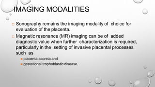
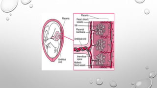
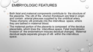
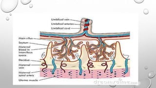
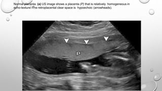
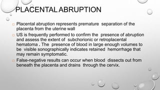
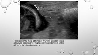
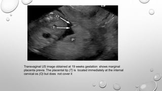
IMAGING OF THE PLACENTA
Dr. Vrishit Saraswat
I Yr. Resident
(M.D. Radio)
IMPORTANCE
 Abnormalities of the placenta are important to recognize
owing to the potential for maternal and fetal morbidity and
mortality. Pathologic conditions of the placenta include
 Placental causes of hemorrhage,
 Gestational trophoblastic disease,
 Retained products of conception,
 Nontrophoblastic placental tumors, metastases, and
 Cystic lesions..
IMAGING MODALITIES
 Sonography remains the imaging modality of choice for
evaluation of the placenta.
 Magnetic resonance (MR) imaging can be of added
diagnostic value when further characterization is required,
particularly in the setting of invasive placental processes
such as
 placenta accreta and
 gestational trophoblastic disease.
INTRODUCTION
 The placenta is named for its appearance (Greek plakuos,
meaning “flat cake”) and is responsible for the nutritive,
respiratory, and excretory functions of the fetus.
 The placenta is often overlooked in the routine evaluation of a
normal gestation, receiving attention only when an
abnormality is detected.
EMBRYOLOGIC FEATURES
 Both fetal and maternal components contribute to the structure of
the placenta. The villi of the chorion frondosum are fetal in origin
and contain arterial plexuses supplied by the umbilical artery.
These chorionic villi protrude into the intervillous space, where
they are bathed in maternal blood.
 The maternal portion of the placenta is composed of the decidua
placentalis, which lines the intervillous space. Fetal trophoblastic
invasion of the endometrium induces decidual changes. Maternal
decidual septa separate groups of villi within the intervillous
space.
Normal placenta. (a) US image shows a placenta (P) that is relatively homogeneous in
echo-texture. The retroplacental clear space is hypoechoic (arrowheads).
MORPHOLOGY AND
VARIANTS
 Typically, the placenta is located along the anterior or
posterior uterine wall, extending onto the lateral walls.
 Although usually discoid, the placenta can be variable in
morphology. Variant placental shapes include bi-lobed,
succenturiate, circumvallate, and placenta membranacea.
Succenturiate placenta. 2(a) Diagram shows a placenta with a succenturiate lobe. (b) US image shows
a placenta (P) with a succenturiate lobe (S). The main body of the placenta is located along the
posterior uterine wall. A second soft-tissue structure of the same echogenicity but located anteriorly is
the succenturiate lobe.
Bilobed placenta.3(a) Diagram shows a bilobed placenta. (b) US image shows a
bilobed placenta. The two lobes of the placenta (P1 and P2) are separated by a thin
bridge of placental tissue that covers the internal os. In this case, the umbilical cord
(arrowhead) inserts into the bridge of tissue.
Circumvallate placenta.-4a-US image shows a circumvallate placenta. The chorionic plate (the
fetal surface of the placenta) (black arrowheads) is smaller than the basal plate (the surface
interfacing with the uterus), with rolling and shouldering of the placental margins (white
arrowheads). F = fetus.
PLACENTA MEMBRANACEA.
Velamentous insertion of the umbilical cord. Doppler US image shows insertion (I) (white
arrow) of the umbilical cord into a thin membrane of tissue extending from the margin (black
arrow) of the placenta (P).
 The umbilical cord typically inserts centrally, but eccentric and
velamentous (outside the placental margin) insertions also occur .
 Eccentric insertions are cord insertions that are less than 1 cm
from the placental edge. These are distinguished from a
velamentous insertion, where the umbilical cord inserts on the
chorioamniotic membranes rather than on the placental mass.
This membranous insertion results in a variable segment of the
umbilical vessels running between the amnion and the chorion,
unprotected by Wharton jelly
 Placental size is expressed in terms of thickness in the
midportion of the organ and should be between 2 and 4 cm.
 Placental thinning has been described in systemic vascular and
hematologic diseases that result in microinfarctions.
 Thicker placentas (>4 cm) are seen in fetal hydrops,
antepartum infections, maternal diabetes, and maternal
anemia. Placental thickening can be simulated by
myometrial contractions and underlying fibroids.
Chorioamniotic separation. Transverse (a) and sagittal (b) images from obstetric US performed
at 20 weeks gestation show a free-floating membrane (arrowheads) surrounding the fetus (F).
This membrane is the amnion, which is completely separated from the underlying chorion;
there is even separation (arrow) over the surface of the placenta (P).
CHORIOAMNIOTIC SEPARATION
 The placental and fetal membranes (chorion and amnion,
respectively) are separate early in gestation, accounting for the
appearance of the amniotic sac. After approximately 14 weeks
gestation, these membranes fuse and are no longer separately
distinguishable (12).
 In rare cases, chorioamniotic separation can occur later in
gestation. This can be focal or extensive, with the amniotic
membrane becoming either free floating or adherent to the fetus.
Extensive cases pose a risk to the fetus, with increased rates of
both preterm delivery and the development of amniotic bands
(12).
 Chorioamniotic separation is most commonly related to prior
intervention such as amniocentesis or surgery but can occur
sporadically. Sporadic cases have been associated with
increased rates of underlying fetal chromosomal and
developmental abnormalities
 Chorioamniotic separation is usually detected with US and is
visible as a free-floating or adherent membrane surrounding
the fetus. Separation can extend throughout the entire
uterine cavity and over the surface of the placenta.
TWIN GESTATIONS-TWIN PEAK SIGN
Twin peak sign in dichorionic-diamniotic twin gestations. (a) US image of an early twin gestation shows
the separate placentas converging at the insertion of the amniotic membrane (arrowhead), forming the
so-called twin peak that is characteristic of a dichorionic-diamniotic gestation.
(b) Sagittal SSFSE MR image shows similar findings, with the twin peak (*) formed by
the two placentas. Arrowhead = intertwin membrane.
PLACENTAL CAUSES OF ANTEPARTUM
HEMORRHAGE
 Placenta previa and placental abruption account for more
than one-half of cases of antepartum hemorrhage and are
increasing in prevalence as the rate of cesarean section
increases.
Placental hematoma. (a) US image shows a rounded collection of mixed- echogenicity material
(arrowheads) deep to the chorion along the lateral margin of the placenta. There is no internal
Doppler signal to suggest blood flow. This appearance is consistent with a subchorionic hematoma.
PLACENTAL HEMATOMA
 placental hematomas appear as well- circumscribed masses with echogenicity that
varies according to chronicity.
 they are hypoechoic or anechoic in the acute phase, heterogeneously echogenic in
the subacute phase, and anechoic in the chronic phase.
 doppler interrogation should reveal absence of internal blood flow; this finding allows
differentiation of hematomas from other placental masses.
 Placental hematomas are foun d mostly on the fetal side of placenta.
 When large in size , are termed as Breus mole.
US image shows placental abruption. A crescenteric collection of predominantly hypoechoic
fluid lifts the edge of the placenta (P) away from the underlying myometrium (M).
PLACENTALABRUPTION
 Placental abruption represents premature separation of the
placenta from the uterine wall
 US is frequently performed to confirm the presence of abruption
and assess the extent of subchorionic or retroplacental
hematoma . The presence of blood in large enough volumes to
be visible sonographically indicates retained hemorrhage that
may remain symptomatic.
 False-negative results can occur when blood dissects out from
beneath the placenta and drains through the cervix.
Transvaginal US image obtained at 20 weeks gestation shows
a low-lying placenta (P). The placental margin comes to within
0.7 cm of the internal cervical os
Transvaginal US image obtained at 19 weeks gestation shows marginal
placenta previa. The placental tip (T) is located immediately at the internal
cervical os (O) but does not cover it
Transvaginal US image obtained at 19 weeks gestation shows complete placenta
previa. The placenta (P) entirely covers the internal cervical os (O).
PLACENTA PREVIA
 Placenta previa refers to abnormal implantation of the
placenta in the lower uterine segment, overlying or near the
internal cervical os
 Normally, the lower placental edge should be at least 2 cm
from the margin of the internal cervical os.
 The diagnosis of placenta previa should not be made before
15 weeks gestation.
There is vascular flow in a vessel (V) that is closely
applied to the internal cervical os (O).
VASA PREVIA
 Vasa previa refers to the presence of abnormal fetal vessels
within the amniotic membranes that cross the internal cervical
os. These vessels are unsupported by Wharton jelly or
placental tissue and are at risk of rupture when the supporting
membranes rupture; such vessels are also at risk of direct
injury during labor. Rupture of these vessels can lead to
catastrophic fetal hemorrhage.
 The diagnosis of vasa previa is made with Doppler US, which
demonstrates vascular flow within vessels overlying the
internal cervical os .
PLACENTA ACCRETA, INCRETA, AND
PERCRETA
 During the process of placental development and implantation, a defect in
the normal decidua basalis from prior surgery or instrumentation allows
abnormal adherence or penetration of the chorionic villi to or into the
uterine wall .
 The extent of adherence to and invasion of the placental tissue varies:
 Superficial invasion of the basalis layer is termed placenta accreta (approximately
75% of cases);
 deeper invasion of the myometrium is termed placenta increta ; and
 even deeper invasion involving the serosa or adjacent pelvic organs is termed
placenta percreta
 Sonographic features of placenta accreta include
 loss of the normal retroplacental clear space,
 anomalies of the bladder-myometrium interface,
 prominent placental lacunae, and
 increased vascularity at the interface of the uterus and bladder .
 Of these various sonographic features, the presence of
prominent placental lacunae has the highest positive predictive
value. Lacunae are characterized by ill-defined margins, irregular
shape, and turbulent flow.
US images show disruption of the normal hypoechoic myometrium (black
arrowheads) by invading placental tissue (white arrowheads). B = bladder, P =
placenta.
Any input or suggestions are
welcome.
Thanks for listening.
Dr Vrishit Saraswat.

Placental evaluation

  • 1.
    IMAGING OF THEPLACENTA Dr. Vrishit Saraswat I Yr. Resident (M.D. Radio)
  • 2.
    IMPORTANCE  Abnormalities ofthe placenta are important to recognize owing to the potential for maternal and fetal morbidity and mortality. Pathologic conditions of the placenta include  Placental causes of hemorrhage,  Gestational trophoblastic disease,  Retained products of conception,  Nontrophoblastic placental tumors, metastases, and  Cystic lesions..
  • 3.
    IMAGING MODALITIES  Sonographyremains the imaging modality of choice for evaluation of the placenta.  Magnetic resonance (MR) imaging can be of added diagnostic value when further characterization is required, particularly in the setting of invasive placental processes such as  placenta accreta and  gestational trophoblastic disease.
  • 4.
    INTRODUCTION  The placentais named for its appearance (Greek plakuos, meaning “flat cake”) and is responsible for the nutritive, respiratory, and excretory functions of the fetus.  The placenta is often overlooked in the routine evaluation of a normal gestation, receiving attention only when an abnormality is detected.
  • 6.
    EMBRYOLOGIC FEATURES  Bothfetal and maternal components contribute to the structure of the placenta. The villi of the chorion frondosum are fetal in origin and contain arterial plexuses supplied by the umbilical artery. These chorionic villi protrude into the intervillous space, where they are bathed in maternal blood.  The maternal portion of the placenta is composed of the decidua placentalis, which lines the intervillous space. Fetal trophoblastic invasion of the endometrium induces decidual changes. Maternal decidual septa separate groups of villi within the intervillous space.
  • 8.
    Normal placenta. (a)US image shows a placenta (P) that is relatively homogeneous in echo-texture. The retroplacental clear space is hypoechoic (arrowheads).
  • 9.
    MORPHOLOGY AND VARIANTS  Typically,the placenta is located along the anterior or posterior uterine wall, extending onto the lateral walls.  Although usually discoid, the placenta can be variable in morphology. Variant placental shapes include bi-lobed, succenturiate, circumvallate, and placenta membranacea.
  • 11.
    Succenturiate placenta. 2(a)Diagram shows a placenta with a succenturiate lobe. (b) US image shows a placenta (P) with a succenturiate lobe (S). The main body of the placenta is located along the posterior uterine wall. A second soft-tissue structure of the same echogenicity but located anteriorly is the succenturiate lobe.
  • 12.
    Bilobed placenta.3(a) Diagramshows a bilobed placenta. (b) US image shows a bilobed placenta. The two lobes of the placenta (P1 and P2) are separated by a thin bridge of placental tissue that covers the internal os. In this case, the umbilical cord (arrowhead) inserts into the bridge of tissue.
  • 13.
    Circumvallate placenta.-4a-US imageshows a circumvallate placenta. The chorionic plate (the fetal surface of the placenta) (black arrowheads) is smaller than the basal plate (the surface interfacing with the uterus), with rolling and shouldering of the placental margins (white arrowheads). F = fetus.
  • 14.
  • 15.
    Velamentous insertion ofthe umbilical cord. Doppler US image shows insertion (I) (white arrow) of the umbilical cord into a thin membrane of tissue extending from the margin (black arrow) of the placenta (P).
  • 16.
     The umbilicalcord typically inserts centrally, but eccentric and velamentous (outside the placental margin) insertions also occur .  Eccentric insertions are cord insertions that are less than 1 cm from the placental edge. These are distinguished from a velamentous insertion, where the umbilical cord inserts on the chorioamniotic membranes rather than on the placental mass. This membranous insertion results in a variable segment of the umbilical vessels running between the amnion and the chorion, unprotected by Wharton jelly
  • 17.
     Placental sizeis expressed in terms of thickness in the midportion of the organ and should be between 2 and 4 cm.  Placental thinning has been described in systemic vascular and hematologic diseases that result in microinfarctions.  Thicker placentas (>4 cm) are seen in fetal hydrops, antepartum infections, maternal diabetes, and maternal anemia. Placental thickening can be simulated by myometrial contractions and underlying fibroids.
  • 18.
    Chorioamniotic separation. Transverse(a) and sagittal (b) images from obstetric US performed at 20 weeks gestation show a free-floating membrane (arrowheads) surrounding the fetus (F). This membrane is the amnion, which is completely separated from the underlying chorion; there is even separation (arrow) over the surface of the placenta (P).
  • 19.
    CHORIOAMNIOTIC SEPARATION  Theplacental and fetal membranes (chorion and amnion, respectively) are separate early in gestation, accounting for the appearance of the amniotic sac. After approximately 14 weeks gestation, these membranes fuse and are no longer separately distinguishable (12).  In rare cases, chorioamniotic separation can occur later in gestation. This can be focal or extensive, with the amniotic membrane becoming either free floating or adherent to the fetus. Extensive cases pose a risk to the fetus, with increased rates of both preterm delivery and the development of amniotic bands (12).
  • 20.
     Chorioamniotic separationis most commonly related to prior intervention such as amniocentesis or surgery but can occur sporadically. Sporadic cases have been associated with increased rates of underlying fetal chromosomal and developmental abnormalities
  • 21.
     Chorioamniotic separationis usually detected with US and is visible as a free-floating or adherent membrane surrounding the fetus. Separation can extend throughout the entire uterine cavity and over the surface of the placenta.
  • 22.
    TWIN GESTATIONS-TWIN PEAKSIGN Twin peak sign in dichorionic-diamniotic twin gestations. (a) US image of an early twin gestation shows the separate placentas converging at the insertion of the amniotic membrane (arrowhead), forming the so-called twin peak that is characteristic of a dichorionic-diamniotic gestation.
  • 23.
    (b) Sagittal SSFSEMR image shows similar findings, with the twin peak (*) formed by the two placentas. Arrowhead = intertwin membrane.
  • 24.
    PLACENTAL CAUSES OFANTEPARTUM HEMORRHAGE  Placenta previa and placental abruption account for more than one-half of cases of antepartum hemorrhage and are increasing in prevalence as the rate of cesarean section increases.
  • 25.
    Placental hematoma. (a)US image shows a rounded collection of mixed- echogenicity material (arrowheads) deep to the chorion along the lateral margin of the placenta. There is no internal Doppler signal to suggest blood flow. This appearance is consistent with a subchorionic hematoma.
  • 26.
    PLACENTAL HEMATOMA  placentalhematomas appear as well- circumscribed masses with echogenicity that varies according to chronicity.  they are hypoechoic or anechoic in the acute phase, heterogeneously echogenic in the subacute phase, and anechoic in the chronic phase.  doppler interrogation should reveal absence of internal blood flow; this finding allows differentiation of hematomas from other placental masses.  Placental hematomas are foun d mostly on the fetal side of placenta.  When large in size , are termed as Breus mole.
  • 27.
    US image showsplacental abruption. A crescenteric collection of predominantly hypoechoic fluid lifts the edge of the placenta (P) away from the underlying myometrium (M).
  • 28.
    PLACENTALABRUPTION  Placental abruptionrepresents premature separation of the placenta from the uterine wall  US is frequently performed to confirm the presence of abruption and assess the extent of subchorionic or retroplacental hematoma . The presence of blood in large enough volumes to be visible sonographically indicates retained hemorrhage that may remain symptomatic.  False-negative results can occur when blood dissects out from beneath the placenta and drains through the cervix.
  • 29.
    Transvaginal US imageobtained at 20 weeks gestation shows a low-lying placenta (P). The placental margin comes to within 0.7 cm of the internal cervical os
  • 30.
    Transvaginal US imageobtained at 19 weeks gestation shows marginal placenta previa. The placental tip (T) is located immediately at the internal cervical os (O) but does not cover it
  • 31.
    Transvaginal US imageobtained at 19 weeks gestation shows complete placenta previa. The placenta (P) entirely covers the internal cervical os (O).
  • 32.
    PLACENTA PREVIA  Placentaprevia refers to abnormal implantation of the placenta in the lower uterine segment, overlying or near the internal cervical os  Normally, the lower placental edge should be at least 2 cm from the margin of the internal cervical os.  The diagnosis of placenta previa should not be made before 15 weeks gestation.
  • 33.
    There is vascularflow in a vessel (V) that is closely applied to the internal cervical os (O).
  • 34.
    VASA PREVIA  Vasaprevia refers to the presence of abnormal fetal vessels within the amniotic membranes that cross the internal cervical os. These vessels are unsupported by Wharton jelly or placental tissue and are at risk of rupture when the supporting membranes rupture; such vessels are also at risk of direct injury during labor. Rupture of these vessels can lead to catastrophic fetal hemorrhage.
  • 35.
     The diagnosisof vasa previa is made with Doppler US, which demonstrates vascular flow within vessels overlying the internal cervical os .
  • 36.
    PLACENTA ACCRETA, INCRETA,AND PERCRETA  During the process of placental development and implantation, a defect in the normal decidua basalis from prior surgery or instrumentation allows abnormal adherence or penetration of the chorionic villi to or into the uterine wall .  The extent of adherence to and invasion of the placental tissue varies:  Superficial invasion of the basalis layer is termed placenta accreta (approximately 75% of cases);  deeper invasion of the myometrium is termed placenta increta ; and  even deeper invasion involving the serosa or adjacent pelvic organs is termed placenta percreta
  • 37.
     Sonographic featuresof placenta accreta include  loss of the normal retroplacental clear space,  anomalies of the bladder-myometrium interface,  prominent placental lacunae, and  increased vascularity at the interface of the uterus and bladder .  Of these various sonographic features, the presence of prominent placental lacunae has the highest positive predictive value. Lacunae are characterized by ill-defined margins, irregular shape, and turbulent flow.
  • 38.
    US images showdisruption of the normal hypoechoic myometrium (black arrowheads) by invading placental tissue (white arrowheads). B = bladder, P = placenta.
  • 39.
    Any input orsuggestions are welcome. Thanks for listening. Dr Vrishit Saraswat.