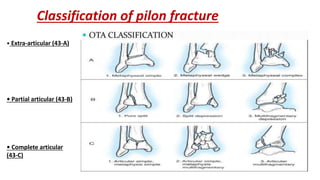
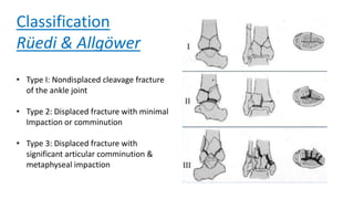
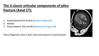
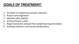
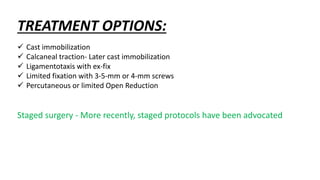
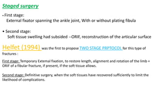
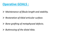
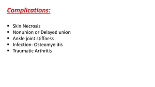
Pilon fracture
Dr. Manish Agrawal
MBBS, D.Ortho, DNB
Historical perspective
The term “ Tibial Pilon “ was first used by DESTOT in
1911,likening the pilon to the pestle.
Pilon is french word for Pestle, an instrument used for
crushing or pounding.
Definition
All fractures of the tibia involving the distal articular surface should be
classified as pilon fractures ,
Exceptfor medial or lateral malleolar fractures and Trimalleolar fractures,
where the posterior malleolar fracture involves < 1/3 of the articular surface.
Anatomy
Tibial pilon = The distal end of the tibia including the articular surface.
Proximal limit of tibial pilon: 8-10 cm from the ankle articular
surface.
Epidemiology
 Pilon fractures account for 7% -10% of all tibia fractures.
 Most pilon fractures are a result of high-energy mechanisms.
Thus, concomitant injuries are common and should be ruled out.
 Most common in 30-40 years of age .
Mechanism of injury
Fracture pattern is dictated by position of foot
and talus at time of impact:
 Plantar flexion Injury: posterior lip
fragment.
 Neutral ankle: anterior and posterior
fragments.
 Dorsiflexion injury: anterior lip fragment.
Clinical Presentation :
Patients typically present Non-ambulatory with
variable gross deformity of the involved distal leg.
CLINICAL EXAMINATION
The extremity should be examined carefully for ----
1. Signs of vascular injury
2. Swelling
3. Fracture blisters
4. Soft-tissue crushing
5. Closed degloving
6. Compartment syndrome.
Blood-filled fracture blisters indicates more extensive cutaneous
damage than blisters filled with clear fluid
RADIOLOGY :
• ROUTINE X-RAYS -
AP, LATERAL, MORTICE VIEW
• CT SCAN - TO KNOW FRACTURE PATTERN AND
INTRAARTICULAR COMMUNITION
Radiographic Evaluation
The 3 important anatomical zones to be
considered in the
decision-making and prognosis:
1. Articular surface
2. Metaphysis
3. Fibula
Classification of pilon fracture
• Extra-articular (43-A)
• Partial articular (43-B)
• Complete articular
(43-C)
Classification
Rüedi & Allgöwer
• Type I: Nondisplaced cleavage fracture
of the ankle joint
• Type 2: Displaced fracture with minimal
Impaction or comminution
• Type 3: Displaced fracture with
significant articular comminution &
metaphyseal impaction
The 3 classic articular components of pilon
fracture (Axial CT):
1. Anterolateral Part of tibia (Chaput fragment)
2. Medial
3. Posterolateral Part of tibia (Volkmann fragment)
These fragments vary in their size and amount of comminution
GOALS OF TREATMENT:
1. To obtain an anatomical articular reduction
2. Restore axial alignment
3. Maintain joint stability
4. Achieve fracture union
5. Regain functional and pain-free weight bearing and motion
6. Avoiding infections and wound complications.
TREATMENT OPTIONS:
 Cast immobilization
 Calcaneal traction- Later cast immobilization
 Ligamentotaxis with ex-fix
 Limited fixation with 3-5-mm or 4-mm screws
 Percutaneous or limited Open Reduction
Staged surgery - More recently, staged protocols have been advocated
Staged surgery
• First stage:
External fixator spanning the ankle joint, With or without plating fibula
• Second stage:
Soft tissue swelling had subsided --ORIF, reconstruction of the articular surface
Helfet (1994) was the first to propose TWO STAGE PRPTOCOL for this type of
fractures :
First stage: Temporary Extemal fixation, to restore length, alignment and rotation of the limb +
ORIF of a fibular fracture, if present, if the soft tissue allows.
Second stage: Definitive surgery, when the soft tissues have recovered sufficiently to limit the
likelihood of complications.
Operative GOALS :
 Maintenance of fibula length and stability.
 Restoration of tibial articular surface.
 Bone grafting of metaphyseal defects.
 Buttressing of the distal tibia.
Complications:
 Skin Necrosis
 Nonunion or Delayed union
 Ankle joint stiffness
 Infection- Osteomyelitis
 Traumatic Arthritis
Pilon fracture

Pilon fracture

  • 1.
    Pilon fracture Dr. ManishAgrawal MBBS, D.Ortho, DNB
  • 2.
    Historical perspective The term“ Tibial Pilon “ was first used by DESTOT in 1911,likening the pilon to the pestle. Pilon is french word for Pestle, an instrument used for crushing or pounding.
  • 3.
    Definition All fractures ofthe tibia involving the distal articular surface should be classified as pilon fractures , Exceptfor medial or lateral malleolar fractures and Trimalleolar fractures, where the posterior malleolar fracture involves < 1/3 of the articular surface.
  • 4.
    Anatomy Tibial pilon =The distal end of the tibia including the articular surface. Proximal limit of tibial pilon: 8-10 cm from the ankle articular surface.
  • 5.
    Epidemiology  Pilon fracturesaccount for 7% -10% of all tibia fractures.  Most pilon fractures are a result of high-energy mechanisms. Thus, concomitant injuries are common and should be ruled out.  Most common in 30-40 years of age .
  • 6.
    Mechanism of injury Fracturepattern is dictated by position of foot and talus at time of impact:  Plantar flexion Injury: posterior lip fragment.  Neutral ankle: anterior and posterior fragments.  Dorsiflexion injury: anterior lip fragment.
  • 7.
    Clinical Presentation : Patientstypically present Non-ambulatory with variable gross deformity of the involved distal leg.
  • 8.
    CLINICAL EXAMINATION The extremityshould be examined carefully for ---- 1. Signs of vascular injury 2. Swelling 3. Fracture blisters 4. Soft-tissue crushing 5. Closed degloving 6. Compartment syndrome.
  • 9.
    Blood-filled fracture blistersindicates more extensive cutaneous damage than blisters filled with clear fluid
  • 10.
    RADIOLOGY : • ROUTINEX-RAYS - AP, LATERAL, MORTICE VIEW • CT SCAN - TO KNOW FRACTURE PATTERN AND INTRAARTICULAR COMMUNITION
  • 11.
    Radiographic Evaluation The 3important anatomical zones to be considered in the decision-making and prognosis: 1. Articular surface 2. Metaphysis 3. Fibula
  • 12.
    Classification of pilonfracture • Extra-articular (43-A) • Partial articular (43-B) • Complete articular (43-C)
  • 13.
    Classification Rüedi & Allgöwer •Type I: Nondisplaced cleavage fracture of the ankle joint • Type 2: Displaced fracture with minimal Impaction or comminution • Type 3: Displaced fracture with significant articular comminution & metaphyseal impaction
  • 14.
    The 3 classicarticular components of pilon fracture (Axial CT): 1. Anterolateral Part of tibia (Chaput fragment) 2. Medial 3. Posterolateral Part of tibia (Volkmann fragment) These fragments vary in their size and amount of comminution
  • 15.
    GOALS OF TREATMENT: 1.To obtain an anatomical articular reduction 2. Restore axial alignment 3. Maintain joint stability 4. Achieve fracture union 5. Regain functional and pain-free weight bearing and motion 6. Avoiding infections and wound complications.
  • 16.
    TREATMENT OPTIONS:  Castimmobilization  Calcaneal traction- Later cast immobilization  Ligamentotaxis with ex-fix  Limited fixation with 3-5-mm or 4-mm screws  Percutaneous or limited Open Reduction Staged surgery - More recently, staged protocols have been advocated
  • 17.
    Staged surgery • Firststage: External fixator spanning the ankle joint, With or without plating fibula • Second stage: Soft tissue swelling had subsided --ORIF, reconstruction of the articular surface Helfet (1994) was the first to propose TWO STAGE PRPTOCOL for this type of fractures : First stage: Temporary Extemal fixation, to restore length, alignment and rotation of the limb + ORIF of a fibular fracture, if present, if the soft tissue allows. Second stage: Definitive surgery, when the soft tissues have recovered sufficiently to limit the likelihood of complications.
  • 18.
    Operative GOALS : Maintenance of fibula length and stability.  Restoration of tibial articular surface.  Bone grafting of metaphyseal defects.  Buttressing of the distal tibia.
  • 19.
    Complications:  Skin Necrosis Nonunion or Delayed union  Ankle joint stiffness  Infection- Osteomyelitis  Traumatic Arthritis