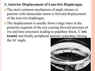
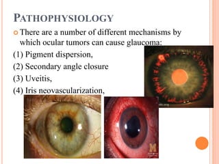
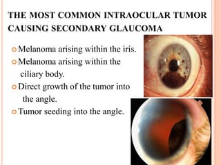
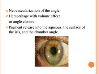
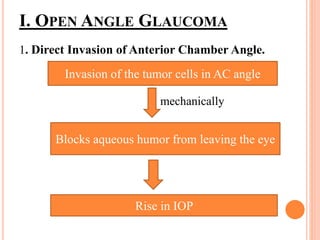
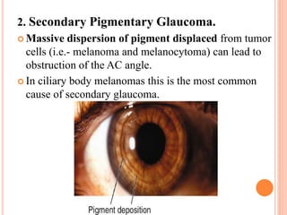
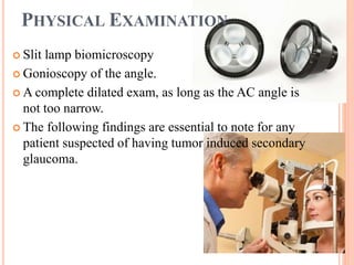
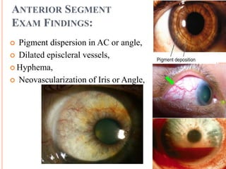
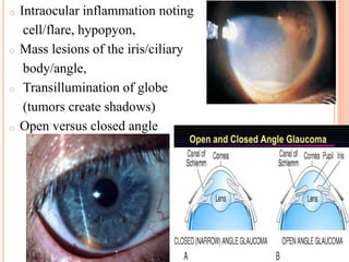
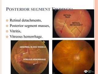
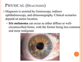
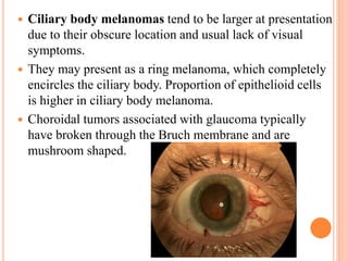
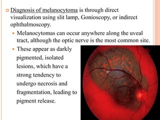
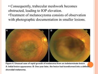
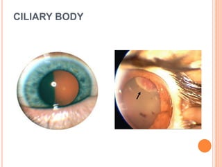
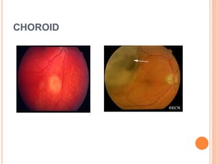
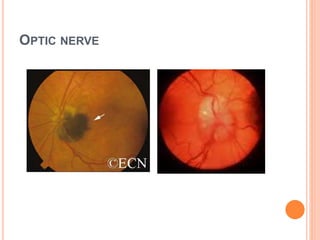
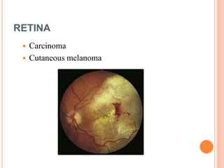
GLAUCOMA IN INTRAOCULAR
TUMOUR
By- Shweta .S. Maurya
3nd year B. Optometry
Institute For Technology And Management
GLAUCOMA
 Glaucoma is the
second leading cause
of blindness.
 Glaucoma is actually
a group of diseases.
The most common
type is hereditary.
INTRAOCULAR TUMORS
 Intraocular tumors are rare causes of glaucoma.
 The epidemiology, prognosis, and mechanism of
action depend on the specific tumor type.
 The tumor may not be directly visible in some
instances, and different methods of indirect
visualization may need to be used.
PATHOPHYSIOLOGY
 There are a number of different mechanisms by
which ocular tumors can cause glaucoma:
(1) Pigment dispersion,
(2) Secondary angle closure
(3) Uveitis,
(4) Iris neovascularization,
(5) Choroidal detachment,
(6) Suprachoroidal hemorrhage, and
(7) Anterior displacement of lens-iris diaphragm.
THE MOST COMMON INTRAOCULAR TUMOR
CAUSING SECONDARY GLAUCOMA
 Melanoma arising within the iris.
 Melanoma arising within the
ciliary body.
 Direct growth of the tumor into
the angle.
 Tumor seeding into the angle.
 Neovascularization of the angle,
 Hemorrhage with volume effect
or angle closure,
 Pigment release into the aqueous, the surface of
the iris, and the chamber angle.
PATHOPHYSIOLOGY
I. OPEN ANGLE GLAUCOMA
1. Direct Invasion of Anterior Chamber Angle.
mechanically
Invasion of the tumor cells in AC angle
Blocks aqueous humor from leaving the eye
Rise in IOP
2. Secondary Pigmentary Glaucoma.
 Massive dispersion of pigment displaced from tumor
cells (i.e.- melanoma and melanocytoma) can lead to
obstruction of the AC angle.
 In ciliary body melanomas this is the most common
cause of secondary glaucoma.
3. Melanomalytic Glaucoma.
 Melanomalytic glaucoma is secondary to the
release of pigment from tumor cells which
subsequently get taken up by macrophages.
 Based on studies , these pigment-laden
macrophages cause blockage of the trabecular
meshwork which leads to increased IOP
 Trabecular meshwork seeding (melanomalytic). Slit-
lamp photography shows collections of episcleral
perilimbal pigment deposition (arrows).
 NEXT
 Episcleral venous hypertension due to traumatic
carotid-cavernous fistula ”
4. Increased Episcleral Venous Pressure.
 Aqueous humor outflow is dependent on the pressure
gradient between IOP and episcleral venous pressure.
 Orbital tumors or extraocular extension of an intraocular
tumor can cause an increase in episcleral venous pressure
through direct compression.
 Due to this pressure gradient
reduction from the elevated
episcleral pressure, there is a
rise in IOP often causing a
secondary glaucoma.
II. CLOSED ANGLE GLAUCOMA
1. Uveitic.
 Inflammation secondary to intraocular
tumors can cause peripheral anterior synechiae, which
can lead to closure of the angle and elevation in IOP.
 In patients with uveal melanomas and uveitis, secondary
angle closure is common from peripheral anterior
synechiae.
 Metastatic tumors also have been shown to have
significant inflammation leading to peripheral anterior
synechiae.
2. Secondary Neovascularization.
 Patients with intraocular tumors can often develop
neovascularization of the iris and angle, which can lead to
angle closure glaucoma.
 This is commonly true in tumors which cause a chronic
RD ,leading to ischemia of the tissue and resultant
secondary neovascularization.
 Metastatic cutaneous melanomas
also are highly associated with
neovascularization.
 Neovascularization can also
present as a complication of
tumor treatment.
3. Anterior Displacement of Lens-Iris Diaphragm.
 The most common mechanism of angle closure in
patients with intraocular tumor is forward displacement
of the lens-iris diaphragm.
 The displacement is usually from a large mass in the
posterior segment of the eye causing forward pressure of
iris and lens structures leading to pupillary block, C iris
bombé and finally peripheral anterior synechiae closing
the AC angle.
DIAGNOSIS
HISTORY
 The workup begins with a thorough history, which should
be performed in all patients with elevated IOP.
A thorough history includes all of the following:
• Complete history of present illness (including current and
past symptoms)
• Past medical and ocular histories
• Past surgical history (including radiation treatments)
• Medication list (including eye drops)
• Family history (especially cancer history)
• Complete review of systems
• Social history (including drugs, tobacco use, alcohol and
occupation)
SYMPTOMS
 The symptoms that the patient experience depend greatly
on the type and location of the malignancy as well as the
amount of ocular involvement.
 The most frequently recorded symptoms are blurred
vision, pain, redness and floaters.
 Some patients, however, will not complain of any
symptoms.
 Glaucoma symptoms are dependent on the speed at which
the pressure rises.
 With acute glaucoma from angle closure, decreased
vision, halos around lights, ocular pain, and nausea may
be present.
 With chronic glaucoma with progressive angle closure
or open angles, no ocular symptoms related to the
elevation of IOP may be present.
 With iris melanoma, a hyperchromic heterochromia
may be present.
 With a ciliary body melanoma, a distorted pupil or
vision change from lenticular astigmatism may occur.
 With choroid melanoma, the visual symptoms may be
decreased vision or a change in the peripheral vision,
depending on the location of the tumor.
PHYSICAL EXAMINATION
 Slit lamp biomicroscopy
 Gonioscopy of the angle.
 A complete dilated exam, as long as the AC angle is
not too narrow.
 The following findings are essential to note for any
patient suspected of having tumor induced secondary
glaucoma.
ANTERIOR SEGMENT
EXAM FINDINGS:
 Pigment dispersion in AC or angle,
 Dilated episcleral vessels,
 Hyphema,
 Neovascularization of Iris or Angle,
o Intraocular inflammation noting
cell/flare, hypopyon,
o Mass lesions of the iris/ciliary
body/angle,
o Transillumination of globe
(tumors create shadows)
o Open versus closed angle
POSTERIOR SEGMENT FINDINGS:
 Retinal detachments,
 Posterior segment masses,
 Vitritis,
 Vitreous hemorrhage.
PHYSICAL (DIAGNOSIS)
 Diagnosis is assisted by Gonioscopy, indirect
ophthalmoscopy, and ultrasonography. Clinical scenarios
depend on tumor location.
 Iris melanoma can occur as either diffuse or well-
circumscribed forms, with the former being less common
and more malignant.
 Ciliary body melanomas tend to be larger at presentation
due to their obscure location and usual lack of visual
symptoms.
 They may present as a ring melanoma, which completely
encircles the ciliary body. Proportion of epithelioid cells
is higher in ciliary body melanoma.
 Choroidal tumors associated with glaucoma typically
have broken through the Bruch membrane and are
mushroom shaped.
 Diagnosis of melanocytoma is through direct
visualization using slit lamp, Gonioscopy, or indirect
ophthalmoscopy.
 Melanocytomas can occur anywhere along the uveal
tract, although the optic nerve is the most common site.
 These appear as darkly
pigmented, isolated
lesions, which have a
strong tendency to
undergo necrosis and
fragmentation, leading to
pigment release.
• Consequently, trabecular meshwork becomes
obstructed, leading to IOP elevation.
•Treatment of melanocytoma consists of observation
with photographic documentation in smaller lesions.
 Benign iris melanocytic lesions (Iris nevi) are difficult
to grossly distinguish from malignant lesions of the iris.
 These occur as small, discrete, flat, or slightly raised
nodules with variable pigmentation.
 Another study identified 5 factors associated with
higher risk of malignancy, as follows:
 (1) Diameter greater than 3 mm,
 (2)Pigment dispersion,
 (3) Prominent tumor vascularity,
 (4) Increased intraocular pressure, and
 (5) Tumor-related ocular symptoms.
 Although iris nevi rarely cause a pathologic process,
diffuse growth can cause elevated IOP by direct extension
into the trabecular meshwork.
 The treatment of an iris nevus consists of observation with
photographic documentation in smaller lesions.
 Tumors of the retina (retinoblastoma)
 The tumor may assume an endophytic or exophytic
configuration. In the former configuration, tumor cells
may invade into the vitreous and anterior chambers.
 Neovascularization is associated with angiogenic factor
production from the tumor or ischemia due to large
retinal vasculature involvement.
 Subsequent neovascularization of the iris with angle
closure accounts for 73% of glaucoma.
 Pupillary block leading to angle closure from large
tumors and infiltration of the trabecular meshwork
account for the remaining cases.
RETINOBLASTOMA
OPHTHALMIC IMAGING
 1. B-Scan Ultrasonography (B-Scan)
 If there is opacification of the media obstructing the
examiner’s view.
 It is helpful in taking measurements of the posterior
segment tumors like uveal melanomas and looking for
concurrent RD , vitreous hemorrhage or vitritis.
 2. Ultrasound Biomicroscopy (UBM)
 To further characterize tumors in the anterior segment,
ultrasound biomicroscopy can be performed.
 UBM uses a high frequency wavelength which allows for
accurate measurements of anterior lesions which is
helpful for diagnosis (solid versus cystic masses).
CAUSES
Intraocular tumors that may cause elevated
IOP may be of 3 types
1. Melanocytoma(benign)
2. Malignant melanoma
3. Metastatic (spreading )
IT CAN ARISE FROM
IRIS
CILIARY BODY
CHOROID
OPTIC NERVE
RETINA
 Carcinoma
 Cutaneous melanoma
METASTATIC TUMORS
 The 2 most common sites of primary tumor metastatic
to the eye are breast and lung cancers
 The posterior uvea is the most common site of
metastases, but glaucoma more often is associated with
anterior metastases.
 Mechanism of glaucoma includes direct invasion of the
trabecular meshwork by tumor cells, secondary angle
closure by anterior displacement of the lens-iris
diaphragm, and neovascularization of AC angle.
 Squamous cell carcinoma may produce a sheet of tumor
cells.
 Glaucoma may be the initial presentation in some
patients.
 Medical management of IOP coupled with radiation
or chemotherapy may be useful.
 Enucleation is reserved for blind, painful eyes.
PHAKOMATOSES
 Phakomatoses are a group of hamartomatous disorders
in which abnormal proliferation of tissues occurs in their
normal locations.
 These tumors primarily affect the eye, skin, and nervous
system, although other systems may be affected to a
lesser degree.
 Neurofibromatosis is a common autosomal dominant
systemic disorder divided into types 1 and 2
 Glaucoma usually is unilateral and associated with
eyelid thickening and contour abnormalities.
 Different mechanisms for elevated IOP have been
proposed, as follows:
(1) Direct infiltration of the angle tissue,
(2)Secondary angle closure due to ciliary body and
choroid thickening,
(3)Fibrovascular membrane
formation over the angle.
MISCELLANEOUS TUMORS
 The eye may become involved in acute and chronic
lymphocytic leukemia and affect the anterior and posterior
segment.
 Elevated IOP results from outflow obstruction secondary to
iritis, hyphema, hypopyon, or leukemic infiltration of the
trabecular meshwork and Schlemm canal, as well as episcleral
tissue, which may involve aqueous veins.
 Management is
primarily through
medical means to
control IOP and to
address the underlying
leukemia through
appropriate means.
 Multiple myeloma may cause ciliary body cysts,
which may lead to secondary glaucoma arising from
lens dislocation or anterior displacement of the iris
root.
 Large cell lymphoma may mimic uveitis and may
infiltrate the uveal tract, leading to secondary angle
closure.
 Juvenile xanthogranuloma is a benign histiocytic
tumor affecting the skin and eye in young children.
• A salmon colored iris tumor is associated with
spontaneous hyphema in children .
• Radiation therapy to the iris tissue has helped to
resolve the iris tumor and glaucoma.
DIFFERENTIAL DIAGNOSES
1. Glaucoma, Angle Closure, Acute
2. Primary Angle-Closure Glaucoma
3. Hyphema Glaucoma
4. Lens-Particle Glaucoma
5. Neovascular Glaucoma
6. Pigmentary Glaucoma
7. Primary Open-Angle Glaucoma
8. Pseudo exfoliation Glaucoma
9. Unilateral Glaucoma
10. Uveitic Glaucoma
Leukemia
 Involvement of the eye can occur in just under one-
third of cases of systemic leukemia. Secondary
glaucoma is generally caused by AC angle obstruction
from leukemic cells
Retinoblastoma
 Retinoblastoma is the most common primary
intraocular tumor in children and most commonly
presents with strabismus and leukocoria. Patients also
rarely can have a red, painful eye either from
inflammation or elevated IOP.
DIAGNOSTIC PROCEDURES
 If there is still uncertainty with the diagnosis after a full
history, physical exam and imaging then diagnostic
sampling of the lesion can be performed for most tumors.
 The use of fine needle aspiration biopsy with either a
25-guage needle or vitrector can be performed .
PROCEDURES
 For tissue diagnosis, fine-needle biopsy, aqueous
aspiration, or excisional biopsy is needed.
 Diagnosis of iris nevus is aided by fluorescein
angiography of iris, aqueous aspiration, or biopsy.
MANAGEMENT
MANAGEMENT
 For intraocular tumors with secondary glaucoma
elimination of the viable tumor cells is of primary
importance.
 Control of IOP control is secondary and can be
done either medically or surgically.
MEDICAL THERAPY
 Treatment of the secondary glaucoma should begin with
topical eye drops (alpha-agonists, beta-blockers and
carbonic anhydrase inhibitors) which decrease aqueous
humor production.
 If the pressure remains uncontrolled on topical medication
then oral hypotensive agents like acetazolamide or
methazolamide should be tried.
 For metastases with multiple lesions in the eye,
systemically chemotherapy can play a large role. In eyes
that have poor visual potential and elevated pressure,
treatment for the secondary glaucoma should only be given
if pain is present.
MEDICAL CARE
 Glaucoma management itself begins with medical therapy,
concurrent with treatment of the intraocular tumor through
surgery, radiation, chemotherapy, or a combination of these
treatments.
1. Standard methods of IOP control should be used, although
success rates with these topical medications will necessarily
be low.
2. Anti-vascular endothelial growth factor (anti-VEGF) therapy
may be helpful in those cases where neovascularization plays
a role in the pathogenesis of the glaucoma.
3. In systemic lymphoma, leukemia, and metastatic
malignancies, treatment will often include systemic
chemotherapy and radiation, with current regimens favoring
the use of chemotherapy first.
SURGERY
 Sometimes treatment of the underlying tumors with
plaque brachytherapy, external beam radiation,
chemotherapy or radiation can improve the IOP.
 For certain tumors like large uveal melanomas,
retinoblastoma or if the eye is painful with no visual
potential then primary enucleation may be the
appropriate option.
SURGICAL CARE
 Surgical options to control intraocular pressure must be
tempered by the need for preventing extraocular tumor
spread.
 For smaller tumors, observation is warranted until
growth is documented.
 Iridectomy are options for removing smaller tumors.
 For anterior tumors, argon laser to tumor-free areas is
an option.
 More posterior tumors may require local resection,
photocoagulation, or episcleral radiopaque therapy.
 Enucleation is also an option.
 For melanocytoma, laser or surgical excision of the
lesion may decrease the pigment load and decrease the
IOP.
 Glaucoma usually is associated with advanced stages of
Retinoblastoma, and enucleation may need to be
discussed.
 Any surgical intervention must be tempered by the risk
of extra ocular spread.
 Blind eyes should not undergo incisional glaucoma
surgery because of the risk of spreading the tumor to
outside the globe.
PROGNOSIS
 With early detection, the outlook is improved.
 The prognosis for the patient depends upon the type
of tumor, the amount of involvement within the eye,
as well as the treatment received.
 The primary goal for any treatment is to control the
tumor first and then control the IOP.
 Management of any intraocular tumor must be done
concurrently with an oncologist to account for
systemic disease.
 Complications
Even with early detection and aggressive therapy for
intraocular tumors, loss of the eye or death can occur.
A significant number of patients will develop side effects
from treatment of the original tumor with brachytherapy
which can lead to neovascularization and secondary angle-
closure glaucoma.
 Consultations
Glaucoma specialist or oncologist
 Patient Education
Follow-up care is important.
THANK YOU FOR LISTENING,
HOPE YOU LEARNED
SOMETHING NEW.

Glaucoma In Intra Ocular Tumour

  • 1.
    GLAUCOMA IN INTRAOCULAR TUMOUR By-Shweta .S. Maurya 3nd year B. Optometry Institute For Technology And Management
  • 2.
    GLAUCOMA  Glaucoma isthe second leading cause of blindness.  Glaucoma is actually a group of diseases. The most common type is hereditary.
  • 3.
    INTRAOCULAR TUMORS  Intraoculartumors are rare causes of glaucoma.  The epidemiology, prognosis, and mechanism of action depend on the specific tumor type.  The tumor may not be directly visible in some instances, and different methods of indirect visualization may need to be used.
  • 4.
    PATHOPHYSIOLOGY  There area number of different mechanisms by which ocular tumors can cause glaucoma: (1) Pigment dispersion, (2) Secondary angle closure (3) Uveitis, (4) Iris neovascularization,
  • 5.
    (5) Choroidal detachment, (6)Suprachoroidal hemorrhage, and (7) Anterior displacement of lens-iris diaphragm.
  • 6.
    THE MOST COMMONINTRAOCULAR TUMOR CAUSING SECONDARY GLAUCOMA  Melanoma arising within the iris.  Melanoma arising within the ciliary body.  Direct growth of the tumor into the angle.  Tumor seeding into the angle.
  • 7.
     Neovascularization ofthe angle,  Hemorrhage with volume effect or angle closure,  Pigment release into the aqueous, the surface of the iris, and the chamber angle.
  • 8.
  • 9.
    I. OPEN ANGLEGLAUCOMA 1. Direct Invasion of Anterior Chamber Angle. mechanically Invasion of the tumor cells in AC angle Blocks aqueous humor from leaving the eye Rise in IOP
  • 10.
    2. Secondary PigmentaryGlaucoma.  Massive dispersion of pigment displaced from tumor cells (i.e.- melanoma and melanocytoma) can lead to obstruction of the AC angle.  In ciliary body melanomas this is the most common cause of secondary glaucoma.
  • 11.
    3. Melanomalytic Glaucoma. Melanomalytic glaucoma is secondary to the release of pigment from tumor cells which subsequently get taken up by macrophages.  Based on studies , these pigment-laden macrophages cause blockage of the trabecular meshwork which leads to increased IOP
  • 12.
     Trabecular meshworkseeding (melanomalytic). Slit- lamp photography shows collections of episcleral perilimbal pigment deposition (arrows).  NEXT  Episcleral venous hypertension due to traumatic carotid-cavernous fistula ”
  • 13.
    4. Increased EpiscleralVenous Pressure.  Aqueous humor outflow is dependent on the pressure gradient between IOP and episcleral venous pressure.  Orbital tumors or extraocular extension of an intraocular tumor can cause an increase in episcleral venous pressure through direct compression.  Due to this pressure gradient reduction from the elevated episcleral pressure, there is a rise in IOP often causing a secondary glaucoma.
  • 14.
    II. CLOSED ANGLEGLAUCOMA 1. Uveitic.  Inflammation secondary to intraocular tumors can cause peripheral anterior synechiae, which can lead to closure of the angle and elevation in IOP.  In patients with uveal melanomas and uveitis, secondary angle closure is common from peripheral anterior synechiae.  Metastatic tumors also have been shown to have significant inflammation leading to peripheral anterior synechiae.
  • 15.
    2. Secondary Neovascularization. Patients with intraocular tumors can often develop neovascularization of the iris and angle, which can lead to angle closure glaucoma.  This is commonly true in tumors which cause a chronic RD ,leading to ischemia of the tissue and resultant secondary neovascularization.  Metastatic cutaneous melanomas also are highly associated with neovascularization.  Neovascularization can also present as a complication of tumor treatment.
  • 16.
    3. Anterior Displacementof Lens-Iris Diaphragm.  The most common mechanism of angle closure in patients with intraocular tumor is forward displacement of the lens-iris diaphragm.  The displacement is usually from a large mass in the posterior segment of the eye causing forward pressure of iris and lens structures leading to pupillary block, C iris bombé and finally peripheral anterior synechiae closing the AC angle.
  • 17.
  • 18.
    HISTORY  The workupbegins with a thorough history, which should be performed in all patients with elevated IOP. A thorough history includes all of the following: • Complete history of present illness (including current and past symptoms) • Past medical and ocular histories • Past surgical history (including radiation treatments) • Medication list (including eye drops) • Family history (especially cancer history) • Complete review of systems • Social history (including drugs, tobacco use, alcohol and occupation)
  • 19.
    SYMPTOMS  The symptomsthat the patient experience depend greatly on the type and location of the malignancy as well as the amount of ocular involvement.  The most frequently recorded symptoms are blurred vision, pain, redness and floaters.  Some patients, however, will not complain of any symptoms.  Glaucoma symptoms are dependent on the speed at which the pressure rises.
  • 20.
     With acuteglaucoma from angle closure, decreased vision, halos around lights, ocular pain, and nausea may be present.  With chronic glaucoma with progressive angle closure or open angles, no ocular symptoms related to the elevation of IOP may be present.  With iris melanoma, a hyperchromic heterochromia may be present.  With a ciliary body melanoma, a distorted pupil or vision change from lenticular astigmatism may occur.  With choroid melanoma, the visual symptoms may be decreased vision or a change in the peripheral vision, depending on the location of the tumor.
  • 21.
    PHYSICAL EXAMINATION  Slitlamp biomicroscopy  Gonioscopy of the angle.  A complete dilated exam, as long as the AC angle is not too narrow.  The following findings are essential to note for any patient suspected of having tumor induced secondary glaucoma.
  • 22.
    ANTERIOR SEGMENT EXAM FINDINGS: Pigment dispersion in AC or angle,  Dilated episcleral vessels,  Hyphema,  Neovascularization of Iris or Angle,
  • 23.
    o Intraocular inflammationnoting cell/flare, hypopyon, o Mass lesions of the iris/ciliary body/angle, o Transillumination of globe (tumors create shadows) o Open versus closed angle
  • 24.
    POSTERIOR SEGMENT FINDINGS: Retinal detachments,  Posterior segment masses,  Vitritis,  Vitreous hemorrhage.
  • 25.
    PHYSICAL (DIAGNOSIS)  Diagnosisis assisted by Gonioscopy, indirect ophthalmoscopy, and ultrasonography. Clinical scenarios depend on tumor location.  Iris melanoma can occur as either diffuse or well- circumscribed forms, with the former being less common and more malignant.
  • 26.
     Ciliary bodymelanomas tend to be larger at presentation due to their obscure location and usual lack of visual symptoms.  They may present as a ring melanoma, which completely encircles the ciliary body. Proportion of epithelioid cells is higher in ciliary body melanoma.  Choroidal tumors associated with glaucoma typically have broken through the Bruch membrane and are mushroom shaped.
  • 27.
     Diagnosis ofmelanocytoma is through direct visualization using slit lamp, Gonioscopy, or indirect ophthalmoscopy.  Melanocytomas can occur anywhere along the uveal tract, although the optic nerve is the most common site.  These appear as darkly pigmented, isolated lesions, which have a strong tendency to undergo necrosis and fragmentation, leading to pigment release.
  • 28.
    • Consequently, trabecularmeshwork becomes obstructed, leading to IOP elevation. •Treatment of melanocytoma consists of observation with photographic documentation in smaller lesions.
  • 29.
     Benign irismelanocytic lesions (Iris nevi) are difficult to grossly distinguish from malignant lesions of the iris.  These occur as small, discrete, flat, or slightly raised nodules with variable pigmentation.  Another study identified 5 factors associated with higher risk of malignancy, as follows:  (1) Diameter greater than 3 mm,  (2)Pigment dispersion,  (3) Prominent tumor vascularity,  (4) Increased intraocular pressure, and  (5) Tumor-related ocular symptoms.
  • 30.
     Although irisnevi rarely cause a pathologic process, diffuse growth can cause elevated IOP by direct extension into the trabecular meshwork.  The treatment of an iris nevus consists of observation with photographic documentation in smaller lesions.
  • 31.
     Tumors ofthe retina (retinoblastoma)  The tumor may assume an endophytic or exophytic configuration. In the former configuration, tumor cells may invade into the vitreous and anterior chambers.  Neovascularization is associated with angiogenic factor production from the tumor or ischemia due to large retinal vasculature involvement.  Subsequent neovascularization of the iris with angle closure accounts for 73% of glaucoma.  Pupillary block leading to angle closure from large tumors and infiltration of the trabecular meshwork account for the remaining cases.
  • 32.
  • 33.
  • 34.
     1. B-ScanUltrasonography (B-Scan)  If there is opacification of the media obstructing the examiner’s view.  It is helpful in taking measurements of the posterior segment tumors like uveal melanomas and looking for concurrent RD , vitreous hemorrhage or vitritis.
  • 35.
     2. UltrasoundBiomicroscopy (UBM)  To further characterize tumors in the anterior segment, ultrasound biomicroscopy can be performed.  UBM uses a high frequency wavelength which allows for accurate measurements of anterior lesions which is helpful for diagnosis (solid versus cystic masses).
  • 36.
    CAUSES Intraocular tumors thatmay cause elevated IOP may be of 3 types 1. Melanocytoma(benign) 2. Malignant melanoma 3. Metastatic (spreading )
  • 37.
    IT CAN ARISEFROM IRIS
  • 38.
  • 39.
  • 40.
  • 41.
  • 42.
    METASTATIC TUMORS  The2 most common sites of primary tumor metastatic to the eye are breast and lung cancers  The posterior uvea is the most common site of metastases, but glaucoma more often is associated with anterior metastases.  Mechanism of glaucoma includes direct invasion of the trabecular meshwork by tumor cells, secondary angle closure by anterior displacement of the lens-iris diaphragm, and neovascularization of AC angle.  Squamous cell carcinoma may produce a sheet of tumor cells.
  • 43.
     Glaucoma maybe the initial presentation in some patients.  Medical management of IOP coupled with radiation or chemotherapy may be useful.  Enucleation is reserved for blind, painful eyes.
  • 44.
    PHAKOMATOSES  Phakomatoses area group of hamartomatous disorders in which abnormal proliferation of tissues occurs in their normal locations.  These tumors primarily affect the eye, skin, and nervous system, although other systems may be affected to a lesser degree.
  • 45.
     Neurofibromatosis isa common autosomal dominant systemic disorder divided into types 1 and 2  Glaucoma usually is unilateral and associated with eyelid thickening and contour abnormalities.  Different mechanisms for elevated IOP have been proposed, as follows: (1) Direct infiltration of the angle tissue, (2)Secondary angle closure due to ciliary body and choroid thickening, (3)Fibrovascular membrane formation over the angle.
  • 46.
  • 47.
    MISCELLANEOUS TUMORS  Theeye may become involved in acute and chronic lymphocytic leukemia and affect the anterior and posterior segment.  Elevated IOP results from outflow obstruction secondary to iritis, hyphema, hypopyon, or leukemic infiltration of the trabecular meshwork and Schlemm canal, as well as episcleral tissue, which may involve aqueous veins.  Management is primarily through medical means to control IOP and to address the underlying leukemia through appropriate means.
  • 48.
     Multiple myelomamay cause ciliary body cysts, which may lead to secondary glaucoma arising from lens dislocation or anterior displacement of the iris root.  Large cell lymphoma may mimic uveitis and may infiltrate the uveal tract, leading to secondary angle closure.  Juvenile xanthogranuloma is a benign histiocytic tumor affecting the skin and eye in young children. • A salmon colored iris tumor is associated with spontaneous hyphema in children . • Radiation therapy to the iris tissue has helped to resolve the iris tumor and glaucoma.
  • 49.
    DIFFERENTIAL DIAGNOSES 1. Glaucoma,Angle Closure, Acute 2. Primary Angle-Closure Glaucoma 3. Hyphema Glaucoma 4. Lens-Particle Glaucoma 5. Neovascular Glaucoma 6. Pigmentary Glaucoma 7. Primary Open-Angle Glaucoma 8. Pseudo exfoliation Glaucoma 9. Unilateral Glaucoma 10. Uveitic Glaucoma
  • 50.
    Leukemia  Involvement ofthe eye can occur in just under one- third of cases of systemic leukemia. Secondary glaucoma is generally caused by AC angle obstruction from leukemic cells Retinoblastoma  Retinoblastoma is the most common primary intraocular tumor in children and most commonly presents with strabismus and leukocoria. Patients also rarely can have a red, painful eye either from inflammation or elevated IOP.
  • 51.
    DIAGNOSTIC PROCEDURES  Ifthere is still uncertainty with the diagnosis after a full history, physical exam and imaging then diagnostic sampling of the lesion can be performed for most tumors.  The use of fine needle aspiration biopsy with either a 25-guage needle or vitrector can be performed .
  • 52.
    PROCEDURES  For tissuediagnosis, fine-needle biopsy, aqueous aspiration, or excisional biopsy is needed.  Diagnosis of iris nevus is aided by fluorescein angiography of iris, aqueous aspiration, or biopsy.
  • 53.
  • 54.
    MANAGEMENT  For intraoculartumors with secondary glaucoma elimination of the viable tumor cells is of primary importance.  Control of IOP control is secondary and can be done either medically or surgically.
  • 55.
    MEDICAL THERAPY  Treatmentof the secondary glaucoma should begin with topical eye drops (alpha-agonists, beta-blockers and carbonic anhydrase inhibitors) which decrease aqueous humor production.  If the pressure remains uncontrolled on topical medication then oral hypotensive agents like acetazolamide or methazolamide should be tried.  For metastases with multiple lesions in the eye, systemically chemotherapy can play a large role. In eyes that have poor visual potential and elevated pressure, treatment for the secondary glaucoma should only be given if pain is present.
  • 56.
    MEDICAL CARE  Glaucomamanagement itself begins with medical therapy, concurrent with treatment of the intraocular tumor through surgery, radiation, chemotherapy, or a combination of these treatments. 1. Standard methods of IOP control should be used, although success rates with these topical medications will necessarily be low. 2. Anti-vascular endothelial growth factor (anti-VEGF) therapy may be helpful in those cases where neovascularization plays a role in the pathogenesis of the glaucoma. 3. In systemic lymphoma, leukemia, and metastatic malignancies, treatment will often include systemic chemotherapy and radiation, with current regimens favoring the use of chemotherapy first.
  • 57.
    SURGERY  Sometimes treatmentof the underlying tumors with plaque brachytherapy, external beam radiation, chemotherapy or radiation can improve the IOP.  For certain tumors like large uveal melanomas, retinoblastoma or if the eye is painful with no visual potential then primary enucleation may be the appropriate option.
  • 58.
    SURGICAL CARE  Surgicaloptions to control intraocular pressure must be tempered by the need for preventing extraocular tumor spread.  For smaller tumors, observation is warranted until growth is documented.  Iridectomy are options for removing smaller tumors.  For anterior tumors, argon laser to tumor-free areas is an option.  More posterior tumors may require local resection, photocoagulation, or episcleral radiopaque therapy.  Enucleation is also an option.
  • 59.
     For melanocytoma,laser or surgical excision of the lesion may decrease the pigment load and decrease the IOP.  Glaucoma usually is associated with advanced stages of Retinoblastoma, and enucleation may need to be discussed.  Any surgical intervention must be tempered by the risk of extra ocular spread.  Blind eyes should not undergo incisional glaucoma surgery because of the risk of spreading the tumor to outside the globe.
  • 60.
    PROGNOSIS  With earlydetection, the outlook is improved.  The prognosis for the patient depends upon the type of tumor, the amount of involvement within the eye, as well as the treatment received.  The primary goal for any treatment is to control the tumor first and then control the IOP.  Management of any intraocular tumor must be done concurrently with an oncologist to account for systemic disease.
  • 61.
     Complications Even withearly detection and aggressive therapy for intraocular tumors, loss of the eye or death can occur. A significant number of patients will develop side effects from treatment of the original tumor with brachytherapy which can lead to neovascularization and secondary angle- closure glaucoma.
  • 62.
     Consultations Glaucoma specialistor oncologist  Patient Education Follow-up care is important.
  • 63.
    THANK YOU FORLISTENING, HOPE YOU LEARNED SOMETHING NEW.