Downloaded 623 times
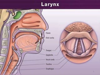
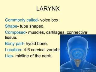
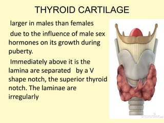
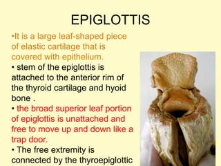
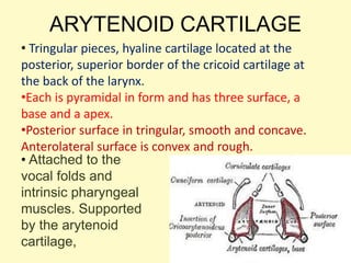
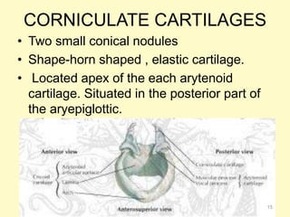
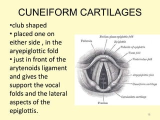
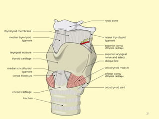
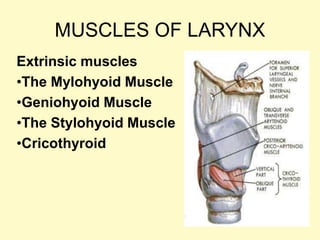
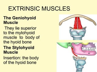
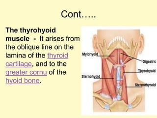
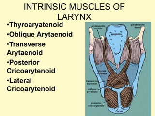
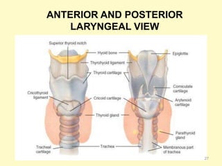
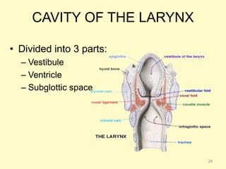
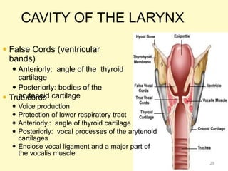
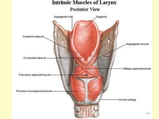
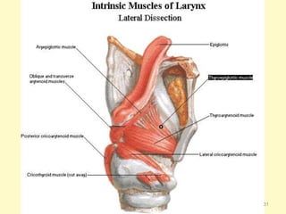
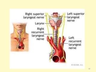
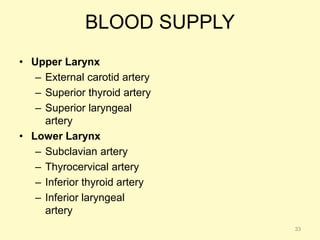

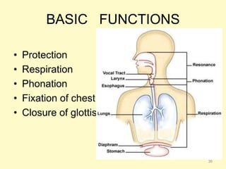

The document provides information on the anatomy and physiology of the larynx. It discusses the larynx's location in the neck, its composition of cartilages, muscles, and ligaments. The key functions of the larynx are identified as protection of the respiratory tract, respiration, phonation (voice production), fixation of the chest, and closure of the glottis. Diagrams are included showing the anterior and posterior views of the larynx cavities and structures.


![APPROACH TO FEVER IN PEDIATRICS[1].pptTT](https://cdn.slidesharecdn.com/ss_thumbnails/approachtofeverinpediatrics1-260125081456-d559e079-thumbnail.jpg?width=640&height=640&fit=bounds)


