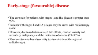
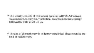
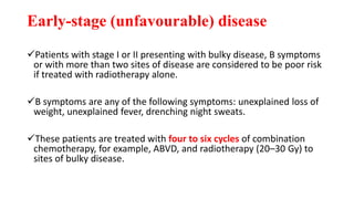
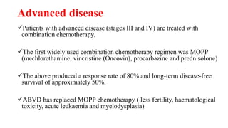
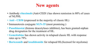
Hodgkin’s
Lymphoma
RVS Chaitanya Koppala
Lymphoma (?) accounts for approximately 3% of new cases of cancer
reported in the UK each year.
The primary cancerous cell of origin is the lymphocyte; as a result,
there is often considerable overlap between lymphomas and lymphoid
leukaemias.
Lymphomas are subdivided into two main categories:
Hodgkin's lymphoma (HL)
Non-Hodgkin's lymphoma (NHL).
Both HL and NHL can be further classified based on histology.
The site of malignancy is usually a lymph node.
Extranodal disease, most frequently of the stomach, skin, oral cavity
and pharynx, small intestine and CNS, can occur and is more common
in NHL than HL.
Hodgkin's lymphoma
Hodgkin's disease, now known as HL, was first described by Thomas
Hodgkin in 1832.
HL accounts for 30% of all lymphomas and has an incidence in the
UK of 2.2 /100,000/women and 3.3/100,000/men.
It is predominantly a disease of young adults, having a peak incidence
between the ages of 15 and 35 years.
Aetiology
The cause of HL is unknown
Epstein–Barr (glandular fever) virus has been identified in 50% of HL
cases.
Certain associations like genetic link with HL ; for example, same-sex
siblings of patients with HL have a 10 times higher risk of developing the
disease.
Patients with reduced immunity, for example, AIDS
Pathology
The diagnosis is made by histological examination of an excised
lymph node biopsy.
The characteristic pathological finding in HL is the identification of a
large, abnormal bi-nucleate lymphocyte called a Reed-Sternberg cell.
HL as classified by the World Health Organization (WHO) has two
distinct entities:
Classic HL
Nodular lymphocyte-predominant Hodgkin's lymphoma (NLPHL).
Classic Hodgkin's is further subdivided into
four histological types:
Nodular sclerosis: this is the most common type in the UK, predominant
in young adults and females, and has an excellent prognosis
Mixed cellularity: this is second most common type of classic HL and
more common in males (70% male)
Lymphocyte depleted: this carries a poor prognosis and is more common
in HIV-positive individuals
Lymphocyte rich: this is a rare type of classic HL
NLPHL accounts for 5% of HL cases and is more common in men.
Signs and symptoms:
Painless enlargement of lymph nodes
About 40% of patients will present with fever, night sweats and/or weight
loss.
Malaise
Itching (25%)
Pain at the site of enlarged nodes.
Bone pain (skeletal involvement)
Primary involvement of the gut, central nervous system or bone marrow is
rare.
Breathlessness.
Viral and fungal infections.
Laboratory findings
Normochromic
Normocytic anaemia
Raised ESR
Eosinophilia.
Leucocytosis due to an increase in neutrophils.
Advanced disease is associated with lymphopenia (lymphocytes <0.6
× 109 L−1).
Plasma lactate dehydrogenase (LDH) is raised in 30–40% of patients
at diagnosis and has been associated with a poor prognosis.
Investigations and staging
 Once the diagnosis has been made on biopsy
 Further investigations are needed to
Assess disease activity and the
Extent of its spread through the lymphoid system.
 staging is essential for assessing prognosis, with cure rates for
localised tumours (stage I or II) being much higher than those for
widespread disease (stage IV).
The tests required to establish the stage include a
Complete history
Physical examination
FBC
Urea and electrolytes (U and Es)
Chest X-ray and computed tomography (CT)
Erythrocyte sedimentation rate (ESR)
Serum LDH
Liver function tests (LFTs)
Positron emission tomography (PET) can be used to detect active residual
disease.
Management
HL is potentially curable and, in general, sensitive to both chemotherapy
and radiotherapy
The two main goals of treatment are to
Maximize the likelihood of cure
Minimizing the risk of late toxicity such as infertility.
Stage of disease is the biggest factor in treatment choice and outcome.
The management determined by the stage of the disease
Localised NLPHL frequently involves one isolated lymph node and tends
to be indolent (slow growing).
If there are no risk factors, it can be treated with IFRT alone; all other
types are treated as advanced (stage III or IV) classic HL.
In Europe, the treatment of classic HL is determined by whether the
disease is staged as
Early favourable disease,
Early unfavourable disease,
Advanced disease or Relapsed
Early-stage (favourable) disease
The cure rate for patients with stages I and IIA disease is greater than
90%.
Patients with stages I and IIA disease may be cured with radiotherapy
alone
However, due to radiation-related late effects, cardiac toxicity and
secondary malignancy and the incidence of relapse (25–30%),
Most receive combined modality treatment (chemotherapy and
radiotherapy).
This usually consists of two to four cycles of ABVD (Adriamycin
(doxorubicin), bleomycin, vinblastine, dacarbazine) chemotherapy
followed by IFRT of 20–30 Gy
The aim of chemotherapy is to destroy subclinical disease outside the
field of radiotherapy.
Early-stage (unfavourable) disease
Patients with stage I or II presenting with bulky disease, B symptoms
or with more than two sites of disease are considered to be poor risk
if treated with radiotherapy alone.
B symptoms are any of the following symptoms: unexplained loss of
weight, unexplained fever, drenching night sweats.
These patients are treated with four to six cycles of combination
chemotherapy, for example, ABVD, and radiotherapy (20–30 Gy) to
sites of bulky disease.
Advanced disease
Patients with advanced disease (stages III and IV) are treated with
combination chemotherapy.
The first widely used combination chemotherapy regimen was MOPP
(mechlorethamine, vincristine (Oncovin), procarbazine and prednisolone)
The above produced a response rate of 80% and long-term disease-free
survival of approximately 50%.
ABVD has replaced MOPP chemotherapy ( less fertility, haematological
toxicity, acute leukaemia and myelodysplasia)
Management
If the Hodgkin disease was in the upper
body, (?) given to the mantle field,
which included lymph node areas in the
neck, chest, and under the arms.
If the Hodgkin disease occurs below
the diaphragam inverted Y field (?)
therapy included the lymph nodes in the
upper abdomen, the spleen, and the
lymph nodes in the pelvis
Combination chemotherapy regimens effective
in the treatment of Hodgkin's lymphoma
Salvage therapy for relapsed disease
Relapsed disease refers to disease progression after completion of
primary treatment which resulted in a complete remission.
Depending on previous treatment, options include
Salvage radiotherapy,
Salvage chemotherapy
High-dose chemotherapy with autologous stem cell support.
In this procedure, stem cells are collected from the patient and
returned following high-dose chemotherapy.
Patients who relapse after initial radiotherapy alone have a good
chance of cure with combination chemotherapy, at least equal to that
of patients initially treated with chemotherapy for advanced disease.
Occasionally, radiotherapy is used if the disease is localised and
previously non-irradiated..
Salvage chemotherapy regimens effective in the
treatment of lymphoma
New agents
• Antibody rituximab (Anti-CD20 ) has shown remission in 80% of cases
of NLPHL
• Anti - CD30 (expressed in the majority of classic HL)
• Immunotoxin conjugate SGN-35 (most promising )
• Panobinostat (histone deacetylases inhibitor), has been granted orphan
drug designation for the treatment of HL.
• Gemcitabine has shown activity in relapsed classic HL with response
rates up to 79%
• Bortezomib and lenalidomide for relapsed HL(licensed for myeloma)

Phamacotherapy for Hodgkin's lymphoma

  • 1.
  • 2.
    Lymphoma (?) accountsfor approximately 3% of new cases of cancer reported in the UK each year. The primary cancerous cell of origin is the lymphocyte; as a result, there is often considerable overlap between lymphomas and lymphoid leukaemias.
  • 3.
    Lymphomas are subdividedinto two main categories: Hodgkin's lymphoma (HL) Non-Hodgkin's lymphoma (NHL). Both HL and NHL can be further classified based on histology. The site of malignancy is usually a lymph node. Extranodal disease, most frequently of the stomach, skin, oral cavity and pharynx, small intestine and CNS, can occur and is more common in NHL than HL.
  • 4.
    Hodgkin's lymphoma Hodgkin's disease,now known as HL, was first described by Thomas Hodgkin in 1832. HL accounts for 30% of all lymphomas and has an incidence in the UK of 2.2 /100,000/women and 3.3/100,000/men. It is predominantly a disease of young adults, having a peak incidence between the ages of 15 and 35 years.
  • 5.
    Aetiology The cause ofHL is unknown Epstein–Barr (glandular fever) virus has been identified in 50% of HL cases. Certain associations like genetic link with HL ; for example, same-sex siblings of patients with HL have a 10 times higher risk of developing the disease. Patients with reduced immunity, for example, AIDS
  • 6.
    Pathology The diagnosis ismade by histological examination of an excised lymph node biopsy. The characteristic pathological finding in HL is the identification of a large, abnormal bi-nucleate lymphocyte called a Reed-Sternberg cell. HL as classified by the World Health Organization (WHO) has two distinct entities: Classic HL Nodular lymphocyte-predominant Hodgkin's lymphoma (NLPHL).
  • 7.
    Classic Hodgkin's isfurther subdivided into four histological types: Nodular sclerosis: this is the most common type in the UK, predominant in young adults and females, and has an excellent prognosis Mixed cellularity: this is second most common type of classic HL and more common in males (70% male) Lymphocyte depleted: this carries a poor prognosis and is more common in HIV-positive individuals Lymphocyte rich: this is a rare type of classic HL NLPHL accounts for 5% of HL cases and is more common in men.
  • 8.
    Signs and symptoms: Painlessenlargement of lymph nodes About 40% of patients will present with fever, night sweats and/or weight loss. Malaise Itching (25%) Pain at the site of enlarged nodes. Bone pain (skeletal involvement) Primary involvement of the gut, central nervous system or bone marrow is rare. Breathlessness. Viral and fungal infections.
  • 9.
    Laboratory findings Normochromic Normocytic anaemia RaisedESR Eosinophilia. Leucocytosis due to an increase in neutrophils. Advanced disease is associated with lymphopenia (lymphocytes <0.6 × 10⁹ L−1). Plasma lactate dehydrogenase (LDH) is raised in 30–40% of patients at diagnosis and has been associated with a poor prognosis.
  • 10.
    Investigations and staging Once the diagnosis has been made on biopsy  Further investigations are needed to Assess disease activity and the Extent of its spread through the lymphoid system.  staging is essential for assessing prognosis, with cure rates for localised tumours (stage I or II) being much higher than those for widespread disease (stage IV).
  • 11.
    The tests requiredto establish the stage include a Complete history Physical examination FBC Urea and electrolytes (U and Es) Chest X-ray and computed tomography (CT) Erythrocyte sedimentation rate (ESR) Serum LDH Liver function tests (LFTs) Positron emission tomography (PET) can be used to detect active residual disease.
  • 12.
    Management HL is potentiallycurable and, in general, sensitive to both chemotherapy and radiotherapy The two main goals of treatment are to Maximize the likelihood of cure Minimizing the risk of late toxicity such as infertility. Stage of disease is the biggest factor in treatment choice and outcome.
  • 13.
    The management determinedby the stage of the disease Localised NLPHL frequently involves one isolated lymph node and tends to be indolent (slow growing). If there are no risk factors, it can be treated with IFRT alone; all other types are treated as advanced (stage III or IV) classic HL. In Europe, the treatment of classic HL is determined by whether the disease is staged as Early favourable disease, Early unfavourable disease, Advanced disease or Relapsed
  • 14.
    Early-stage (favourable) disease Thecure rate for patients with stages I and IIA disease is greater than 90%. Patients with stages I and IIA disease may be cured with radiotherapy alone However, due to radiation-related late effects, cardiac toxicity and secondary malignancy and the incidence of relapse (25–30%), Most receive combined modality treatment (chemotherapy and radiotherapy).
  • 15.
    This usually consistsof two to four cycles of ABVD (Adriamycin (doxorubicin), bleomycin, vinblastine, dacarbazine) chemotherapy followed by IFRT of 20–30 Gy The aim of chemotherapy is to destroy subclinical disease outside the field of radiotherapy.
  • 16.
    Early-stage (unfavourable) disease Patientswith stage I or II presenting with bulky disease, B symptoms or with more than two sites of disease are considered to be poor risk if treated with radiotherapy alone. B symptoms are any of the following symptoms: unexplained loss of weight, unexplained fever, drenching night sweats. These patients are treated with four to six cycles of combination chemotherapy, for example, ABVD, and radiotherapy (20–30 Gy) to sites of bulky disease.
  • 17.
    Advanced disease Patients withadvanced disease (stages III and IV) are treated with combination chemotherapy. The first widely used combination chemotherapy regimen was MOPP (mechlorethamine, vincristine (Oncovin), procarbazine and prednisolone) The above produced a response rate of 80% and long-term disease-free survival of approximately 50%. ABVD has replaced MOPP chemotherapy ( less fertility, haematological toxicity, acute leukaemia and myelodysplasia)
  • 19.
  • 20.
    If the Hodgkindisease was in the upper body, (?) given to the mantle field, which included lymph node areas in the neck, chest, and under the arms. If the Hodgkin disease occurs below the diaphragam inverted Y field (?) therapy included the lymph nodes in the upper abdomen, the spleen, and the lymph nodes in the pelvis
  • 21.
    Combination chemotherapy regimenseffective in the treatment of Hodgkin's lymphoma
  • 23.
    Salvage therapy forrelapsed disease Relapsed disease refers to disease progression after completion of primary treatment which resulted in a complete remission. Depending on previous treatment, options include Salvage radiotherapy, Salvage chemotherapy High-dose chemotherapy with autologous stem cell support. In this procedure, stem cells are collected from the patient and returned following high-dose chemotherapy.
  • 24.
    Patients who relapseafter initial radiotherapy alone have a good chance of cure with combination chemotherapy, at least equal to that of patients initially treated with chemotherapy for advanced disease. Occasionally, radiotherapy is used if the disease is localised and previously non-irradiated..
  • 25.
    Salvage chemotherapy regimenseffective in the treatment of lymphoma
  • 26.
    New agents • Antibodyrituximab (Anti-CD20 ) has shown remission in 80% of cases of NLPHL • Anti - CD30 (expressed in the majority of classic HL) • Immunotoxin conjugate SGN-35 (most promising ) • Panobinostat (histone deacetylases inhibitor), has been granted orphan drug designation for the treatment of HL. • Gemcitabine has shown activity in relapsed classic HL with response rates up to 79% • Bortezomib and lenalidomide for relapsed HL(licensed for myeloma)