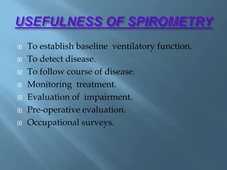
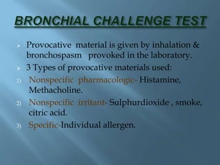
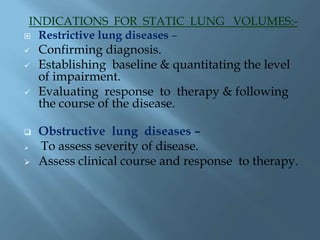
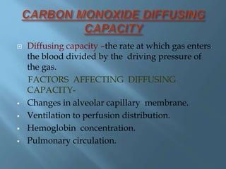
This document discusses various pulmonary function tests used to assess lung function, detect respiratory diseases, and evaluate treatment responses. It describes tests such as spirometry, which measures volumes of air inhaled and exhaled; bronchial provocation tests, which provoke bronchospasm; measurements of static lung volumes; carbon monoxide diffusing capacity; and exercise testing. The tests provide quantitative information about airflow obstruction, restriction, gas exchange ability, and cardiopulmonary response to exercise. Indications for the tests include diagnosing and monitoring lung diseases and impairments.
2. Aspects of pulmonary function:
Perfusion- Blood flow.
Diffusion- Movement of O2 & CO2.
Ventilation- Air exchange btwn alveolar
spaces & the atmosphere.
4. Assessment of various aspects of pulmonary
physiology.
Detect & quantify the respiratory disease.
Evaluation of the disease & its response to
therapy.
Provide valuable clinical information.
7. Measures the volume of air inspired or expired &
records the time –the volume change occurs.
Mechanical signal →electrical signal→records the
amt of gas breathed in & out→ SPIROGRAM.
2 major categories:-
mechanical electronic or flow sensing
or
volume-displacement
8. o Determines the effectiveness of various
mechanical forces involved in lung & chest
wall movement.
o The values obtained provide quantitative
information abt the degree of obstruction to
expiratory airflow or the degree of restriction
of inspired air.
9. Metal container with 2 chambers.
Outer chamber(water chamber) has a floating
drum.
Drum is counter balanced by weight-string.
Pen attached to counter wt-writes on calibrated
paper , fixed to a recording device.
Inner chamber inverted with small hole at the
top& long metal tube passes through it.
Top end of the tube penetrates into outer
chamber above level of water & other end is
mouthpiece.
Respiration-closed nose by nose clip(5-30min).
10. The forced expiratory volumes exhaled within
1,2,3 seconds – timed vital
capacities(FEV1,FEV2 & FEV3 res).
FEF25-75 –forced expiratory flow btwn 25%
&75%.
Measurements are useful for evaluating a
patient’s response to bronchodilators.
If FEV1 <80% of predicted or FEF25-75% is
<60% of predicted, bronchodilators are
administered –nebulizer; spirometry is
repeated.
11. Measured (actual) spirometry values are
compared with predicted values- age , height,
weight & gender; expressed as a % of predicted
value.(>80% normal limits).
During expiration floating drum moves up ,
counter wt comes down , downward deflection
on the graph.
Inspiration the drum moves down, counter wt
comes up, upward deflection on the graph.
Spirometry test –measured twice both before
and after given a bronchodilator.
Improvement in measurements means patient
will respond well.
12. DISADVANTAGE
• Only for single use due to CO2 accumulation &
O2 cannot be supplied.
• Residual Volume, Functional Residual
Capacity and Total Lung Capacity cannot be
measured.
13. To establish baseline ventilatory function.
To detect disease.
To follow course of disease.
Monitoring treatment.
Evaluation of impairment.
Pre-operative evaluation.
Occupational surveys.
14. Provocative material is given by inhalation &
bronchospasm provoked in the laboratory.
3 Types of provocative materials used:
1) Nonspecific pharmacologic- Histamine,
Methacholine.
2) Nonspecific irritant- Sulphurdioxide , smoke,
citric acid.
3) Specific-Individual allergen.
15. 20% fall in FEV1 from basal values – positive
response.
Histamine solution by nebulisation - 8mg/ml.
INDICATIONS FOR TESTING:-
To identify patients with hypersensitive or
hyper reactive airways- diagnosing asthma.
To identify specific provocative factors.
Research tool to study pathophysiology of
acute reversible bronchospasm.
16. 3 Basic techniques for measurement of lung
volumes:-
Gas dilution methods.
Body plethysmography.
Radiographic techniques.
17. INDICATIONS FOR STATIC LUNG VOLUMES:-
Restrictive lung diseases –
Confirming diagnosis.
Establishing baseline & quantitating the level
of impairment.
Evaluating response to therapy & following
the course of the disease.
Obstructive lung diseases –
To assess severity of disease.
Assess clinical course and response to therapy.
18. Diffusing capacity –the rate at which gas enters
the blood divided by the driving pressure of
the gas.
FACTORS AFFECTING DIFFUSING
CAPACITY-
Changes in alveolar capillary membrane.
Ventilation to perfusion distribution.
Hemoglobin concentration.
Pulmonary circulation.
19. Reduced diffusing capacity is seen in:-
Anemia, multiple pulmonary emboli, emphysema,
pulmonary resection interstitial lung disease
&severe bronchospasm.
INDICATIONS FOR CO DIFFUSING
CAPACITY:-
Unexplained dyspnoea due to early interstitial
lung disease.
Differentiating emphysema from chronic
bronchitis & asthma.
Diagnosis & follow up of patients with interstitial
lung disease.
Diagnosis of recurrent multiple pulmonary emboli.
20. Alveolar oxygen tension is calculated & arterial
oxygen tension measured by blood gas
estimation.
Difference btwn the two gives a measurement
of alveolar to arterial oxygen gradient. In
normal →5-15 mm Hg.
Increased AAOG is due to 3 mechanisms:-
i. Ventilation perfusion mismatch.
ii. Increased right to left shunt.
iii. Diffusion block.
21. Alveolar arterial oxygen gradient are measured
after exercise.
Gradient reduces but if it increases it indicates
ventilation perfusion mismatch.
Absolute shunt fraction is also calculated
when AAG are measured 10 min after 100%
oxygen.
Increased gradient after 100% oxygen with an
increased shunt, indicate presence of right to
left shunt.
22. Cardiopulmonary stress test ,with the addition
pulmonary factors are also evaluated during
exercise.
Evaluate the response of the cardiovascular &
respiratory systems to exercise, allows
measurement of gas exchange.
It categorizes disorders that limit exercise
tolerance by documenting their
pathophysiology.
Allows for an objective assessment of the
patients symptoms , accurate prescription.
23. A MANUAL OF LBORATORY &
DIAGNOSTIC TESTS ,8th EDITION.
A PATHOPHYSIOLOGIC APPROACH BY
JOSEPH .T. DIPIRO.
APPLIED THERAPEUTICS,8th EDITION.
PULMONARY FUNCTION TESTS BY RAJIV
.S. MATHUR
M.D(Med),D.N.B.(Resp.Med),CHEST
PHYSICIAN,JASLOK HOSPITAL,BOMBAY.
http://www.webmed.com/asthma/guide/lun
g-function-tests-asthma.