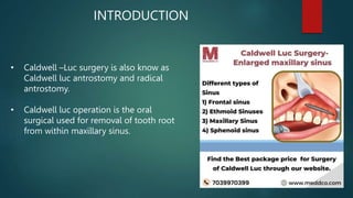
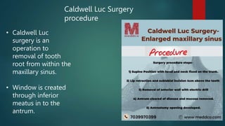
1. Caldwell Luc surgery (CWS) is over 120 years old and remains an important procedure for ENT surgeons. It involves making a window in the anterior maxillary bone to access and treat diseases of the maxillary sinus.
2. Indications for CWS include chronic maxillary sinusitis, cysts and polyps in the sinus, and benign or malignant tumors. Contraindications are acute infections and performing it in children due to risk of damaging developing teeth.
3. The procedure involves making a U-shaped incision over the canine fossa, removing bone to create a window, excising the sinus lining and lesions, debriding, irrigating, placing a drain, and packing with
This document discusses cysts of the oral and maxillofacial region. It defines true cysts as pathological cavities lined by epithelium and containing fluid, and pseudo cysts as cavities not lined by epithelium that may contain fluid. Cysts are classified based on their origin (odontogenic vs non-odontogenic) and location. Diagnosis involves history, clinical examination, radiographic evaluation and sometimes aspiration biopsy or surgical biopsy. Treatment options include enucleation, marsupialization, or a combination depending on the cyst size and location.
Dr. Ahmed M. Adawy, Professor Emeritus, Dep. Oral & Maxillofacial Surgery. Former Dean, Faculty of Dental Medicine
Al-Azhar University. There are four pairs of air sinuses making the boundaries of the nasal cavity. Maxillary sinus is the largest air cell. Anatomy and physiology of the maxillary sinus are given. Maxillary sinusitis is an inflammation of the sinus. Odontogenic causes represent nearly 30% of the etiology. Clinical and radiographic examinations are discussed together with treatment plan.
Oro-antral fistula is a rare complication of surgery at the posterior maxillary region. Several techniques for closure are presented. Additionally, information about sinus lift procedure is given.
This document provides an overview of cysts of the oral and maxillofacial region. It defines cysts and discusses their classification, parts, pathogenesis and mechanisms of enlargement. It also describes key cysts such as dentigerous cysts, odontogenic keratocysts and eruption cysts in detail, covering their definitions, locations, clinical and radiographic features, histology, differential diagnosis and complications. Dentigerous and odontogenic keratocysts are the most common epithelial cysts of the jaws.
The document discusses salivary gland disorders. It begins with definitions and classifications of salivary glands. It then discusses the anatomy, functions, and disorders of the parotid, submandibular, and sublingual salivary glands. Diagnostic aids are outlined including clinical history, physical examination, imaging such as CT, MRI, ultrasound and sialography. Cystic conditions of the minor salivary glands such as mucoceles are also summarized. Disorders are classified and inflammatory, obstructive, neoplastic and other conditions are described.
A cyst is an epithelium-lined sac containing fluid or semisolid material. In the formation of a cyst, the epithelial cells first proliferate and later undergo degeneration and liquefaction. The liquefied material exerts equal pressure on the walls of the cyst from within. Cysts grow by expansion and thus displace the adjacent teeth by pressure. May can produce expansion of the cortical bone. On a radiograph, the radiolucency of a cyst is usually bordered by a radiopaque periphery of dense sclerotic bone. The radiolucency may be unilocular or multilocular. Odontogenic cysts are those which arise from the epithelium associated with the development of teeth. The source of epithelium is from the enamel organ, the reduced enamel epithelium, the cell rests of Malassez or the remnants of the dental lamina.
1) Ameloblastoma is a benign, locally invasive odontogenic tumor of enamel organ-type tissue that is the second most common odontogenic tumor.
2) It typically presents as a painless swelling in the mandible and is classified based on histological and clinical features into subtypes including follicular, plexiform, unicystic, and peripheral ameloblastoma.
3) Treatment involves surgical resection such as segmental resection for large tumors due to the high recurrence risk with more conservative treatments like curettage or enucleation.
This document discusses Le Fort fractures and their management. It begins by describing the three areas that make up the facial skeleton: upper third, lower third, and middle third. It then provides detailed descriptions and classifications of Le Fort I, II, and III fractures based on the location and direction of the fracture lines. For each type of fracture, it outlines the characteristic signs, symptoms, and clinical features both externally and internally. It also discusses other midface fractures and dentoalveolar fractures. In summary, the document provides an in-depth overview of Le Fort fractures, including their anatomical basis, classification, and clinical presentation.
The document describes the submandibular and retromandibular surgical approaches. The submandibular approach involves making a 1.5-2 cm incision inferior to the mandible and dissecting through the layers of the skin, subcutaneous tissue, platysma muscle, and pterygomasseteric sling. The retromandibular approach uses a vertical incision 2 cm posterior to the mandibular ramus and dissects through the same layers to the pterygomasseteric sling. Both approaches give access below the mandible for surgical procedures.
This document provides information on fascial spaces of the head and neck region. It begins by defining fascial spaces as clefts or compartments containing connective tissue. It then classifies the spaces based on mode of involvement and clinical significance. Several key fascial spaces are described in detail, including boundaries, contents, etiology, clinical features, and spread of infection. These include the buccal, sublingual, submandibular, pterygomandibular, masseteric, temporal, lateral pharyngeal, and retropharyngeal spaces. The objectives and microbiology of odontogenic infections are also summarized.
This document discusses odontogenic keratocysts (OKCs), a type of jaw cyst. It covers the classification, causes, histopathology, clinical features, radiographic features, differential diagnosis, treatment principles, and surgical treatment options for OKCs. OKCs most commonly occur in the mandibular molar and ramus areas and are often radiolucent and multilocular in appearance on radiographs. Treatment options include wide surgical excision or marsupialization to prevent recurrence of these cysts which have a high rate of recurrence compared to other jaw cysts.
This document discusses condylar fractures of the mandible. It begins by providing background on condylar fracture development, anatomy, surgical anatomy, blood supply, nerve supply and muscle attachments. It then covers the etiology, associated injuries, mechanisms of injury and various classification systems for condylar fractures. The document outlines the diagnosis process including history, clinical examination and radiographic imaging. It concludes by discussing treatment approaches, focusing on the aims of surgery and indications for conservative versus surgical management.
This document discusses odontogenic tumors, specifically ameloblastoma. It provides details on the classification, clinical features, histologic features, treatment and prognosis of ameloblastoma. Key points include:
- Ameloblastoma is the most common odontogenic tumor and occurs most often in the mandible. It is typically benign but locally invasive.
- Radiographically, it appears as a well-circumscribed radiolucent lesion that can be unilocular or multilocular.
- Treatment options range from curettage to marginal resection, with the goal of obtaining clear margins of at least 1cm. Wide excision is necessary in the maxilla due to risk of local invasion.
The document discusses various approaches for reducing fractures of the zygomatic arch. It describes the temporal (Gillies) approach which involves a temporal incision to access the arch. The trans-oral (Keen) approach uses a lateral maxillary vestibular incision for a more direct approach. Quinn's approach and the towel clip technique are also indirect approaches described for reducing depressed zygomatic arch fractures. A bi-coronal incision provides direct visualization of fractures involving multiple facial bones.
This document discusses mandibular fractures, including:
- The anatomy and common sites of fracture in the mandible.
- Various classification systems used to describe fracture location and complexity.
- Clinical signs seen with mandibular fractures like swelling, step deformities, and malocclusion.
- Radiographic tools like panoramic x-rays, CT scans, and occlusal views used to diagnose and characterize fractures.
- Principles of managing mandibular fractures through techniques like open reduction and internal fixation.
Mucocele and ranula are lesions caused by the extravasation of mucus from salivary glands into surrounding tissues. Mucoceles are commonly caused by trauma that severs or obstructs salivary ducts, allowing mucus to pool in surrounding tissues. Ranulas specifically occur on the floor of the mouth associated with sublingual or submandibular gland ducts. Histologically, they consist of mucus-filled cavities surrounded by granulation tissue and inflammatory cells. Treatment involves complete surgical excision to prevent recurrence.
1) The nasopalatine duct cyst originates from epithelial remnants of the nasopalatine duct and most commonly presents as a well-defined radiolucency in the midline of the anterior maxilla near the incisive foramen.
2) A 35-year-old male presented with a painless swelling over the palate that was diagnosed as a nasopalatine duct cyst based on radiographic and histological features.
3) The cyst was treated by surgical enucleation and recurrence is uncommon.
Sialolithiasis and its management in oral and maxillofacial surgeryArjun Shenoy
Sialolithiasis refers to calcified structures that develop within the salivary glands or ductal system. The document discusses the pathogenesis, diagnosis and treatment of sialolithiasis. It notes that 80-92% of sialoliths occur in the submandibular gland, which has an abundant calcium concentration and alkaline pH that promotes stone formation. Diagnosis involves imaging like sialography, ultrasound or CT scan to detect radiopaque stones. Treatment options include surgical removal of stones, sialoendoscopy or shockwave lithotripsy depending on the size and location of the sialolith.
This document discusses various spaces in the mandible that can become infected from dental infections, including the submental, sublingual, submandibular, masseteric, pterygomandibular, and temporal spaces. It describes the anatomy and boundaries of each space, potential causes of infection, clinical signs and symptoms, and surgical approaches for incision and drainage. Infections can spread between spaces if not properly treated.
The document discusses the management of jaw tumors, including diagnosis, treatment planning, and surgical excision principles. Diagnosis involves history, examination, biopsy, and imaging. Treatment planning considers tumor factors like location, size, and histology. Surgical excision modalities range from conservative enucleation to more aggressive resection, depending on the tumor's aggressiveness, location, size, and duration. The goal is to completely remove the tumor while preserving adjacent structures.
This document discusses oroantral communications and fistulas. It defines them as abnormal connections between the oral and maxillary sinus cavities. Causes include tooth extraction, tumors, cysts, and trauma. Signs and symptoms may include unpleasant taste/odor, fluid/food reflux into the nose, and air leakage. Examination involves inspection, suctioning the socket, and radiographs. Management includes immediate closure attempts and antibiotics to prevent sinusitis. Surgical techniques like buccal and palatal flaps are used for larger defects or fistulas based on factors like location, size, and presence of infection.
This document discusses various types of odontogenic cysts. It begins with introducing cysts in general and then classifies odontogenic cysts based on etiology and tissue of origin. Several specific types of odontogenic cysts are then described in more detail, including their clinical features, radiographic features, and differential diagnosis. These include dentigerous cysts, eruption cysts, odontogenic keratocysts, gingival cysts of newborn and adult, lateral periodontal cysts, calcifying odontogenic cysts, periapical cysts, residual cysts, and paradental cysts.
The document discusses various tumors of the jaw bones, including benign and malignant tumors. It provides classifications for odontogenic tumors, which originate from tooth-forming tissues, and non-odontogenic tumors. Specific benign jaw tumors mentioned include ameloblastoma, calcifying epithelial odontogenic tumor (CEOT), adenomatoid odontogenic tumor (AOT), odontoma, and cementoblastoma. Ameloblastoma is described as a locally invasive benign epithelial odontogenic neoplasm with a strong tendency to recur. Surgical treatment options aim to completely remove the tumor while preserving normal tissue.
Cosmetic Dentistry is generally used to refer to any dental work that improves the appearance of a person's teeth, gums and/or bite. It primarily focuses on improvement dental esthetics in color, position, shape, size, alignment and overall smile appearance.
http://sachdevadentalcare.com/
The Indian Dental Academy is the Leader in continuing dental education , training dentists in all aspects of dentistry and
offering a wide range of dental certified courses in different formats.for more details please visit
www.indiandentalacademy.com
Diseases of maxillary sinus /certified fixed orthodontic courses by Indian d...Indian dental academy
The Indian Dental Academy is the Leader in continuing dental education , training dentists in all aspects of dentistry and offering a wide range of dental certified courses in different formats.
Aggarwal Dental Clinic is a leading dental center in Delhi that specializes in root canal treatment and other procedures. Root canal therapy involves removing infected pulp from the tooth, cleaning and shaping the root canals, and filling the canals to prevent future bacterial infection. The procedure is performed over multiple visits, including drilling out the pulp, cleaning and enlarging the canals, filling them temporarily, replacing the temporary filling with a permanent one along with supports if needed, and placing a crown. New technologies have made root canals faster and more accurate, using magnification, automated instrumentation, and advanced filling methods. Aggarwal Dental Clinic has state-of-the-art equipment like digital x-
The endo-crown is a conservative restoration for endodontically treated teeth that have lost significant coronal structure. It involves preparing the tooth with a 2mm occlusal reduction and cylindrical cavity into the pulp chamber. The endo-crown is then bonded into the cavity as a single ceramic piece, providing strength while preserving tooth structure compared to traditional crowns. Studies have shown endo-crowns distribute stresses similarly to natural teeth and provide effective, long-lasting restorations for molars with extensive decay or fractures.
The document discusses the role of periodontal flap surgeries in correcting periodontal pockets. It describes how a periodontal flap provides visibility and access to the bone and root surfaces. The goals of flap surgery are to gain access for root preparation, establish favorable gingival contours, facilitate oral hygiene, lengthen clinical crowns, and promote periodontal regeneration. Indications for flap surgery include deep pockets, furcation involvement, and persistent inflammation. Contraindications include poor health and poor oral hygiene. The document outlines principles of periodontal surgery and details techniques for various flap incisions and designs used to treat different areas.
Alternatives to conventional cavity preparation in paedodonticsSana Mateen Munshi
Introduction to ART, Air Abrasion, Air Polishing, Ozone Therapy, Chemo-mechanical caries removal and Caries Infiltration procedures in Dentistry with indications, advantages and disadvantages.
This document provides an introduction to dental radiographs and digital radiography. It discusses the basics of radiation and how dental radiographs are formed. It describes common types of intraoral and extraoral radiographs along with their indications. Proper radiation protection and image receptor types are covered. Digital radiography is introduced along with its advantages over traditional film-based radiography. Common patient questions regarding radiation safety are also addressed.
ACHIEVE YOUTHFUL EYES WITH PERMANENT SOLUTIONS FOR SAGGING EYELIDS IN DUBAI.pptxImrantahir57
here, we will explore the most effective and long-lasting solution for sagging eyelids: eyelid surgery. Specifically, we will delve into the benefits of eyelid surgery in Dubai, where skilled surgeons and state-of-the-art facilities make it an ideal destination for such procedures.
The document discusses diagnostic wax-ups, which are dental procedures used to plan treatments. It defines diagnostic wax-ups as replicas of proposed dental treatment plans created on diagnostic casts. The document outlines the importance of diagnostic wax-ups, including improving diagnosis and treatment planning, minimizing corrections needed, enhancing temporary solutions, and facilitating communication between dental professionals and patients. Recent advances discussed include digital wax-ups created from 3D scans, which allow viewing treatment plans digitally.
Minor Oral Surgical Procedures / oral surgery courses training by indian dent...Indian dental academy
Welcome to Indian Dental Academy
The Indian Dental Academy is the Leader in continuing dental education , training dentists in all aspects of dentistry and offering a wide range of dental certified courses in different formats.
Indian dental academy has a unique training program & curriculum that provides students with exceptional clinical skills and enabling them to return to their office with high level confidence and start treating patients
State of the art comprehensive training-Faculty of world wide repute &Very affordable.
Rhinoplasty, or nose reshaping surgery, carries several risks if not performed properly. Potential risks include infection, bleeding, swelling, bruising, and deformities such as a collapsed bridge or pinched tip if too much cartilage is removed. Recovery takes several weeks as swelling and bruising subside. Patients are instructed to keep their head elevated and avoid strenuous activity during the initial recovery period.
Minimal invasive dentistry in caris management.pptxMuddaAbdo1
This document discusses principles of minimal invasive dentistry for caries management. Minimal invasive dentistry aims to detect and treat dental caries early through conservative methods to preserve tooth structure. It describes techniques like ART which uses hand instruments to remove caries and restore teeth with glass ionomer cement. Other techniques discussed include tunnel/slot preparations, air abrasion, laser caries removal, and repairing old restorations where possible instead of replacement. The document emphasizes the importance of diagnosing caries severity and using the least invasive treatment options based on the ICDAS classification.
Doctors & Services in Emirates Hospital Jumeirah ClinicRavi Akella
This document provides summaries of the specialties and services offered at Emirates Hospital in Dubai. It outlines the departments of dermatology, ENT, general surgery, gynecology, internal medicine, orthopedics, pediatrics, and plastic surgery. For each department, it lists the specialty areas and names the consulting physicians, their board certifications and countries of qualification. The document aims to inform patients of the comprehensive medical services available at this hospital.
Information technology project on IT infrastructureKumar Kumar
The document provides an overview of the IT infrastructure at Sharp Sight Centre, an eye care organization with multiple locations. It discusses the ERP system used, Lekhisoft, which integrates key modules like patient registration, appointment scheduling, billing, administration, and reports. It also outlines the facilities, services, and commitments of Sharp Sight Centre related to safety, expertise, technology, and more. Overall, the ERP system manages clinical, administrative, and financial operations across Sharp Sight Centre's hospitals and optical outlets.
Brief introduction to the latest innovations that are used at dentistry, where equipment used are fully digitized and computerized, with the differences between using conventional methods and digital equipment in dentistry.
Main equipment to be discussed are dental imaging systems and CAD/CAM systems
A plastic surgery to reconstruct form, shape of nose tip, narrow the nostril spans, aesthetic enhancement of the nose.Reconstructive rhinoplasty allows a surgeon to correct congenital deformity,collapsed dorsum, intranasal drug abuse, trauma and also failed primary rhinoplasty-which require restoration of form and nasal appearance.
share - Lions, tigers, AI and health misinformation, oh my!.pptxTina Purnat
• Pitfalls and pivots needed to use AI effectively in public health
• Evidence-based strategies to address health misinformation effectively
• Building trust with communities online and offline
• Equipping health professionals to address questions, concerns and health misinformation
• Assessing risk and mitigating harm from adverse health narratives in communities, health workforce and health system
- Video recording of this lecture in English language: https://youtu.be/kqbnxVAZs-0
- Video recording of this lecture in Arabic language: https://youtu.be/SINlygW1Mpc
- Link to download the book free: https://nephrotube.blogspot.com/p/nephrotube-nephrology-books.html
- Link to NephroTube website: www.NephroTube.com
- Link to NephroTube social media accounts: https://nephrotube.blogspot.com/p/join-nephrotube-on-social-media.html
Adhd Medication Shortage Uk - trinexpharmacy.comreignlana06
The UK is currently facing a Adhd Medication Shortage Uk, which has left many patients and their families grappling with uncertainty and frustration. ADHD, or Attention Deficit Hyperactivity Disorder, is a chronic condition that requires consistent medication to manage effectively. This shortage has highlighted the critical role these medications play in the daily lives of those affected by ADHD. Contact : +1 (747) 209 – 3649 E-mail : sales@trinexpharmacy.com
Integrating Ayurveda into Parkinson’s Management: A Holistic ApproachAyurveda ForAll
Explore the benefits of combining Ayurveda with conventional Parkinson's treatments. Learn how a holistic approach can manage symptoms, enhance well-being, and balance body energies. Discover the steps to safely integrate Ayurvedic practices into your Parkinson’s care plan, including expert guidance on diet, herbal remedies, and lifestyle modifications.
These lecture slides, by Dr Sidra Arshad, offer a quick overview of the physiological basis of a normal electrocardiogram.
Learning objectives:
1. Define an electrocardiogram (ECG) and electrocardiography
2. Describe how dipoles generated by the heart produce the waveforms of the ECG
3. Describe the components of a normal electrocardiogram of a typical bipolar lead (limb II)
4. Differentiate between intervals and segments
5. Enlist some common indications for obtaining an ECG
6. Describe the flow of current around the heart during the cardiac cycle
7. Discuss the placement and polarity of the leads of electrocardiograph
8. Describe the normal electrocardiograms recorded from the limb leads and explain the physiological basis of the different records that are obtained
9. Define mean electrical vector (axis) of the heart and give the normal range
10. Define the mean QRS vector
11. Describe the axes of leads (hexagonal reference system)
12. Comprehend the vectorial analysis of the normal ECG
13. Determine the mean electrical axis of the ventricular QRS and appreciate the mean axis deviation
14. Explain the concepts of current of injury, J point, and their significance
Study Resources:
1. Chapter 11, Guyton and Hall Textbook of Medical Physiology, 14th edition
2. Chapter 9, Human Physiology - From Cells to Systems, Lauralee Sherwood, 9th edition
3. Chapter 29, Ganong’s Review of Medical Physiology, 26th edition
4. Electrocardiogram, StatPearls - https://www.ncbi.nlm.nih.gov/books/NBK549803/
5. ECG in Medical Practice by ABM Abdullah, 4th edition
6. Chapter 3, Cardiology Explained, https://www.ncbi.nlm.nih.gov/books/NBK2214/
7. ECG Basics, http://www.nataliescasebook.com/tag/e-c-g-basics
Rasamanikya is a excellent preparation in the field of Rasashastra, it is used in various Kushtha Roga, Shwasa, Vicharchika, Bhagandara, Vatarakta, and Phiranga Roga. In this article Preparation& Comparative analytical profile for both Formulationon i.e Rasamanikya prepared by Kushmanda swarasa & Churnodhaka Shodita Haratala. The study aims to provide insights into the comparative efficacy and analytical aspects of these formulations for enhanced therapeutic outcomes.
Osteoporosis - Definition , Evaluation and Management .pdfJim Jacob Roy
Osteoporosis is an increasing cause of morbidity among the elderly.
In this document , a brief outline of osteoporosis is given , including the risk factors of osteoporosis fractures , the indications for testing bone mineral density and the management of osteoporosis
Local Advanced Lung Cancer: Artificial Intelligence, Synergetics, Complex Sys...Oleg Kshivets
Overall life span (LS) was 1671.7±1721.6 days and cumulative 5YS reached 62.4%, 10 years – 50.4%, 20 years – 44.6%. 94 LCP lived more than 5 years without cancer (LS=2958.6±1723.6 days), 22 – more than 10 years (LS=5571±1841.8 days). 67 LCP died because of LC (LS=471.9±344 days). AT significantly improved 5YS (68% vs. 53.7%) (P=0.028 by log-rank test). Cox modeling displayed that 5YS of LCP significantly depended on: N0-N12, T3-4, blood cell circuit, cell ratio factors (ratio between cancer cells-CC and blood cells subpopulations), LC cell dynamics, recalcification time, heparin tolerance, prothrombin index, protein, AT, procedure type (P=0.000-0.031). Neural networks, genetic algorithm selection and bootstrap simulation revealed relationships between 5YS and N0-12 (rank=1), thrombocytes/CC (rank=2), segmented neutrophils/CC (3), eosinophils/CC (4), erythrocytes/CC (5), healthy cells/CC (6), lymphocytes/CC (7), stick neutrophils/CC (8), leucocytes/CC (9), monocytes/CC (10). Correct prediction of 5YS was 100% by neural networks computing (error=0.000; area under ROC curve=1.0).
ABDOMINAL TRAUMA in pediatrics part one.drhasanrajab
Abdominal trauma in pediatrics refers to injuries or damage to the abdominal organs in children. It can occur due to various causes such as falls, motor vehicle accidents, sports-related injuries, and physical abuse. Children are more vulnerable to abdominal trauma due to their unique anatomical and physiological characteristics. Signs and symptoms include abdominal pain, tenderness, distension, vomiting, and signs of shock. Diagnosis involves physical examination, imaging studies, and laboratory tests. Management depends on the severity and may involve conservative treatment or surgical intervention. Prevention is crucial in reducing the incidence of abdominal trauma in children.
3. INTRODUCTION
• Caldwell –Luc surgery is also know as
Caldwell luc antrostomy and radical
antrostomy.
• Caldwell luc operation is the oral
surgical used for removal of tooth root
from within maxillary sinus.
4. Symptoms
• Chronic sinusitis can be
caused by an infection,
growths in the sinuses
or swelling of the lining
of your sinuses.
• Difficulty breathing
through your nose, and
pain and swelling
around your eyes,
cheeks, nose or
forehead.
5. Caldwell Luc Surgery
procedure
• Caldwell Luc
surgery is an
operation to
removal of tooth
root from within the
maxillary sinus.
• Window is created
through inferior
meatus in to the
antrum.
7. About Meddco
• Compare Prices of surgical Diagnostic Test And other healthcare services more than 16000 hospitals.
• Meddco .com is India first Digital pricing online platform, where price Transparency is the key.
• You can find Related to Caldwell Luc surgery.