SlideShare a Scribd company logo
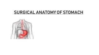
SURGICAL ANATOMY OF STOMACH
CONTENTS
• INTRODUCTION and FUNCTIONS
• LOCATION
• PARTS
• MICROSCOPIC STRUCTURE
• INTERNAL APPEARANCE
• BLOOD SUPPLY
• VENOUS DRAINAGE
• LYMPHATIC DRAINAGE
• INNERVATION
• LIGAMENTS OR PERITONEAL
REFLECTIONS
INTRODUCTION
• Stomach is a muscular bag forming the widest and most distensible part of
the digestive tube.
• Begins as a dilatation in tubular embryonic foregut during fifth week of
gestation.
• By seventh week,it descends,rotates and further dilates with a
disproportionate elongation of greater curvature into its normal anatomic
shape and position.
• MAIN FUNCTIONS OF THE STOMACH ARE:
• Forms a reservoir for the food.
• Mixes food with gastric secretions to form a semi fluid substance
called CHYME.
• Controls the the rate of delivery of chyme into the small intestine to
allow proper digestion and absorption in the small intestine.
• Hcl secreted by the gastric glands breaks down the food & destroys
bacteria present in the food and drink.
• Castle’s intrinsic factor present in the gastric juice helps in the
absorption of vitamin B12 in the small intestine.
• Lies obliquely in upper and left part of the abdomen occupying the
epigastric,umbilical and left hypochondriac regions.
• Most of it lies under the cover of the left coastal margin and the ribs.
• Shape depends upon the degree of distension and that of the surrounding
viscera eg colon.
• When empty,it is J shaped(vertical),when partially distended,it is pyriform in
shape.
• In short and obese persons ,it is more horizontal as a steer horn shape.
LOCATION,SHAPE,SIZE AND CAPACITY
• Low and elongated in Tall and weak persons.
• The shape of the stomach is studied by radiographic examination using barium
meal .Generally 4 types of shape of the stomach are seen in barium meal X ray
reverse L shaped, J shaped,semilunar shaped and Steer horn shaped
• It is 25cms long,capacity of 30ml at birth,1 litre at puberty, 1 ½ to 2 litres or more
in adults.
PARTS
• CARDIAC PART-CONTAINS FUNDUS AND BODY
• FUNDUS-Upper convex dome shaped part and projects above and to the left of the
esophageal opening(cardiac orifice) and lies in contact with the left dome of
diaphragm.It is floppy and distensible.
• Superiorly ,the fundus usually reaches at the level of the left 5th intercostal space
just below the nipple, hence gastric pain sometimes imitate the pain of angina
pectoris.
• The cardiac notch lies between the fundus and esophagus.
• ANGLE OF HIS –Formed by the fundus with the left margin of the esophagus.
• BODY-Lies between fundus and pyloric antrum.It is the largest portion,also
referred to as the corpus.
• TRAUBE SPACE- it is a topographic area overlying the fundus of the stomach
which is tympanic on percussion. It is bounded superiorly by the lower border
of the left lung ,inferiorly by the Left costal margin, on the left side by the
lateral end of the spleen and on the right side by the lower border of the left
lobe of the liver.
• PYLORIC PART-Divided into pyloric antrum and pyloric canal.
• PYLORIC ANTRUM-Separated from the pyloric canal by an inconstant
sulcus,sulcus intermedius present on greater curvature.it is about 3 inches
long and leads into the pyloric canal.
• PYLORIC CANAL-Narrow and tubular part measuring one inch in length.It lies
on the head and neck of the pancreas. It terminates at pyloric orifice.
MICROSCOPIC STRUCTURE
• The wall of the stomach contains of 4 coats, from outside inwards, these are
serous, muscular, sub mucous and mucous layer.
• The serous coat is formed by the peritoneum.
• Muscular coat consists of 3 layers of unstriped muscles ,outer longitudinal
,middle circular and inner oblique.
• Close to the pyloric end ,the longitudinal muscle coat separates into superficial
and deep fibres. The deep fibres turn inwards at the pylorus and join with the
circular muscle coat to help form the pyloric sphincter.
• The major sphincteric component of the pyloric sphincter is derived from the
circular muscle coat and its minor dilator component is derived from the
longitudinal muscle coat.
• The sub mucous coat consists of loose areolar tissue.
• Mucous membrane is thick soft and velvety. It presents a number of
temporary folds(rugae) which disappear when the stomach is distended.
• The mucous membrane of the stomach is lined by simple columnar epithelium
which forms simple tubular glands.
• Glands in the cardiac region secrete mucus.
• Glands in the fundus and body contains neck cells which secrete mucus.
• Parietal or oxyntic cells which secrete hydrochloric acid and castle’s
intrinsic factor.
• Chief cells secrete pepsinogen
• Glands in the pyloric region secrete mucus.
• Ecl cells secrete histamine ,key factor in driving gastric acid secretion.
• D cells secrete somatostatin,which have negative regulatory role.
• G cells ,present in the gastric antrum produces gastrin.
INTERNAL APPEARANCES
• During endoscopic examination the stomach is typically partially distended by air.
• The cardiac orifice and the lowest portion of the abdominal esophagus viewed from
above are typically closed at rest by tonic contraction of the lower esophageal sphincter.
• Viewed retrogradely from within the distended stomach, the cardiac orifice lies medial
to the fundus of the stomach.
• In a partially distended stomach,the mucosa of the body and to the lesser extent the
fundus is thrown into the longitudinal folds called rugae.
• They are longitudinal along the lesser curvature and irregular in the remaining part,the
rugae are flattened and when the stomach is distended.
• GASTRIC CANAL OR MAGENTRASSE: Longitudinal furrow that forms
temporarily during swallowing between the longitudinal folds of the mucosa
along the lesser curvature.
• The gastric canal forms due to firm attachment of the gastric mucosa to the
underlying muscular layer which does not have an oblique layer at this site.
• This canal allows a rapid passage of the swallowed liquids along the lesser
curvature to the lower part before it spreads to the other part of the stomach.
• Thus the lesser curvature is subject to maximum insult of the swallowed spicy
food and the irritable liquids such as alcohol which makes it vulnerable for the
gastric ulceration.
• The area gastricae within the antrum are small undulations of the mucosal
surface that give a subtle cobblestone appearance on double contrast barium
meal examination.
• The few folds present in the antrum when the stomach is relaxed disappear
with distension,the antrum adjacent to the pyloric canal ,the prepyloric antrum
has a smooth mucosal surface
EXTERNAL FEATURES
• It has 2 orifices,2 curvatures and 2 surfaces.
• CARDIAC ORIFICE- the esophagus opens into the stomach at the Cardiac
orifice,lies behind the left 7th costal cartilage at the level of T11.It marks the level
at which curvatures begin.
• PYLORIC ORIFICE-Opens into proximal end of the duodenum,1.5cms to right of
the median plane at the lower border of L1.has an anatomical pyloric sphincter
formed by the thickening of the circular coat of the muscle assisted by a deep set
of longitudinal muscle coat.
• LESSER CURVATURE-Forms the medial border of the stomach,descends from
the medial side of esophagus in front of the decussating fibres of the right crus
of the diaphragm,curves downwards and to the right and ends at the pylorus.It
contains right and left gastric vessels, and provides attachment to the lesser
omentum.
• Most dependant part of curvature is marked by INCISURA ANGULARIS.
• GREATER CURVATURE-It is two to three times longer than the lesser curvature,starts
from the cardiac notch formed between the lateral border of the abdominal
esophagus and the fundus of the stomach,and arches upwards,posterolaterally and
to the left.Its highest convexity,the apex of the fundus is approximately at the level of
6th rib anteriorly.from there,it sweeps anteriorly and inferiorly, to end at the pylorus in
transpyloric plane at lower border of first lumbar vertebra, and forms the left border
of the stomach.It provides attachment to the greater omentum,gastrosplenic
ligament,gastrophrenic ligament.
• ANTERIOR OR ANTEROSUPERIOR SURFACE-Entirely covered by peritoneum ,faces
forwards and upwards.
• POSTERIOR OR POSTEROINFERIOR SURFACE- Faces backwards and downwards.
RELATIONS
• RELATIONS OF THE ANTERIOR SURFACE:
• On the right side this surface is related to the gastric impression of the left
lobe of the liver and near the pylorus to the quadrate lobe of the liver.
• The left half of this surface is related to the diaphragm and rib cage.
• The lower half is related to the anterior abdominal wall.
• RELATIONS OF THE POSTERIOR SURFACE:
• This surface is related to a number of structures on the posterior abdominal
wall which collectively form the ‘stomach bed’.
• These structures are:
• Left kidney
• Left suprarenal gland
• Pancreas
• Transverse mesocolon
• Left colic flexure
• Splenic artery
• Spleen
BLOOD SUPPLY
• CELIAC TRUNK –MOST OF THE BLOOD SUPPLY.
• RIGHT GASTRIC ARTERY-BRANCH OF HEPATIC ARTERY PROPER.
• LEFT GASTRIC ARTERY-ARISES DIRECTLY FROM CELIAC TRUNK.
• IN 15-20% PEOPLE,ABERRANT LEFT HEPATIC ARTERY ORIGINATES FROM IT.
• LIGATION OCCASIONALLY RESULTS IN ACUTE LEFT SIDED ISCHEMIA.
• RIGHT GASTRIC ARTERY AND LEFT GASTRIC ARTERY LIES ALONG LESSER
CURVATURE
• RIGHT GASTROEPIPLOIC ARTERY-BRANCH OF GASTRODUODENAL ARTERY,
ends by anastomosing with the left gastroepiploic artery although this
anastomosis is variably developed and maybe absent.
• LEFT GASTRO EPIPLOIC ARTERY- Largest branch of splenic artery. It gives off
gastric branches to the fundus of the stomach through the gastrosplenic
ligament and to the body of the stomach through the gastrocolic omentum.
• A particularly large epiploic branch commonly originates close to the origin of
the left gastroepiploic artery, descends in the lateral portion of the greater
omentum and provides a large arterial supply to the lateral half of the
omentum.
• POSTERIOR GASTRIC ARTERY- posterior gastric artery supplying the posterior
wall of the upper part of the gastric body is commonly present ,it usually arises
from this splenic artery,usually from its midsection.
• SHORT GASTRIC ARTERY-Branch of splenic artery-supplies fundus of the
stomach on its greater curvature.
• THE PYLORIC ARTERIES are branches of the right gastric and right
gastroepiploic artery.
• They pierce the duodenal wall around its entire circumference just distal to the
pyloric sphincter and reach the submucosal layer, here they divide into 2 or 3
rami that run towards the stomach in the submucosa of the pyloric canal and
terminate in the mucosa of the pyloric antrum.
• They anastomose with the sub mucosal arteries in the duodenum and gastric
antrum ,close to their origin and termination respectively.
• Extensive anastamotic connection ensures that stomach will survive even if 3
out of 4 arteries are ligated.
• In patients requiring replacement of esophagus for severe congenital
anomalies or after a resection for cancer, a gastric tube supplied by left
gastroepiploic artery can be fashioned from the greater curvature of the
stomach and anastamosed to the cervical esophagus.
• The right gastroepiploic artery has been used for coronary artery
revascularization in some centers and this may pose a particular hazard if the
patient is subsequently required surgery for gastric cancer.
• Abnormalities of the intramural vascularity of the stomach are a rare cause of
the acute upper GI haemorrhage .The so called Dieulafoy lesion occurs most
often near the lesser curvature within a few centimeters of gastroesophageal
junction and consists of an abnormally large artery that penetrates the
muscular coat of the stomach runs a tortuous coarse in the sub mucosa and
protrudes through a small mucosal defect where it is vulnerable to rupture.
VENOUS DRAINAGE
• LEFT GASTRIC VEIN - Drains the upper body and fundus of the stomach,
drains into PORTAL VEIN.
• RIGHT GASTRIC VEIN- lies anterior to stomach ,drains into PORTAL VEIN.
• RIGHT GASTROEPIPLOIC VEIN - Drains greater omentum ,distal body and
antrum of stomach, drains into SUPERIOR MESENTERIC VEIN.
• LEFT GASTROEPIPLOIC VEIN Drains both anterior and posterior surface of
the body of the stomach and the adjacent greater omentum via multiple
tributaries ,drains INTO SPLENIC VEIN.
• SHORT GASTRIC VEINS drains gastric fundus in the upper part of the greater
curvature into SPLENIC VEIN.
• POSTER GASTRIC VEINS -one or more distinct posterior gastric veins may be
present draining in the middle of the posterior surface of the stomach in to the
SPLENIC VEIN they become particularly prominent in portal hypertension.
• PREPYLORIC VEIN OF MAYO DISTINGUISHES PYLORIC CANAL FROM THE FIRST
PART OF DUODENUM.
LYMPHATIC DRAINAGE
• The knowledge of lymphatic drainage of the stomach is clinically very important as the
gastric cancer spreads through lymph vessels.
• The lymphatic drainage of the stomach parallels the vasculature and drains into four
zones of lymph nodes.
• 1.SUPERIOR GASTRIC GROUP-Drains lymph from the upper lesser curvature into the
left gastric and paracardial nodes.
• 2.SUPRAPYLORIC GROUP -Drains the antral segment on the lesser curvature of the
stomach into right suprapancreatic nodes.
• 3.PANCREATICOSPLENIC GROUP-Drain lymph high on the greater curvature into the
left gastroepiploic and splenic nodes.
• 4.INFERIOR GASTRIC AND SUBPYLORIC GROUP OF NODES -Drains lymph along the
right gastroepiploic vascular pedicle.
• All these lymph nodes drain into the celiac group and into the thoracic duct.
• Gastric ca. Spreads through the lymphatic vessels to left supraclavicular lymph
nodes.
• Enlarged and palpable left supraclavicular node is called virchows node and
this sign is called troisier’s sign
• Cancer cells reach the supraclavicular lymph nodes through the thoracic duct.
INNERVATION
• Extrinsic innervationof stomach is parasympathetic via vagus and sympatheticvia celiac
plexus.
• Sympathetic fibres are derived from T6 to T10 spinal segments via greater splanchnic
nerves,celiac and hepatic plexuses.
• Sympathetic supply to stomach is vasomotor,motorto pyloric sphincter,inhibitoryto
remaininggastric musculature,serves as chief pathway for pain sensationfrom the stomach.
• Vagus originates in the vagal nucleus in the floor of the fourth ventricle and traverses the neck
in carotid sheath to enter the mediastinum where it divides into severalbranches around the
esophagus.
• These branches coalesce above the esophagealhiatus to form the left and right vagus nerves.
• At the GE junction,left vagus is anterior and right vagus is posterior.
• Anterior vagus gives a
• a) hepatic branch to the liver,
• b)celiac branch which follow the left gastric artery to the celiac plexus
• c) and continues along the lesser curvature as ANTERIOR NERVE OF
LATARJET.
• Posterior or right vagus gives off the first branch-CRIMINAL NERVE OF
GRASSI-supplies gastric fundus causes recurrence of the ulcers when left
undivided.
• Also gives off a branch to celiac plexus and continues posteriorly along the
lesser curvature.
VAGOTOMY
• In peptic ulcer disease,there is increased acid output.
• As acid production is under the control of vagus,vagotomy is done,healing
occur.
• TRUNCAL VAGOTOMY-anterior and post vagal trunks are cut at the lower
end of the esophagus above the origin of hepatic and celiac branches.
• SELECTIVE VAGOTOMY-both the vagus nerves are cut below the origin of
hepatic and celiac branches.
• HIGHLY SELECTIVE VAGOTOMY -is performed by dividing crows feet to the
proximal stomach while preserving the innervation of the antral and pyloric
parts of the stomach.
LIGAMENTS OR PERITONEAL RELATIONS
• Stomach is lined by peritoneum on both its surfaces.
• At the lesser curvature the layers of peritoneum lining the anterior and posterior
surfaces meet and become continuous with the lesser omentum.
• Along the greater curvature ,2 layers meet to form greater omentum.
• HEPATOGASTRIC LIGAMENT-It is the proximal part of lesser omentum,extends
from porta hepatis to the lesser curvature of the stomach.
• Contains left gastric artery and vein,hepatic divisions of anterior vagal
trunk,nerve of latarjet,lymph nodes and vessels.
• HEPATODUODENAL LIGAMENT-Is the distal part of the lesser
omentum,extending from liver to first 2.5cms of the duodenum.
• The free edge envelops the hepatic triad.
• GASTROSPLENIC LIGAMENT-At the hilum of the spleen,visceral peritoneum
joins the right layer of the greater omentum,forms the gastrosplenic ligaments
and splenorenal ligaments,these two form the splenic pedicle.
• Splenic capsule is formed by the visceral peritoneum and is as friable as spleen
itself and excessive traction during the mobilisation of greater curvature might
result in tearing of the capsule,
• GASTROPHRENIC LIGAMENT- It is continuous with the hepatogastric ligament
to the left, has a avascular region through which the surgeons finger can easily
pass through which a penrose drain can be placed easily around the cardia to
pull down the esophagus.
• Upper part is avascular and lower part contains short gastric arteries,veins and
lymph nodes.

More Related Content

What's hot

SURGICAL ANATOMY
SURGICAL ANATOMYSURGICAL ANATOMY
SURGICAL ANATOMY
Meenal Sethi
 
Peritoneum and peritoneal cavity
Peritoneum and peritoneal cavityPeritoneum and peritoneal cavity
Peritoneum and peritoneal cavity
Dr. Noura El Tahawy
 
Tumors of the Appendix.
Tumors of the Appendix.Tumors of the Appendix.
Tumors of the Appendix.
KETAN VAGHOLKAR
 
Radiological anatomy of abdominal spaces ...pathway of tumor and infection s...
Radiological anatomy of abdominal spaces ...pathway of tumor  and infection s...Radiological anatomy of abdominal spaces ...pathway of tumor  and infection s...
Radiological anatomy of abdominal spaces ...pathway of tumor and infection s...
Ahmed Bahnassy
 
Liver anatomy
Liver anatomyLiver anatomy
Liver anatomy
Dr. Rahul Jain
 
Anatomy of oesophagus
Anatomy of oesophagusAnatomy of oesophagus
Anatomy of oesophagus
Mohammed Nishad N
 
ANATOMY OF ESOPHAGUS-Dr.Neeraj Kumar Banoria
ANATOMY OF ESOPHAGUS-Dr.Neeraj Kumar BanoriaANATOMY OF ESOPHAGUS-Dr.Neeraj Kumar Banoria
ANATOMY OF ESOPHAGUS-Dr.Neeraj Kumar Banoriadrnkb2000
 
Liver and billiary anatomy
Liver and billiary anatomyLiver and billiary anatomy
Liver and billiary anatomy
maroofkhanjabasaria
 
Pancreas Congenital Anomalies (agenesis, pancreas divisum, annular pancreas, ...
Pancreas Congenital Anomalies (agenesis, pancreas divisum, annular pancreas, ...Pancreas Congenital Anomalies (agenesis, pancreas divisum, annular pancreas, ...
Pancreas Congenital Anomalies (agenesis, pancreas divisum, annular pancreas, ...
Ainul Basyirah
 
surgical anatomy Inguinal canal anatomy dr.vishnu
surgical anatomy Inguinal canal anatomy dr.vishnusurgical anatomy Inguinal canal anatomy dr.vishnu
surgical anatomy Inguinal canal anatomy dr.vishnu
vishnu mohan
 
Resectional Anatomy of Pancreas
Resectional Anatomy of PancreasResectional Anatomy of Pancreas
Resectional Anatomy of Pancreas
Happykumar Kagathara
 
Omentum.pptx
Omentum.pptxOmentum.pptx
Omentum.pptx
Pradeep Pande
 
Anatomy of small and large intestine
Anatomy of  small and large intestineAnatomy of  small and large intestine
Anatomy of small and large intestine
Dr. Mohammad Mahmoud
 
Peritoneum , Intraperitoneal Spaces
Peritoneum , Intraperitoneal SpacesPeritoneum , Intraperitoneal Spaces
Peritoneum , Intraperitoneal Spaces
Jisa Rafael Rodriguez
 
Surgical anatomy of breasts
Surgical anatomy of breastsSurgical anatomy of breasts
Surgical anatomy of breasts
Ahmed Almumtin
 
Anatomy of Retroperitoneum.
Anatomy of Retroperitoneum.Anatomy of Retroperitoneum.
Anatomy of Retroperitoneum.
Pavan Kumar
 
Introduction abdomen-dr.gosai
Introduction abdomen-dr.gosaiIntroduction abdomen-dr.gosai
Introduction abdomen-dr.gosai
Dr.B.B. Gosai
 
Venous drainage of lower limb ppt
Venous drainage of lower limb pptVenous drainage of lower limb ppt
Venous drainage of lower limb ppt
Aditi Ujjawal
 
Rectum & Anal canal By Prof.Dr.N.Mugunthan
Rectum & Anal canal  By Prof.Dr.N.MugunthanRectum & Anal canal  By Prof.Dr.N.Mugunthan
Rectum & Anal canal By Prof.Dr.N.Mugunthan
MUGUNTHAN Dr.Mugunthan
 
Anatomy of anal canal
Anatomy of anal canalAnatomy of anal canal
Anatomy of anal canal
Ayesha Jalil
 

What's hot (20)

SURGICAL ANATOMY
SURGICAL ANATOMYSURGICAL ANATOMY
SURGICAL ANATOMY
 
Peritoneum and peritoneal cavity
Peritoneum and peritoneal cavityPeritoneum and peritoneal cavity
Peritoneum and peritoneal cavity
 
Tumors of the Appendix.
Tumors of the Appendix.Tumors of the Appendix.
Tumors of the Appendix.
 
Radiological anatomy of abdominal spaces ...pathway of tumor and infection s...
Radiological anatomy of abdominal spaces ...pathway of tumor  and infection s...Radiological anatomy of abdominal spaces ...pathway of tumor  and infection s...
Radiological anatomy of abdominal spaces ...pathway of tumor and infection s...
 
Liver anatomy
Liver anatomyLiver anatomy
Liver anatomy
 
Anatomy of oesophagus
Anatomy of oesophagusAnatomy of oesophagus
Anatomy of oesophagus
 
ANATOMY OF ESOPHAGUS-Dr.Neeraj Kumar Banoria
ANATOMY OF ESOPHAGUS-Dr.Neeraj Kumar BanoriaANATOMY OF ESOPHAGUS-Dr.Neeraj Kumar Banoria
ANATOMY OF ESOPHAGUS-Dr.Neeraj Kumar Banoria
 
Liver and billiary anatomy
Liver and billiary anatomyLiver and billiary anatomy
Liver and billiary anatomy
 
Pancreas Congenital Anomalies (agenesis, pancreas divisum, annular pancreas, ...
Pancreas Congenital Anomalies (agenesis, pancreas divisum, annular pancreas, ...Pancreas Congenital Anomalies (agenesis, pancreas divisum, annular pancreas, ...
Pancreas Congenital Anomalies (agenesis, pancreas divisum, annular pancreas, ...
 
surgical anatomy Inguinal canal anatomy dr.vishnu
surgical anatomy Inguinal canal anatomy dr.vishnusurgical anatomy Inguinal canal anatomy dr.vishnu
surgical anatomy Inguinal canal anatomy dr.vishnu
 
Resectional Anatomy of Pancreas
Resectional Anatomy of PancreasResectional Anatomy of Pancreas
Resectional Anatomy of Pancreas
 
Omentum.pptx
Omentum.pptxOmentum.pptx
Omentum.pptx
 
Anatomy of small and large intestine
Anatomy of  small and large intestineAnatomy of  small and large intestine
Anatomy of small and large intestine
 
Peritoneum , Intraperitoneal Spaces
Peritoneum , Intraperitoneal SpacesPeritoneum , Intraperitoneal Spaces
Peritoneum , Intraperitoneal Spaces
 
Surgical anatomy of breasts
Surgical anatomy of breastsSurgical anatomy of breasts
Surgical anatomy of breasts
 
Anatomy of Retroperitoneum.
Anatomy of Retroperitoneum.Anatomy of Retroperitoneum.
Anatomy of Retroperitoneum.
 
Introduction abdomen-dr.gosai
Introduction abdomen-dr.gosaiIntroduction abdomen-dr.gosai
Introduction abdomen-dr.gosai
 
Venous drainage of lower limb ppt
Venous drainage of lower limb pptVenous drainage of lower limb ppt
Venous drainage of lower limb ppt
 
Rectum & Anal canal By Prof.Dr.N.Mugunthan
Rectum & Anal canal  By Prof.Dr.N.MugunthanRectum & Anal canal  By Prof.Dr.N.Mugunthan
Rectum & Anal canal By Prof.Dr.N.Mugunthan
 
Anatomy of anal canal
Anatomy of anal canalAnatomy of anal canal
Anatomy of anal canal
 

Similar to SURGICAL ANATOMY OF STOMACH [Autosaved].pptx

Development of-WPS Office.pptxHVDSUVHDSUIVDSVDF
Development of-WPS Office.pptxHVDSUVHDSUIVDSVDFDevelopment of-WPS Office.pptxHVDSUVHDSUIVDSVDF
Development of-WPS Office.pptxHVDSUVHDSUIVDSVDF
sgaur356
 
Stomach.pptx
Stomach.pptxStomach.pptx
Stomach.pptx
OtemaJames
 
Digestive system part 2
Digestive system part 2Digestive system part 2
Digestive system part 2
Zainab&Sons
 
Digestive system by dr tayyaba......pptx
Digestive system by dr tayyaba......pptxDigestive system by dr tayyaba......pptx
Digestive system by dr tayyaba......pptx
BIANOOR123
 
Stomach anatomy
Stomach anatomy Stomach anatomy
Stomach anatomy
Mawara Ali
 
Stomach.pdf
Stomach.pdfStomach.pdf
Stomach.pdf
ssuserbf4af22
 
Anatomy of the stomach
Anatomy of the stomach  Anatomy of the stomach
Anatomy of the stomach
Dr. Mohammad Mahmoud
 
9.spleen
9.spleen9.spleen
Lect 3 abddomen
Lect 3 abddomenLect 3 abddomen
Lect 3 abddomen
Ali Qatrawi
 
Anatomy of abdomen to medicine and health student
Anatomy of abdomen to medicine and health studentAnatomy of abdomen to medicine and health student
Anatomy of abdomen to medicine and health student
osamaessa10
 
Peritoneum & Abdominal cavity .pptx
Peritoneum & Abdominal cavity .pptxPeritoneum & Abdominal cavity .pptx
Peritoneum & Abdominal cavity .pptx
ABHIJIT BHOYAR
 
Development of-WPS Office.pptxtvvtvtvvgc
Development of-WPS Office.pptxtvvtvtvvgcDevelopment of-WPS Office.pptxtvvtvtvvgc
Development of-WPS Office.pptxtvvtvtvvgc
sgaur356
 
Anatomy of small and large Bowel PPT.pptx
Anatomy of small and large Bowel PPT.pptxAnatomy of small and large Bowel PPT.pptx
Anatomy of small and large Bowel PPT.pptx
kamrankhan432971
 
Digestive system HAP II.pptx
Digestive system HAP II.pptxDigestive system HAP II.pptx
Digestive system HAP II.pptx
sachinshitole12
 
Development of stomach
Development of stomachDevelopment of stomach
Development of stomach
Farhan Ali
 
Abdomen Presentation anat.ppt
Abdomen Presentation anat.pptAbdomen Presentation anat.ppt
Abdomen Presentation anat.ppt
WinstonM3
 
1-stomach-100331081026-phpapp01 (1).pdf
1-stomach-100331081026-phpapp01 (1).pdf1-stomach-100331081026-phpapp01 (1).pdf
1-stomach-100331081026-phpapp01 (1).pdf
Rithika866837
 
digestive 3.pptxpfanatomy small intestine and
digestive 3.pptxpfanatomy small intestine anddigestive 3.pptxpfanatomy small intestine and
digestive 3.pptxpfanatomy small intestine and
phatimamohamett054
 
Lecture 1 elementary system
Lecture 1 elementary systemLecture 1 elementary system
Lecture 1 elementary system
sazzadulbarihridoy
 

Similar to SURGICAL ANATOMY OF STOMACH [Autosaved].pptx (20)

Development of-WPS Office.pptxHVDSUVHDSUIVDSVDF
Development of-WPS Office.pptxHVDSUVHDSUIVDSVDFDevelopment of-WPS Office.pptxHVDSUVHDSUIVDSVDF
Development of-WPS Office.pptxHVDSUVHDSUIVDSVDF
 
Stomach.pptx
Stomach.pptxStomach.pptx
Stomach.pptx
 
Digestive system part 2
Digestive system part 2Digestive system part 2
Digestive system part 2
 
Digestive system by dr tayyaba......pptx
Digestive system by dr tayyaba......pptxDigestive system by dr tayyaba......pptx
Digestive system by dr tayyaba......pptx
 
Stomach anatomy
Stomach anatomy Stomach anatomy
Stomach anatomy
 
Stomach.pdf
Stomach.pdfStomach.pdf
Stomach.pdf
 
Anatomy of the stomach
Anatomy of the stomach  Anatomy of the stomach
Anatomy of the stomach
 
9.spleen
9.spleen9.spleen
9.spleen
 
Lect 3 abddomen
Lect 3 abddomenLect 3 abddomen
Lect 3 abddomen
 
Anatomy of abdomen to medicine and health student
Anatomy of abdomen to medicine and health studentAnatomy of abdomen to medicine and health student
Anatomy of abdomen to medicine and health student
 
Peritoneum & Abdominal cavity .pptx
Peritoneum & Abdominal cavity .pptxPeritoneum & Abdominal cavity .pptx
Peritoneum & Abdominal cavity .pptx
 
Development of-WPS Office.pptxtvvtvtvvgc
Development of-WPS Office.pptxtvvtvtvvgcDevelopment of-WPS Office.pptxtvvtvtvvgc
Development of-WPS Office.pptxtvvtvtvvgc
 
Anatomy of small and large Bowel PPT.pptx
Anatomy of small and large Bowel PPT.pptxAnatomy of small and large Bowel PPT.pptx
Anatomy of small and large Bowel PPT.pptx
 
Duodenum
DuodenumDuodenum
Duodenum
 
Digestive system HAP II.pptx
Digestive system HAP II.pptxDigestive system HAP II.pptx
Digestive system HAP II.pptx
 
Development of stomach
Development of stomachDevelopment of stomach
Development of stomach
 
Abdomen Presentation anat.ppt
Abdomen Presentation anat.pptAbdomen Presentation anat.ppt
Abdomen Presentation anat.ppt
 
1-stomach-100331081026-phpapp01 (1).pdf
1-stomach-100331081026-phpapp01 (1).pdf1-stomach-100331081026-phpapp01 (1).pdf
1-stomach-100331081026-phpapp01 (1).pdf
 
digestive 3.pptxpfanatomy small intestine and
digestive 3.pptxpfanatomy small intestine anddigestive 3.pptxpfanatomy small intestine and
digestive 3.pptxpfanatomy small intestine and
 
Lecture 1 elementary system
Lecture 1 elementary systemLecture 1 elementary system
Lecture 1 elementary system
 

Recently uploaded

The Electrocardiogram - Physiologic Principles
The Electrocardiogram - Physiologic PrinciplesThe Electrocardiogram - Physiologic Principles
The Electrocardiogram - Physiologic Principles
MedicoseAcademics
 
Basavarajeeyam - Ayurvedic heritage book of Andhra pradesh
Basavarajeeyam - Ayurvedic heritage book of Andhra pradeshBasavarajeeyam - Ayurvedic heritage book of Andhra pradesh
Basavarajeeyam - Ayurvedic heritage book of Andhra pradesh
Dr. Madduru Muni Haritha
 
Best Ayurvedic medicine for Gas and Indigestion
Best Ayurvedic medicine for Gas and IndigestionBest Ayurvedic medicine for Gas and Indigestion
Best Ayurvedic medicine for Gas and Indigestion
Swastik Ayurveda
 
Colonic and anorectal physiology with surgical implications
Colonic and anorectal physiology with surgical implicationsColonic and anorectal physiology with surgical implications
Colonic and anorectal physiology with surgical implications
Dr Maria Tamanna
 
micro teaching on communication m.sc nursing.pdf
micro teaching on communication m.sc nursing.pdfmicro teaching on communication m.sc nursing.pdf
micro teaching on communication m.sc nursing.pdf
Anurag Sharma
 
Local Advanced Lung Cancer: Artificial Intelligence, Synergetics, Complex Sys...
Local Advanced Lung Cancer: Artificial Intelligence, Synergetics, Complex Sys...Local Advanced Lung Cancer: Artificial Intelligence, Synergetics, Complex Sys...
Local Advanced Lung Cancer: Artificial Intelligence, Synergetics, Complex Sys...
Oleg Kshivets
 
Tom Selleck Health: A Comprehensive Look at the Iconic Actor’s Wellness Journey
Tom Selleck Health: A Comprehensive Look at the Iconic Actor’s Wellness JourneyTom Selleck Health: A Comprehensive Look at the Iconic Actor’s Wellness Journey
Tom Selleck Health: A Comprehensive Look at the Iconic Actor’s Wellness Journey
greendigital
 
Chapter 11 Nutrition and Chronic Diseases.pptx
Chapter 11 Nutrition and Chronic Diseases.pptxChapter 11 Nutrition and Chronic Diseases.pptx
Chapter 11 Nutrition and Chronic Diseases.pptx
Earlene McNair
 
Hemodialysis: Chapter 4, Dialysate Circuit - Dr.Gawad
Hemodialysis: Chapter 4, Dialysate Circuit - Dr.GawadHemodialysis: Chapter 4, Dialysate Circuit - Dr.Gawad
Hemodialysis: Chapter 4, Dialysate Circuit - Dr.Gawad
NephroTube - Dr.Gawad
 
Physiology of Special Chemical Sensation of Taste
Physiology of Special Chemical Sensation of TastePhysiology of Special Chemical Sensation of Taste
Physiology of Special Chemical Sensation of Taste
MedicoseAcademics
 
Role of Mukta Pishti in the Management of Hyperthyroidism
Role of Mukta Pishti in the Management of HyperthyroidismRole of Mukta Pishti in the Management of Hyperthyroidism
Role of Mukta Pishti in the Management of Hyperthyroidism
Dr. Jyothirmai Paindla
 
KDIGO 2024 guidelines for diabetologists
KDIGO 2024 guidelines for diabetologistsKDIGO 2024 guidelines for diabetologists
KDIGO 2024 guidelines for diabetologists
د.محمود نجيب
 
Journal Article Review on Rasamanikya
Journal Article Review on RasamanikyaJournal Article Review on Rasamanikya
Journal Article Review on Rasamanikya
Dr. Jyothirmai Paindla
 
Cervical & Brachial Plexus By Dr. RIG.pptx
Cervical & Brachial Plexus By Dr. RIG.pptxCervical & Brachial Plexus By Dr. RIG.pptx
Cervical & Brachial Plexus By Dr. RIG.pptx
Dr. Rabia Inam Gandapore
 
Triangles of Neck and Clinical Correlation by Dr. RIG.pptx
Triangles of Neck and Clinical Correlation by Dr. RIG.pptxTriangles of Neck and Clinical Correlation by Dr. RIG.pptx
Triangles of Neck and Clinical Correlation by Dr. RIG.pptx
Dr. Rabia Inam Gandapore
 
Superficial & Deep Fascia of the NECK.pptx
Superficial & Deep Fascia of the NECK.pptxSuperficial & Deep Fascia of the NECK.pptx
Superficial & Deep Fascia of the NECK.pptx
Dr. Rabia Inam Gandapore
 
Integrating Ayurveda into Parkinson’s Management: A Holistic Approach
Integrating Ayurveda into Parkinson’s Management: A Holistic ApproachIntegrating Ayurveda into Parkinson’s Management: A Holistic Approach
Integrating Ayurveda into Parkinson’s Management: A Holistic Approach
Ayurveda ForAll
 
A Classical Text Review on Basavarajeeyam
A Classical Text Review on BasavarajeeyamA Classical Text Review on Basavarajeeyam
A Classical Text Review on Basavarajeeyam
Dr. Jyothirmai Paindla
 
Knee anatomy and clinical tests 2024.pdf
Knee anatomy and clinical tests 2024.pdfKnee anatomy and clinical tests 2024.pdf
Knee anatomy and clinical tests 2024.pdf
vimalpl1234
 
BRACHYTHERAPY OVERVIEW AND APPLICATORS
BRACHYTHERAPY OVERVIEW  AND  APPLICATORSBRACHYTHERAPY OVERVIEW  AND  APPLICATORS
BRACHYTHERAPY OVERVIEW AND APPLICATORS
Krishan Murari
 

Recently uploaded (20)

The Electrocardiogram - Physiologic Principles
The Electrocardiogram - Physiologic PrinciplesThe Electrocardiogram - Physiologic Principles
The Electrocardiogram - Physiologic Principles
 
Basavarajeeyam - Ayurvedic heritage book of Andhra pradesh
Basavarajeeyam - Ayurvedic heritage book of Andhra pradeshBasavarajeeyam - Ayurvedic heritage book of Andhra pradesh
Basavarajeeyam - Ayurvedic heritage book of Andhra pradesh
 
Best Ayurvedic medicine for Gas and Indigestion
Best Ayurvedic medicine for Gas and IndigestionBest Ayurvedic medicine for Gas and Indigestion
Best Ayurvedic medicine for Gas and Indigestion
 
Colonic and anorectal physiology with surgical implications
Colonic and anorectal physiology with surgical implicationsColonic and anorectal physiology with surgical implications
Colonic and anorectal physiology with surgical implications
 
micro teaching on communication m.sc nursing.pdf
micro teaching on communication m.sc nursing.pdfmicro teaching on communication m.sc nursing.pdf
micro teaching on communication m.sc nursing.pdf
 
Local Advanced Lung Cancer: Artificial Intelligence, Synergetics, Complex Sys...
Local Advanced Lung Cancer: Artificial Intelligence, Synergetics, Complex Sys...Local Advanced Lung Cancer: Artificial Intelligence, Synergetics, Complex Sys...
Local Advanced Lung Cancer: Artificial Intelligence, Synergetics, Complex Sys...
 
Tom Selleck Health: A Comprehensive Look at the Iconic Actor’s Wellness Journey
Tom Selleck Health: A Comprehensive Look at the Iconic Actor’s Wellness JourneyTom Selleck Health: A Comprehensive Look at the Iconic Actor’s Wellness Journey
Tom Selleck Health: A Comprehensive Look at the Iconic Actor’s Wellness Journey
 
Chapter 11 Nutrition and Chronic Diseases.pptx
Chapter 11 Nutrition and Chronic Diseases.pptxChapter 11 Nutrition and Chronic Diseases.pptx
Chapter 11 Nutrition and Chronic Diseases.pptx
 
Hemodialysis: Chapter 4, Dialysate Circuit - Dr.Gawad
Hemodialysis: Chapter 4, Dialysate Circuit - Dr.GawadHemodialysis: Chapter 4, Dialysate Circuit - Dr.Gawad
Hemodialysis: Chapter 4, Dialysate Circuit - Dr.Gawad
 
Physiology of Special Chemical Sensation of Taste
Physiology of Special Chemical Sensation of TastePhysiology of Special Chemical Sensation of Taste
Physiology of Special Chemical Sensation of Taste
 
Role of Mukta Pishti in the Management of Hyperthyroidism
Role of Mukta Pishti in the Management of HyperthyroidismRole of Mukta Pishti in the Management of Hyperthyroidism
Role of Mukta Pishti in the Management of Hyperthyroidism
 
KDIGO 2024 guidelines for diabetologists
KDIGO 2024 guidelines for diabetologistsKDIGO 2024 guidelines for diabetologists
KDIGO 2024 guidelines for diabetologists
 
Journal Article Review on Rasamanikya
Journal Article Review on RasamanikyaJournal Article Review on Rasamanikya
Journal Article Review on Rasamanikya
 
Cervical & Brachial Plexus By Dr. RIG.pptx
Cervical & Brachial Plexus By Dr. RIG.pptxCervical & Brachial Plexus By Dr. RIG.pptx
Cervical & Brachial Plexus By Dr. RIG.pptx
 
Triangles of Neck and Clinical Correlation by Dr. RIG.pptx
Triangles of Neck and Clinical Correlation by Dr. RIG.pptxTriangles of Neck and Clinical Correlation by Dr. RIG.pptx
Triangles of Neck and Clinical Correlation by Dr. RIG.pptx
 
Superficial & Deep Fascia of the NECK.pptx
Superficial & Deep Fascia of the NECK.pptxSuperficial & Deep Fascia of the NECK.pptx
Superficial & Deep Fascia of the NECK.pptx
 
Integrating Ayurveda into Parkinson’s Management: A Holistic Approach
Integrating Ayurveda into Parkinson’s Management: A Holistic ApproachIntegrating Ayurveda into Parkinson’s Management: A Holistic Approach
Integrating Ayurveda into Parkinson’s Management: A Holistic Approach
 
A Classical Text Review on Basavarajeeyam
A Classical Text Review on BasavarajeeyamA Classical Text Review on Basavarajeeyam
A Classical Text Review on Basavarajeeyam
 
Knee anatomy and clinical tests 2024.pdf
Knee anatomy and clinical tests 2024.pdfKnee anatomy and clinical tests 2024.pdf
Knee anatomy and clinical tests 2024.pdf
 
BRACHYTHERAPY OVERVIEW AND APPLICATORS
BRACHYTHERAPY OVERVIEW  AND  APPLICATORSBRACHYTHERAPY OVERVIEW  AND  APPLICATORS
BRACHYTHERAPY OVERVIEW AND APPLICATORS
 

SURGICAL ANATOMY OF STOMACH [Autosaved].pptx

  • 2. CONTENTS • INTRODUCTION and FUNCTIONS • LOCATION • PARTS • MICROSCOPIC STRUCTURE • INTERNAL APPEARANCE • BLOOD SUPPLY • VENOUS DRAINAGE • LYMPHATIC DRAINAGE • INNERVATION • LIGAMENTS OR PERITONEAL REFLECTIONS
  • 3. INTRODUCTION • Stomach is a muscular bag forming the widest and most distensible part of the digestive tube. • Begins as a dilatation in tubular embryonic foregut during fifth week of gestation. • By seventh week,it descends,rotates and further dilates with a disproportionate elongation of greater curvature into its normal anatomic shape and position.
  • 4. • MAIN FUNCTIONS OF THE STOMACH ARE: • Forms a reservoir for the food. • Mixes food with gastric secretions to form a semi fluid substance called CHYME. • Controls the the rate of delivery of chyme into the small intestine to allow proper digestion and absorption in the small intestine. • Hcl secreted by the gastric glands breaks down the food & destroys bacteria present in the food and drink. • Castle’s intrinsic factor present in the gastric juice helps in the absorption of vitamin B12 in the small intestine.
  • 5. • Lies obliquely in upper and left part of the abdomen occupying the epigastric,umbilical and left hypochondriac regions. • Most of it lies under the cover of the left coastal margin and the ribs. • Shape depends upon the degree of distension and that of the surrounding viscera eg colon. • When empty,it is J shaped(vertical),when partially distended,it is pyriform in shape. • In short and obese persons ,it is more horizontal as a steer horn shape. LOCATION,SHAPE,SIZE AND CAPACITY
  • 6.
  • 7. • Low and elongated in Tall and weak persons. • The shape of the stomach is studied by radiographic examination using barium meal .Generally 4 types of shape of the stomach are seen in barium meal X ray reverse L shaped, J shaped,semilunar shaped and Steer horn shaped • It is 25cms long,capacity of 30ml at birth,1 litre at puberty, 1 ½ to 2 litres or more in adults.
  • 8.
  • 9.
  • 10. PARTS • CARDIAC PART-CONTAINS FUNDUS AND BODY • FUNDUS-Upper convex dome shaped part and projects above and to the left of the esophageal opening(cardiac orifice) and lies in contact with the left dome of diaphragm.It is floppy and distensible. • Superiorly ,the fundus usually reaches at the level of the left 5th intercostal space just below the nipple, hence gastric pain sometimes imitate the pain of angina pectoris. • The cardiac notch lies between the fundus and esophagus. • ANGLE OF HIS –Formed by the fundus with the left margin of the esophagus. • BODY-Lies between fundus and pyloric antrum.It is the largest portion,also referred to as the corpus.
  • 11. • TRAUBE SPACE- it is a topographic area overlying the fundus of the stomach which is tympanic on percussion. It is bounded superiorly by the lower border of the left lung ,inferiorly by the Left costal margin, on the left side by the lateral end of the spleen and on the right side by the lower border of the left lobe of the liver. • PYLORIC PART-Divided into pyloric antrum and pyloric canal. • PYLORIC ANTRUM-Separated from the pyloric canal by an inconstant sulcus,sulcus intermedius present on greater curvature.it is about 3 inches long and leads into the pyloric canal. • PYLORIC CANAL-Narrow and tubular part measuring one inch in length.It lies on the head and neck of the pancreas. It terminates at pyloric orifice.
  • 12.
  • 13. MICROSCOPIC STRUCTURE • The wall of the stomach contains of 4 coats, from outside inwards, these are serous, muscular, sub mucous and mucous layer. • The serous coat is formed by the peritoneum. • Muscular coat consists of 3 layers of unstriped muscles ,outer longitudinal ,middle circular and inner oblique. • Close to the pyloric end ,the longitudinal muscle coat separates into superficial and deep fibres. The deep fibres turn inwards at the pylorus and join with the circular muscle coat to help form the pyloric sphincter. • The major sphincteric component of the pyloric sphincter is derived from the circular muscle coat and its minor dilator component is derived from the longitudinal muscle coat.
  • 14. • The sub mucous coat consists of loose areolar tissue. • Mucous membrane is thick soft and velvety. It presents a number of temporary folds(rugae) which disappear when the stomach is distended. • The mucous membrane of the stomach is lined by simple columnar epithelium which forms simple tubular glands. • Glands in the cardiac region secrete mucus. • Glands in the fundus and body contains neck cells which secrete mucus.
  • 15. • Parietal or oxyntic cells which secrete hydrochloric acid and castle’s intrinsic factor. • Chief cells secrete pepsinogen • Glands in the pyloric region secrete mucus. • Ecl cells secrete histamine ,key factor in driving gastric acid secretion. • D cells secrete somatostatin,which have negative regulatory role. • G cells ,present in the gastric antrum produces gastrin.
  • 16.
  • 17. INTERNAL APPEARANCES • During endoscopic examination the stomach is typically partially distended by air. • The cardiac orifice and the lowest portion of the abdominal esophagus viewed from above are typically closed at rest by tonic contraction of the lower esophageal sphincter. • Viewed retrogradely from within the distended stomach, the cardiac orifice lies medial to the fundus of the stomach. • In a partially distended stomach,the mucosa of the body and to the lesser extent the fundus is thrown into the longitudinal folds called rugae. • They are longitudinal along the lesser curvature and irregular in the remaining part,the rugae are flattened and when the stomach is distended.
  • 18. • GASTRIC CANAL OR MAGENTRASSE: Longitudinal furrow that forms temporarily during swallowing between the longitudinal folds of the mucosa along the lesser curvature. • The gastric canal forms due to firm attachment of the gastric mucosa to the underlying muscular layer which does not have an oblique layer at this site. • This canal allows a rapid passage of the swallowed liquids along the lesser curvature to the lower part before it spreads to the other part of the stomach. • Thus the lesser curvature is subject to maximum insult of the swallowed spicy food and the irritable liquids such as alcohol which makes it vulnerable for the gastric ulceration.
  • 19. • The area gastricae within the antrum are small undulations of the mucosal surface that give a subtle cobblestone appearance on double contrast barium meal examination. • The few folds present in the antrum when the stomach is relaxed disappear with distension,the antrum adjacent to the pyloric canal ,the prepyloric antrum has a smooth mucosal surface
  • 20.
  • 21.
  • 22. EXTERNAL FEATURES • It has 2 orifices,2 curvatures and 2 surfaces. • CARDIAC ORIFICE- the esophagus opens into the stomach at the Cardiac orifice,lies behind the left 7th costal cartilage at the level of T11.It marks the level at which curvatures begin. • PYLORIC ORIFICE-Opens into proximal end of the duodenum,1.5cms to right of the median plane at the lower border of L1.has an anatomical pyloric sphincter formed by the thickening of the circular coat of the muscle assisted by a deep set of longitudinal muscle coat. • LESSER CURVATURE-Forms the medial border of the stomach,descends from the medial side of esophagus in front of the decussating fibres of the right crus of the diaphragm,curves downwards and to the right and ends at the pylorus.It contains right and left gastric vessels, and provides attachment to the lesser omentum.
  • 23. • Most dependant part of curvature is marked by INCISURA ANGULARIS. • GREATER CURVATURE-It is two to three times longer than the lesser curvature,starts from the cardiac notch formed between the lateral border of the abdominal esophagus and the fundus of the stomach,and arches upwards,posterolaterally and to the left.Its highest convexity,the apex of the fundus is approximately at the level of 6th rib anteriorly.from there,it sweeps anteriorly and inferiorly, to end at the pylorus in transpyloric plane at lower border of first lumbar vertebra, and forms the left border of the stomach.It provides attachment to the greater omentum,gastrosplenic ligament,gastrophrenic ligament. • ANTERIOR OR ANTEROSUPERIOR SURFACE-Entirely covered by peritoneum ,faces forwards and upwards. • POSTERIOR OR POSTEROINFERIOR SURFACE- Faces backwards and downwards.
  • 24. RELATIONS • RELATIONS OF THE ANTERIOR SURFACE: • On the right side this surface is related to the gastric impression of the left lobe of the liver and near the pylorus to the quadrate lobe of the liver. • The left half of this surface is related to the diaphragm and rib cage. • The lower half is related to the anterior abdominal wall.
  • 25.
  • 26. • RELATIONS OF THE POSTERIOR SURFACE: • This surface is related to a number of structures on the posterior abdominal wall which collectively form the ‘stomach bed’. • These structures are: • Left kidney • Left suprarenal gland • Pancreas • Transverse mesocolon • Left colic flexure • Splenic artery • Spleen
  • 27.
  • 28. BLOOD SUPPLY • CELIAC TRUNK –MOST OF THE BLOOD SUPPLY. • RIGHT GASTRIC ARTERY-BRANCH OF HEPATIC ARTERY PROPER. • LEFT GASTRIC ARTERY-ARISES DIRECTLY FROM CELIAC TRUNK. • IN 15-20% PEOPLE,ABERRANT LEFT HEPATIC ARTERY ORIGINATES FROM IT. • LIGATION OCCASIONALLY RESULTS IN ACUTE LEFT SIDED ISCHEMIA. • RIGHT GASTRIC ARTERY AND LEFT GASTRIC ARTERY LIES ALONG LESSER CURVATURE
  • 29. • RIGHT GASTROEPIPLOIC ARTERY-BRANCH OF GASTRODUODENAL ARTERY, ends by anastomosing with the left gastroepiploic artery although this anastomosis is variably developed and maybe absent. • LEFT GASTRO EPIPLOIC ARTERY- Largest branch of splenic artery. It gives off gastric branches to the fundus of the stomach through the gastrosplenic ligament and to the body of the stomach through the gastrocolic omentum. • A particularly large epiploic branch commonly originates close to the origin of the left gastroepiploic artery, descends in the lateral portion of the greater omentum and provides a large arterial supply to the lateral half of the omentum.
  • 30. • POSTERIOR GASTRIC ARTERY- posterior gastric artery supplying the posterior wall of the upper part of the gastric body is commonly present ,it usually arises from this splenic artery,usually from its midsection. • SHORT GASTRIC ARTERY-Branch of splenic artery-supplies fundus of the stomach on its greater curvature. • THE PYLORIC ARTERIES are branches of the right gastric and right gastroepiploic artery. • They pierce the duodenal wall around its entire circumference just distal to the pyloric sphincter and reach the submucosal layer, here they divide into 2 or 3 rami that run towards the stomach in the submucosa of the pyloric canal and terminate in the mucosa of the pyloric antrum.
  • 31. • They anastomose with the sub mucosal arteries in the duodenum and gastric antrum ,close to their origin and termination respectively. • Extensive anastamotic connection ensures that stomach will survive even if 3 out of 4 arteries are ligated.
  • 32. • In patients requiring replacement of esophagus for severe congenital anomalies or after a resection for cancer, a gastric tube supplied by left gastroepiploic artery can be fashioned from the greater curvature of the stomach and anastamosed to the cervical esophagus. • The right gastroepiploic artery has been used for coronary artery revascularization in some centers and this may pose a particular hazard if the patient is subsequently required surgery for gastric cancer. • Abnormalities of the intramural vascularity of the stomach are a rare cause of the acute upper GI haemorrhage .The so called Dieulafoy lesion occurs most often near the lesser curvature within a few centimeters of gastroesophageal junction and consists of an abnormally large artery that penetrates the muscular coat of the stomach runs a tortuous coarse in the sub mucosa and protrudes through a small mucosal defect where it is vulnerable to rupture.
  • 33.
  • 34.
  • 35.
  • 36.
  • 37. VENOUS DRAINAGE • LEFT GASTRIC VEIN - Drains the upper body and fundus of the stomach, drains into PORTAL VEIN. • RIGHT GASTRIC VEIN- lies anterior to stomach ,drains into PORTAL VEIN. • RIGHT GASTROEPIPLOIC VEIN - Drains greater omentum ,distal body and antrum of stomach, drains into SUPERIOR MESENTERIC VEIN. • LEFT GASTROEPIPLOIC VEIN Drains both anterior and posterior surface of the body of the stomach and the adjacent greater omentum via multiple tributaries ,drains INTO SPLENIC VEIN.
  • 38. • SHORT GASTRIC VEINS drains gastric fundus in the upper part of the greater curvature into SPLENIC VEIN. • POSTER GASTRIC VEINS -one or more distinct posterior gastric veins may be present draining in the middle of the posterior surface of the stomach in to the SPLENIC VEIN they become particularly prominent in portal hypertension. • PREPYLORIC VEIN OF MAYO DISTINGUISHES PYLORIC CANAL FROM THE FIRST PART OF DUODENUM.
  • 39.
  • 40.
  • 41. LYMPHATIC DRAINAGE • The knowledge of lymphatic drainage of the stomach is clinically very important as the gastric cancer spreads through lymph vessels. • The lymphatic drainage of the stomach parallels the vasculature and drains into four zones of lymph nodes. • 1.SUPERIOR GASTRIC GROUP-Drains lymph from the upper lesser curvature into the left gastric and paracardial nodes. • 2.SUPRAPYLORIC GROUP -Drains the antral segment on the lesser curvature of the stomach into right suprapancreatic nodes. • 3.PANCREATICOSPLENIC GROUP-Drain lymph high on the greater curvature into the left gastroepiploic and splenic nodes. • 4.INFERIOR GASTRIC AND SUBPYLORIC GROUP OF NODES -Drains lymph along the right gastroepiploic vascular pedicle. • All these lymph nodes drain into the celiac group and into the thoracic duct.
  • 42. • Gastric ca. Spreads through the lymphatic vessels to left supraclavicular lymph nodes. • Enlarged and palpable left supraclavicular node is called virchows node and this sign is called troisier’s sign • Cancer cells reach the supraclavicular lymph nodes through the thoracic duct.
  • 43.
  • 44. INNERVATION • Extrinsic innervationof stomach is parasympathetic via vagus and sympatheticvia celiac plexus. • Sympathetic fibres are derived from T6 to T10 spinal segments via greater splanchnic nerves,celiac and hepatic plexuses. • Sympathetic supply to stomach is vasomotor,motorto pyloric sphincter,inhibitoryto remaininggastric musculature,serves as chief pathway for pain sensationfrom the stomach. • Vagus originates in the vagal nucleus in the floor of the fourth ventricle and traverses the neck in carotid sheath to enter the mediastinum where it divides into severalbranches around the esophagus. • These branches coalesce above the esophagealhiatus to form the left and right vagus nerves. • At the GE junction,left vagus is anterior and right vagus is posterior.
  • 45. • Anterior vagus gives a • a) hepatic branch to the liver, • b)celiac branch which follow the left gastric artery to the celiac plexus • c) and continues along the lesser curvature as ANTERIOR NERVE OF LATARJET. • Posterior or right vagus gives off the first branch-CRIMINAL NERVE OF GRASSI-supplies gastric fundus causes recurrence of the ulcers when left undivided. • Also gives off a branch to celiac plexus and continues posteriorly along the lesser curvature.
  • 46.
  • 47. VAGOTOMY • In peptic ulcer disease,there is increased acid output. • As acid production is under the control of vagus,vagotomy is done,healing occur. • TRUNCAL VAGOTOMY-anterior and post vagal trunks are cut at the lower end of the esophagus above the origin of hepatic and celiac branches. • SELECTIVE VAGOTOMY-both the vagus nerves are cut below the origin of hepatic and celiac branches. • HIGHLY SELECTIVE VAGOTOMY -is performed by dividing crows feet to the proximal stomach while preserving the innervation of the antral and pyloric parts of the stomach.
  • 48. LIGAMENTS OR PERITONEAL RELATIONS • Stomach is lined by peritoneum on both its surfaces. • At the lesser curvature the layers of peritoneum lining the anterior and posterior surfaces meet and become continuous with the lesser omentum. • Along the greater curvature ,2 layers meet to form greater omentum. • HEPATOGASTRIC LIGAMENT-It is the proximal part of lesser omentum,extends from porta hepatis to the lesser curvature of the stomach. • Contains left gastric artery and vein,hepatic divisions of anterior vagal trunk,nerve of latarjet,lymph nodes and vessels.
  • 49.
  • 50. • HEPATODUODENAL LIGAMENT-Is the distal part of the lesser omentum,extending from liver to first 2.5cms of the duodenum. • The free edge envelops the hepatic triad. • GASTROSPLENIC LIGAMENT-At the hilum of the spleen,visceral peritoneum joins the right layer of the greater omentum,forms the gastrosplenic ligaments and splenorenal ligaments,these two form the splenic pedicle. • Splenic capsule is formed by the visceral peritoneum and is as friable as spleen itself and excessive traction during the mobilisation of greater curvature might result in tearing of the capsule, • GASTROPHRENIC LIGAMENT- It is continuous with the hepatogastric ligament to the left, has a avascular region through which the surgeons finger can easily pass through which a penrose drain can be placed easily around the cardia to pull down the esophagus. • Upper part is avascular and lower part contains short gastric arteries,veins and lymph nodes.