Download as PDF, PPTX
![Lower Limb Root Value Branches Muscles Supplied ⋮ At Least 3 of Each
↯
↯ Effect of Injury
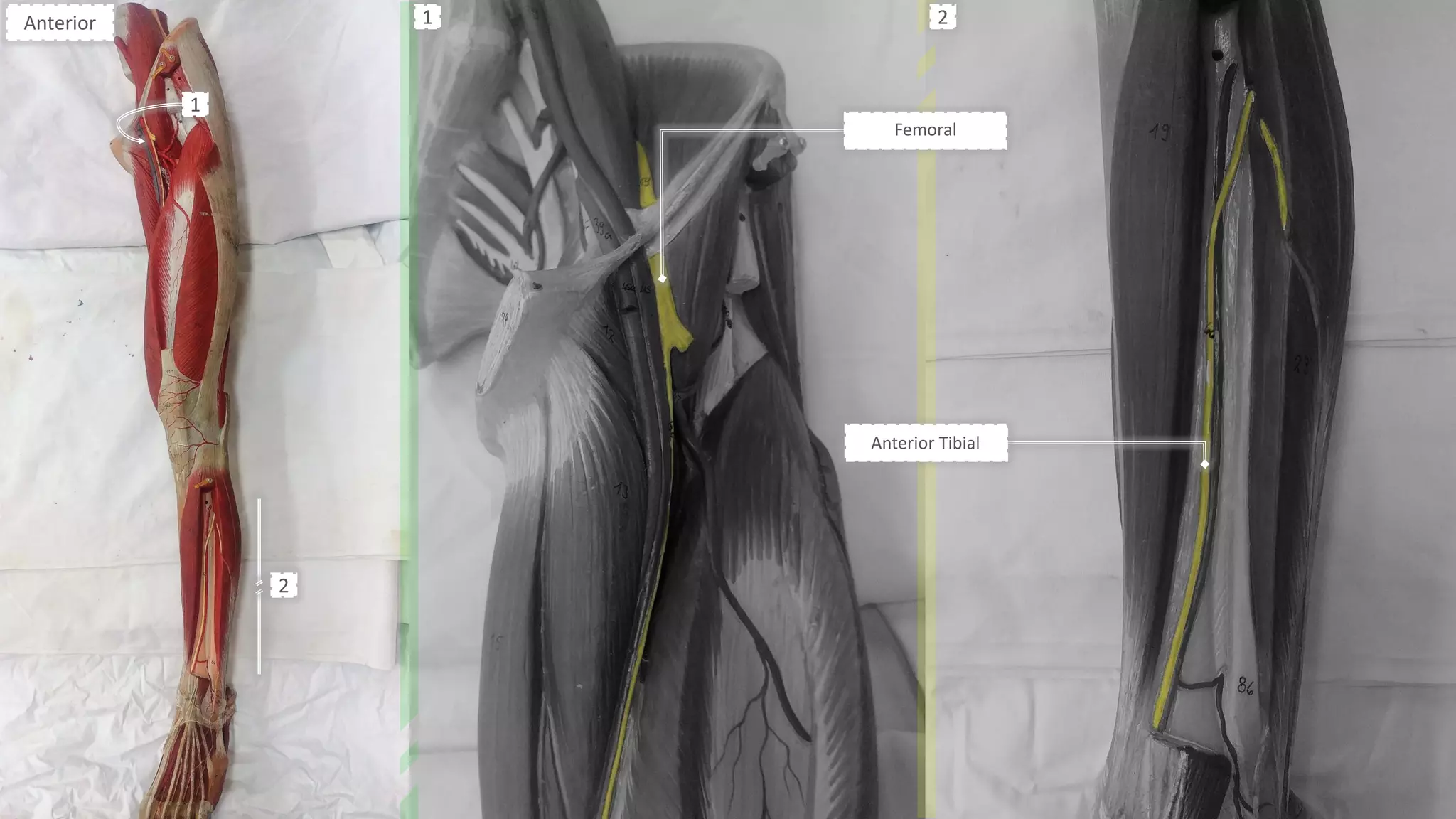
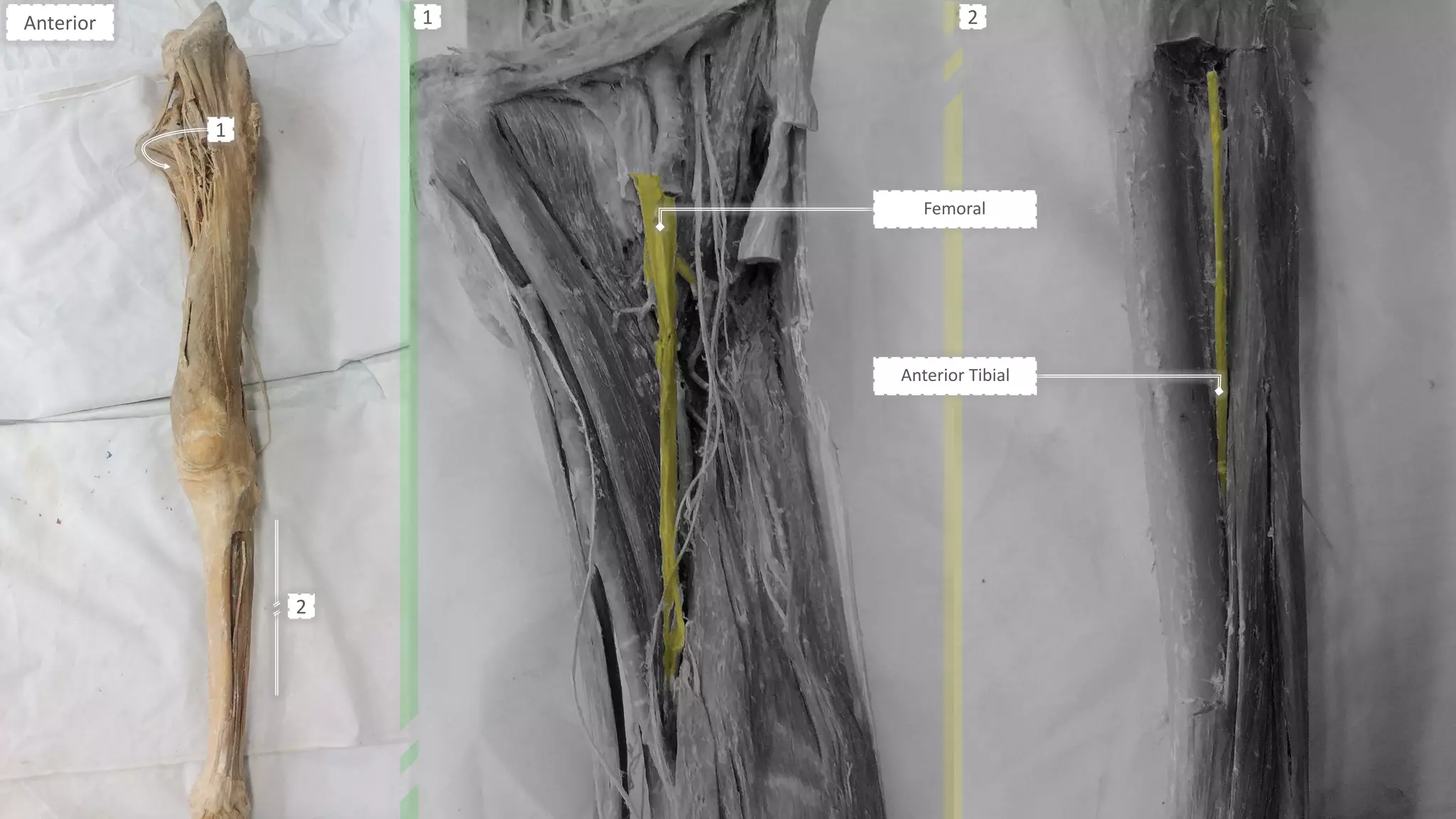
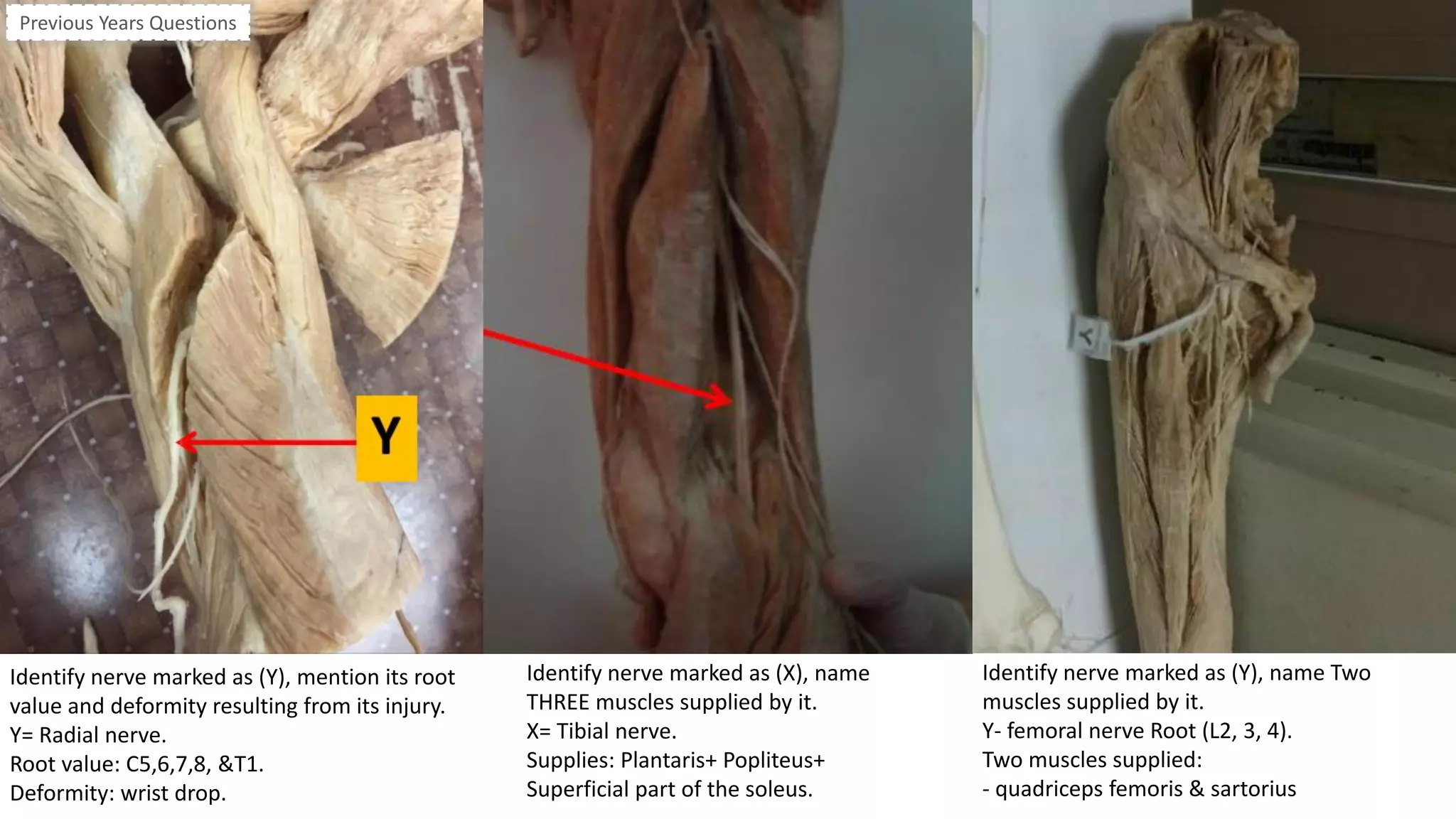
Femoral L2-L4
- Muscular branches⋮
- Intermediate cutaneous N.
- Medial cutaneous N.
- Saphenous N.⤵
[sensation to anteromedial, &
posteromedial surface of leg.]
- Quadriceps Femoris.
- Pectineus.
- Sartorius.
- Iliacus.
K
P
Sa
- Wasting of quadriceps & Loss of knee
extension.
- Sensory loss over anteromedial side of
thigh & medial side of leg & foot.
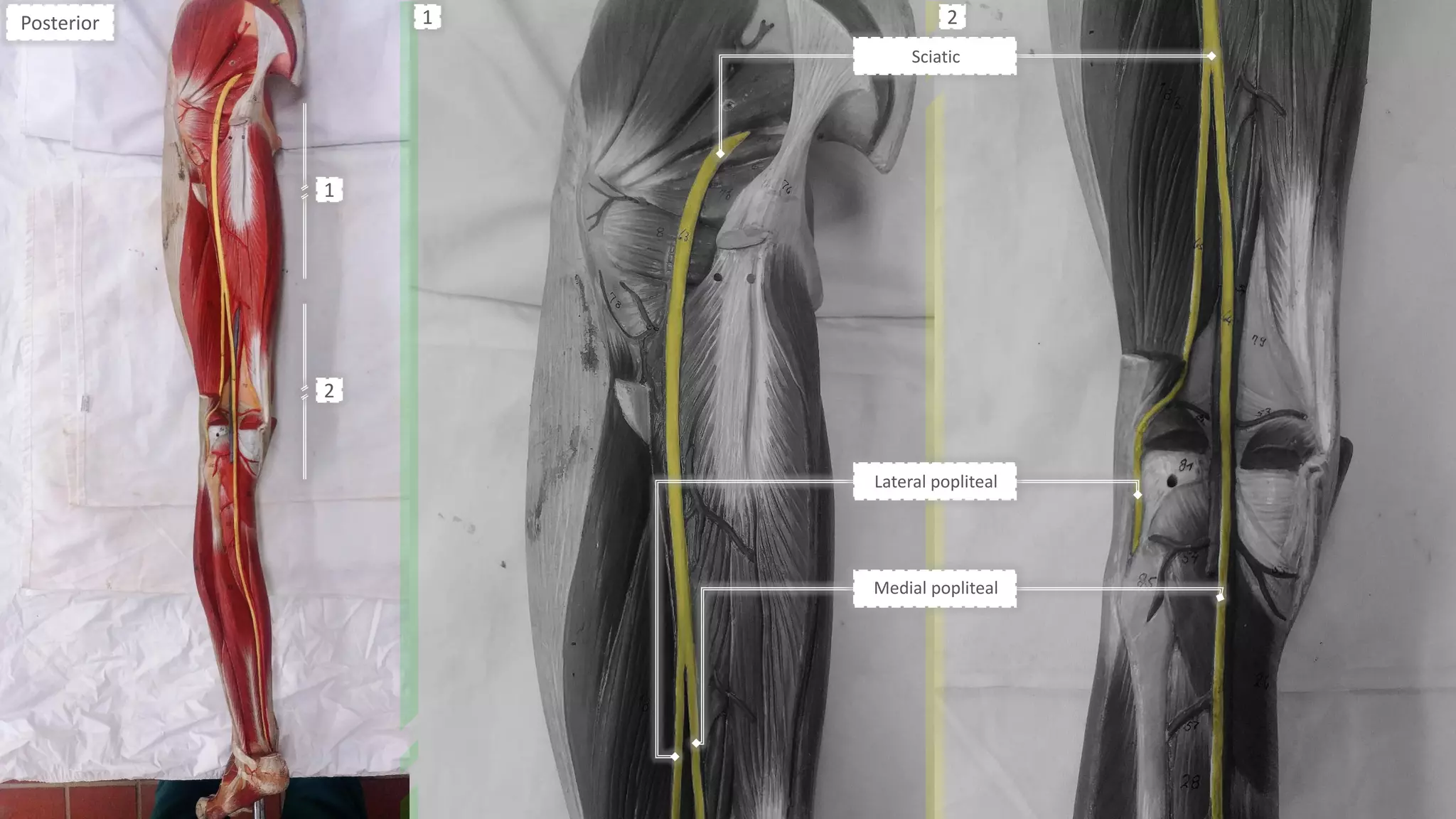
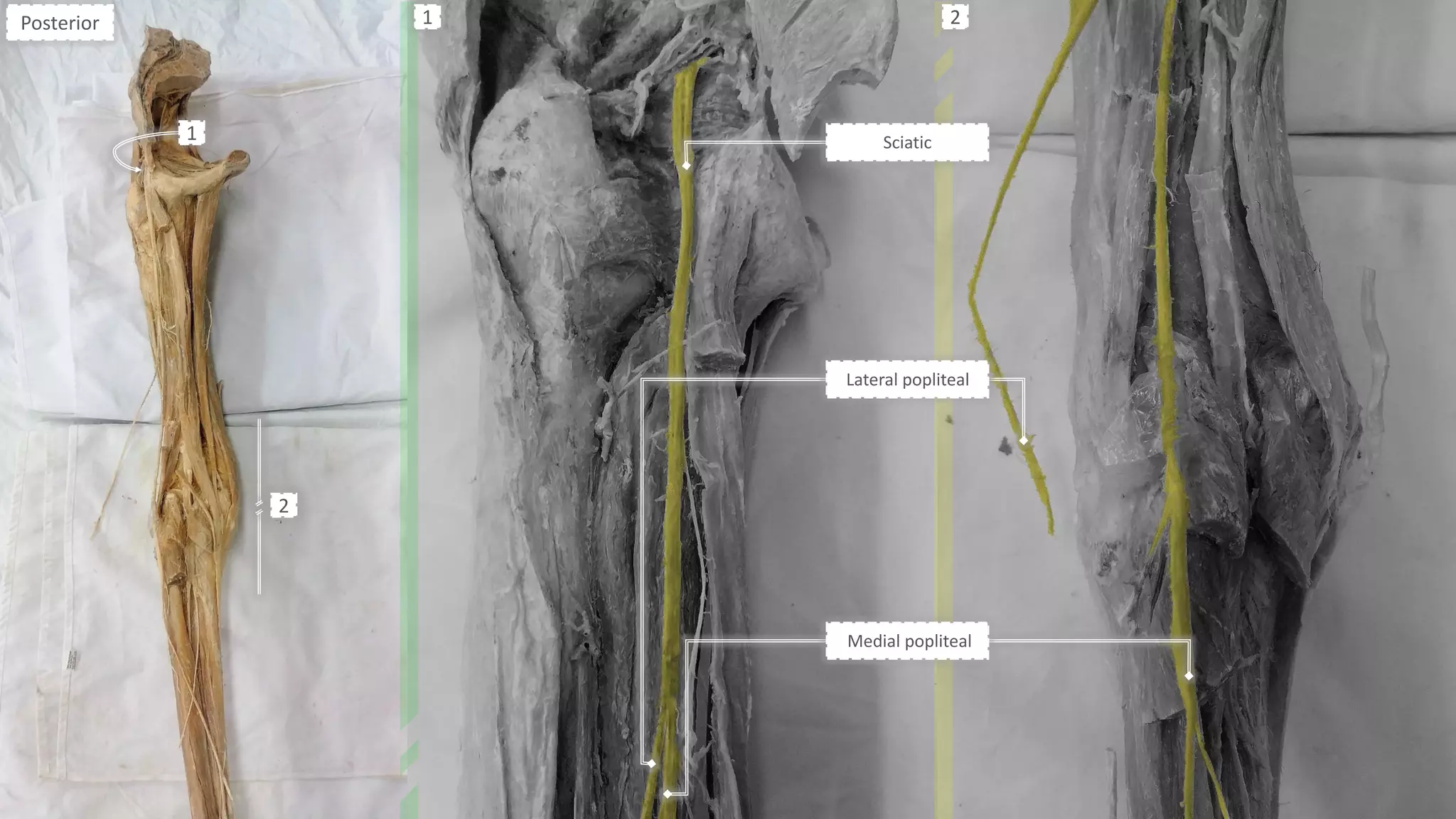
Sciatic L4-S3
- Muscular branches⋮
- Articular to hip joint.
- Medial & Lateral popliteal Ns.
- Semimembranosus.
- Biceps Femoris.
- Semitendinosus.
- Ischial part of Adductor Magnus.
S
P
S
S
- Foot drop.
- Sensory loss below knee Except area
supplied by saphenous N.
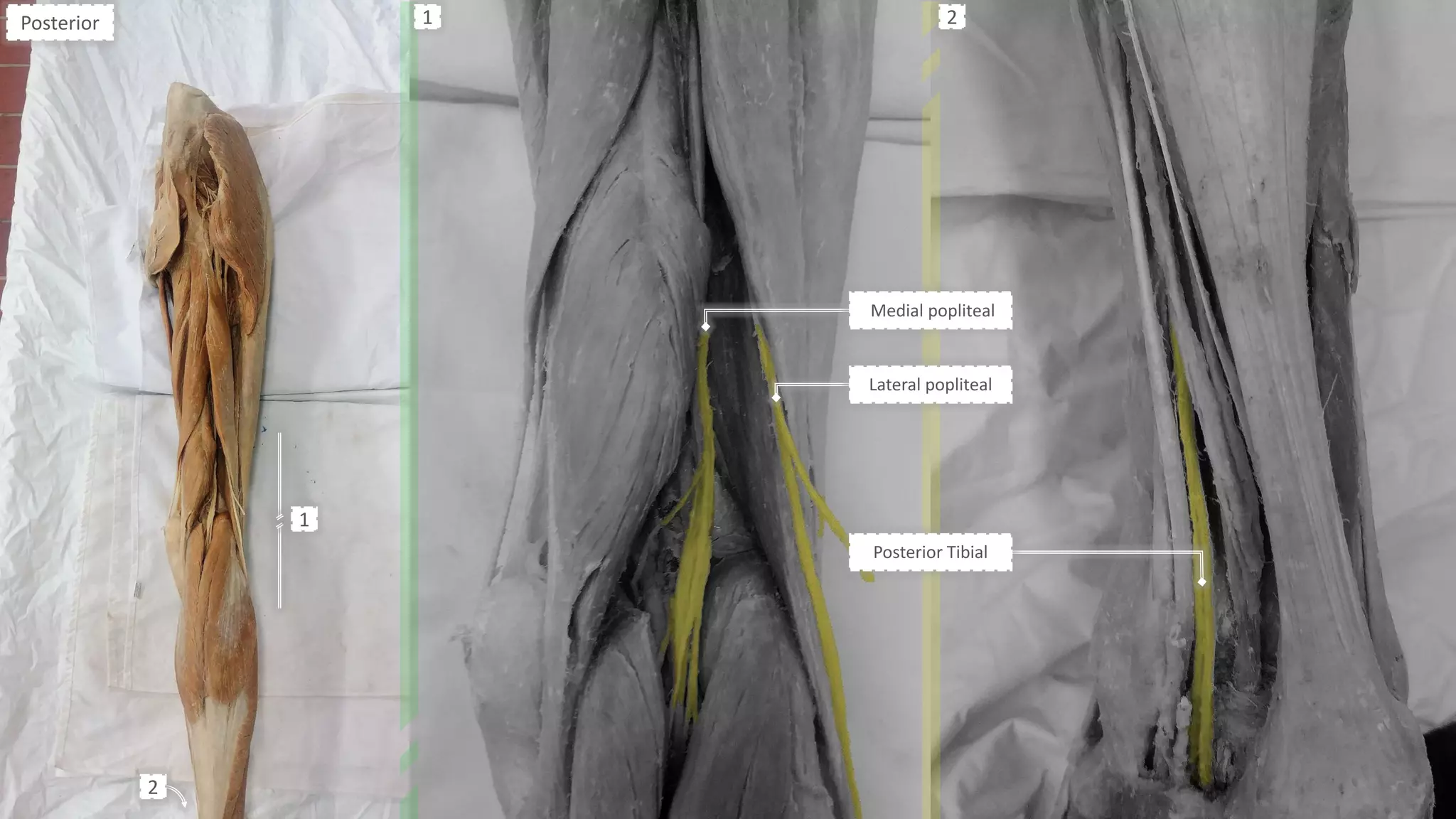
Medial
Popliteal
- Tibial -
L4-S3
- Muscular branches⋮
- Sural N.
- Articular to knee joint.
- Gastrocnemius.
- Popliteus.
- Soleus.
- Plantaris.
G
P
S
- Dorsiflexion & eversion of foot.
- Sensory loss over sole of foot.
Lateral
Popliteal
- Common
Peroneal -
L4-S2
- Lateral cutaneous N. of calf.
- Sural communicating N.
- Articular to knee joint.
- Foot drop
- Sensory loss over anterior & lateral
sides of leg & dorsum of foot except
areas supplies by sural & saphenous Ns.
Anterior
Tibial
L4-S2
- Muscular branches⋮
- Cutaneous branches.
All front of leg muscles & extensor digitorum brevis. - Drop foot with high steppage gait.
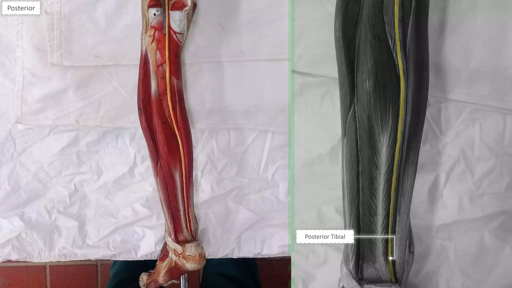
Posterior
Tibial
L4-S3
- Muscular branches⋮
- Cutaneous branches.
- Articular to ankle joint.
- Flexor Hallucis Longus.
- Flexor Digitorum Longus.
- Tibialis Posterior.
- Deep part of Soleus.
FuLLHDTv
- Weak planter flexion.](https://image.slidesharecdn.com/peripheralnervessummary-191108181032/75/Peripheral-nerves-summary-3-2048.jpg)
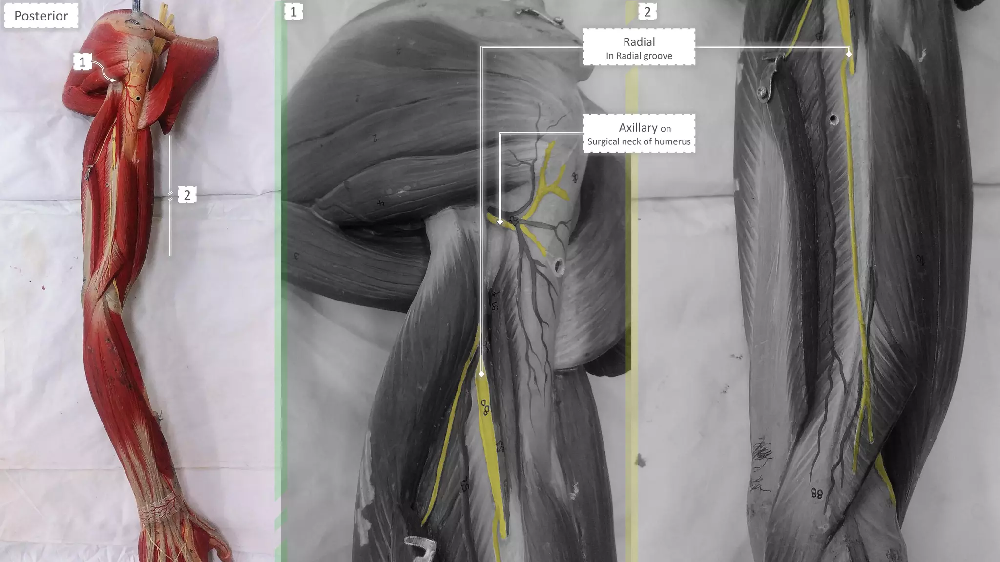
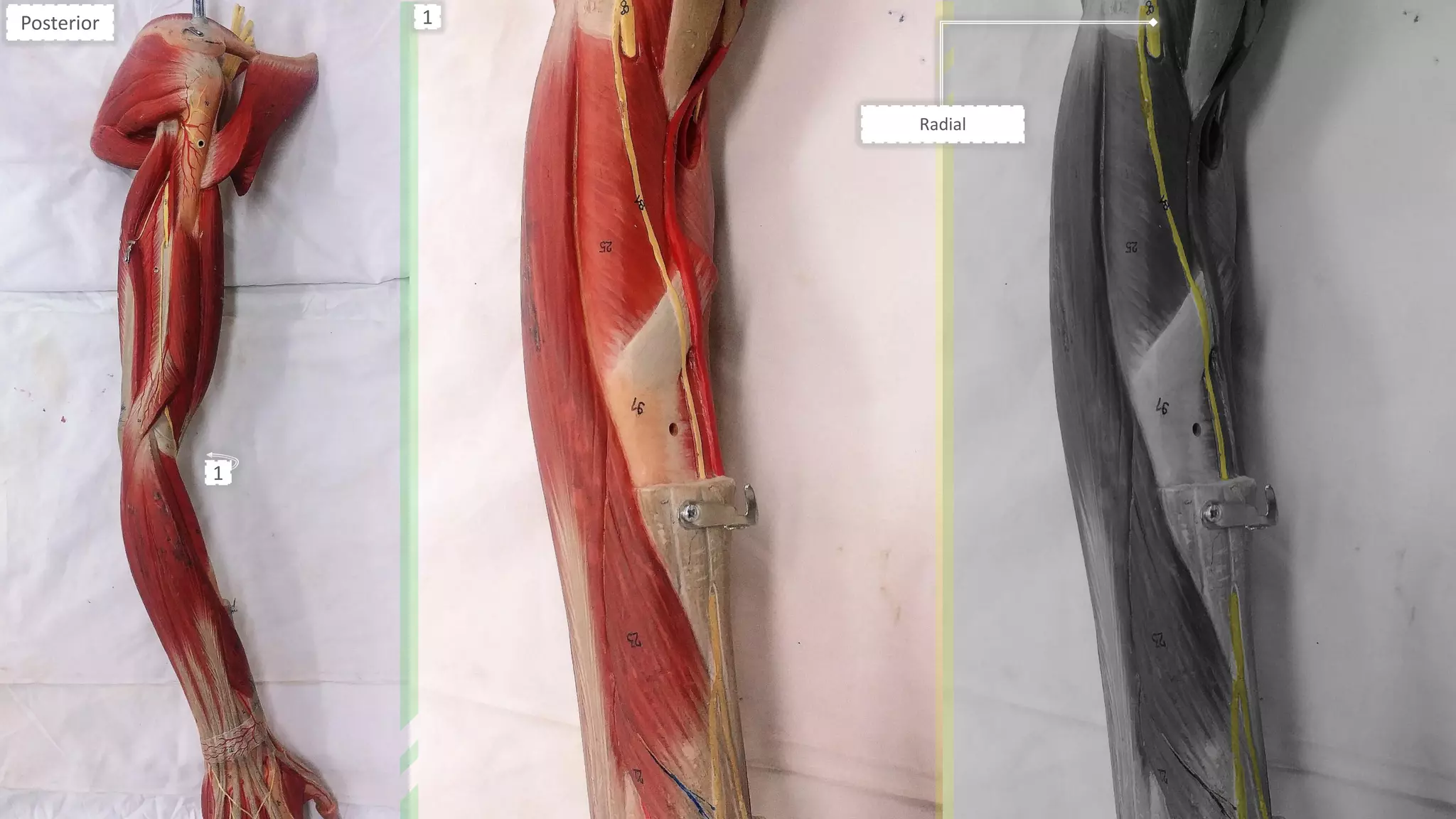
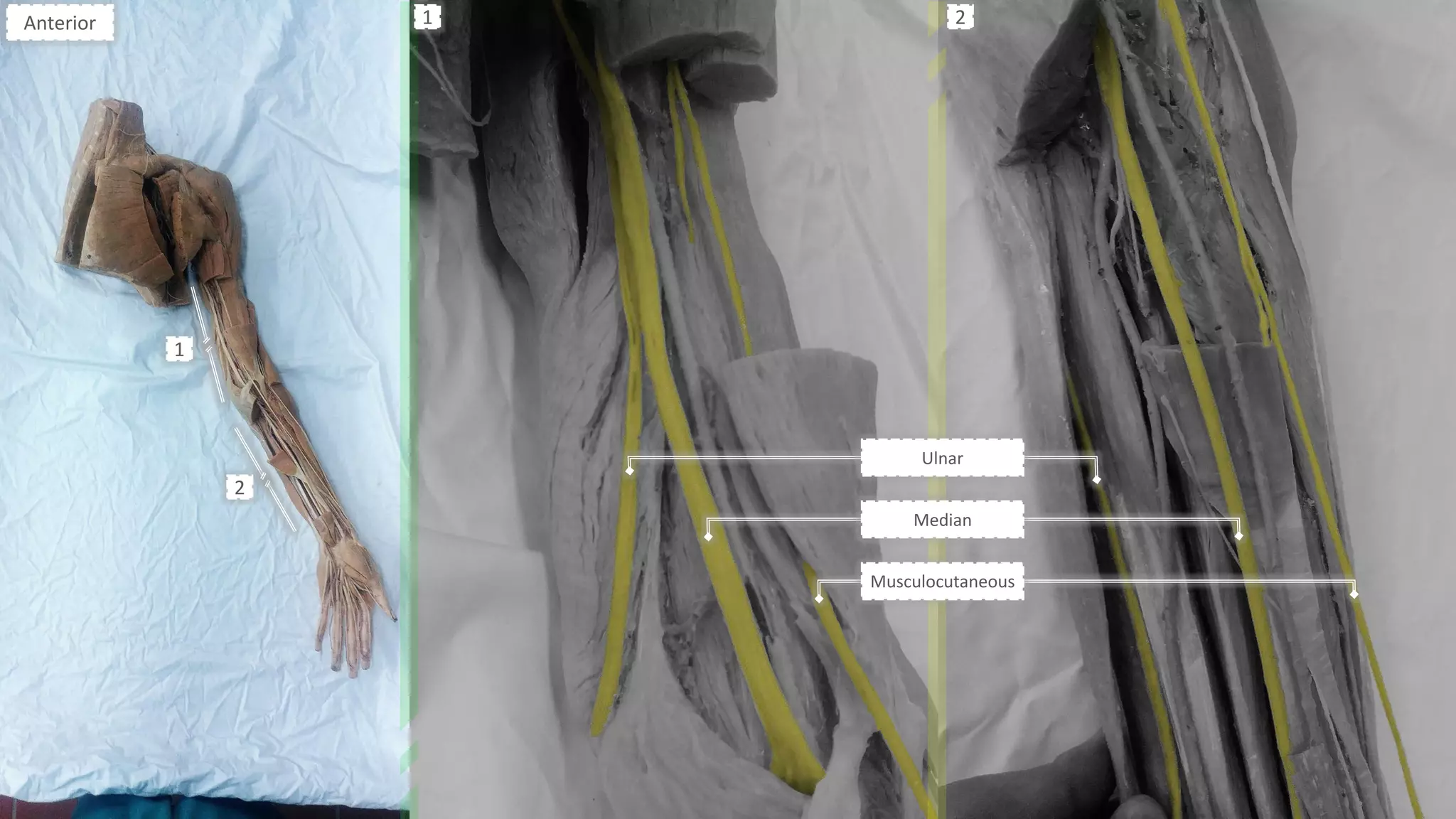
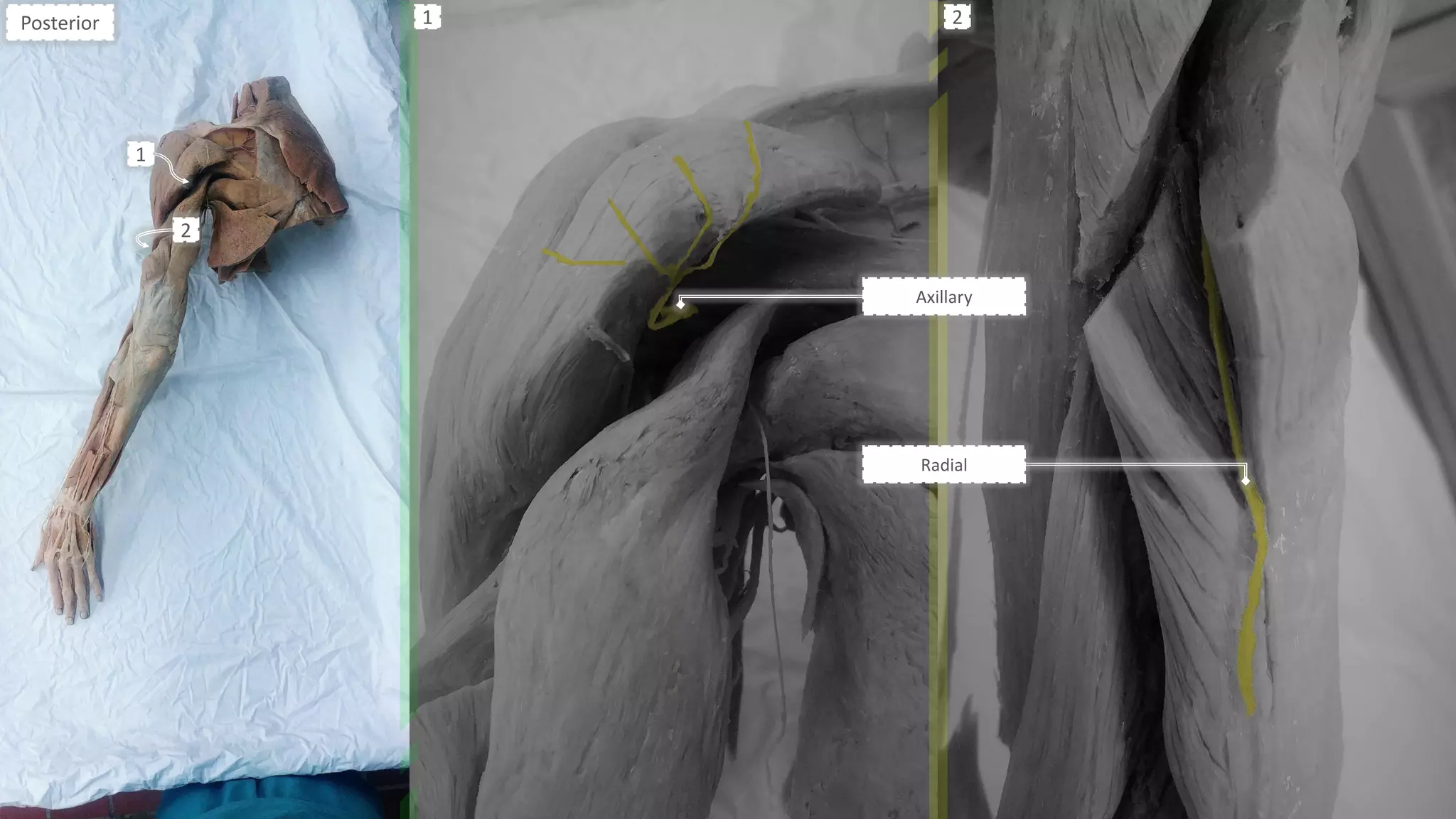
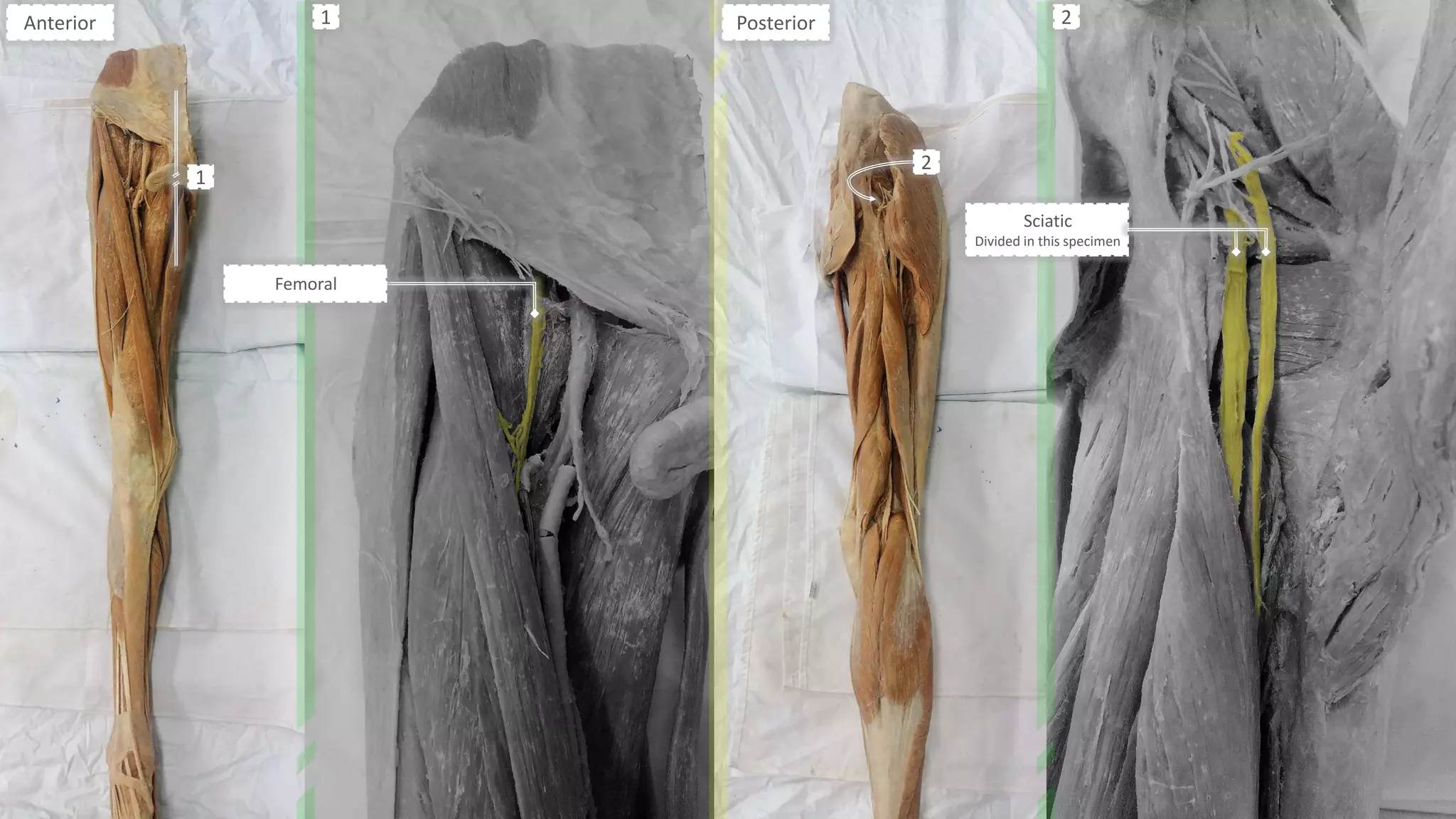

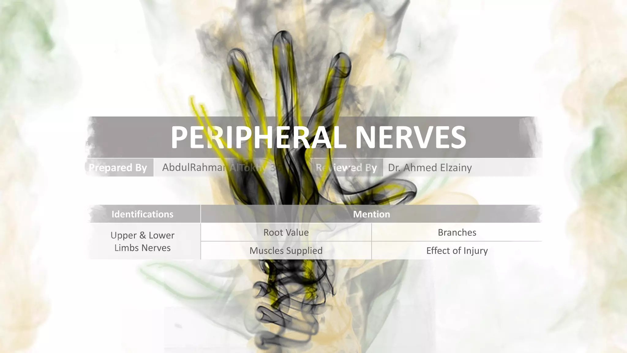
This document provides information on peripheral nerves of the upper and lower limbs, including their root values, branches, muscles supplied, and effects of injury. For each nerve, it lists at least 3 branches, muscles supplied, and describes the effect of injury. Diagrams are included showing the course and distribution of each nerve. Identification questions are presented at the end assessing knowledge of specific nerves, their roots, and muscles supplied.


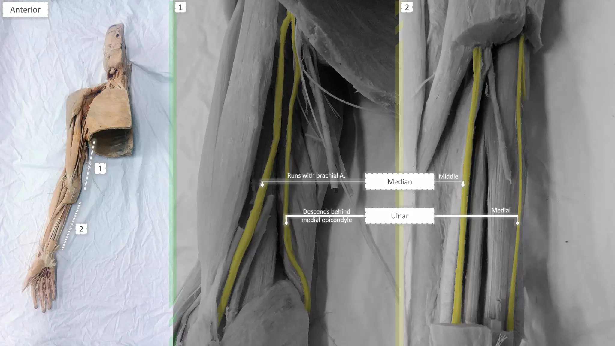
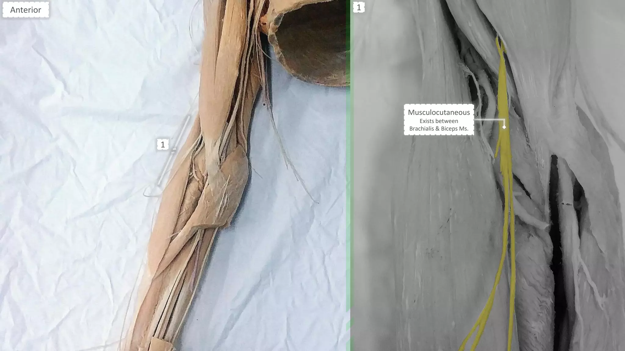
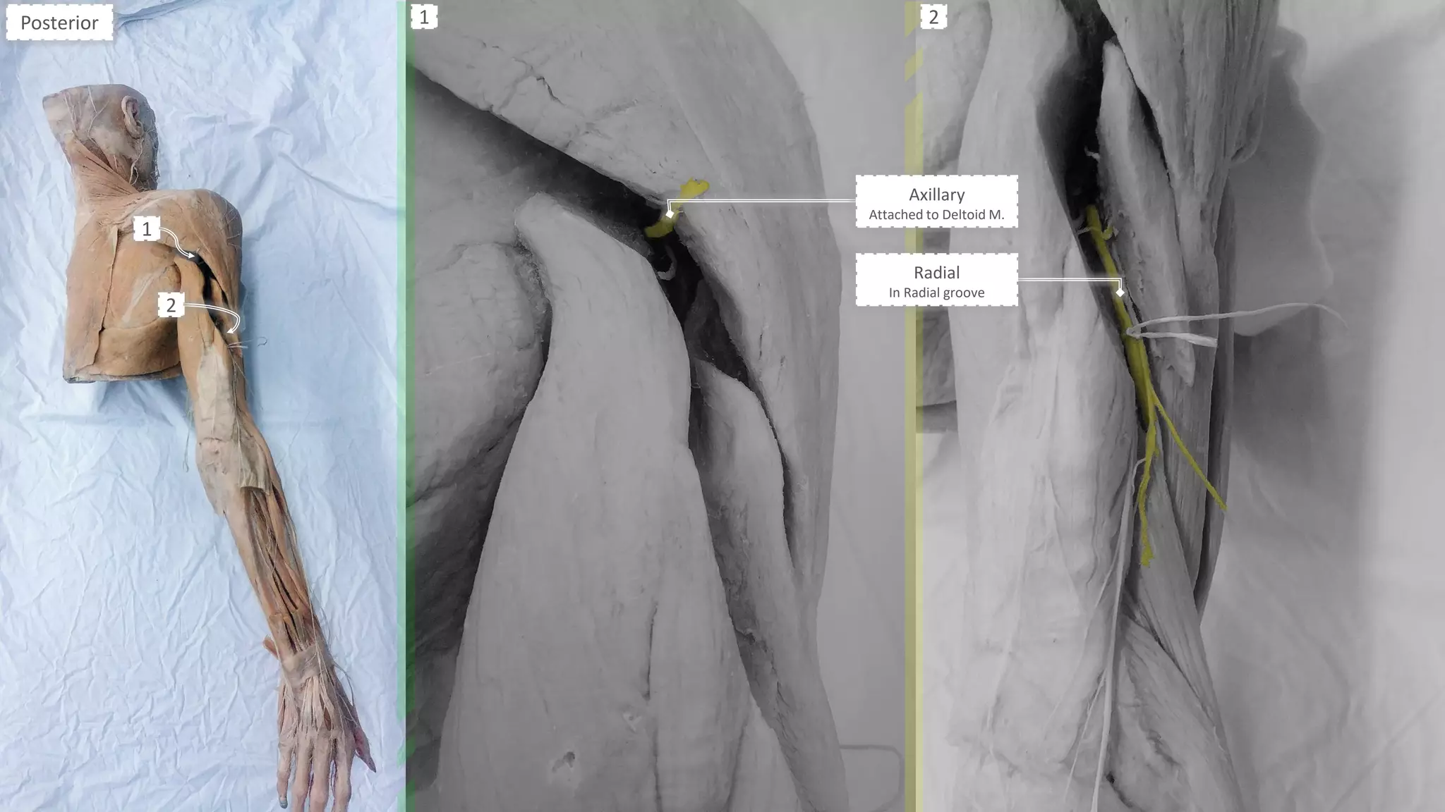
![Peripheral Nerves of Upper Limb [Radial, Median & Ulnar nerve]](https://cdn.slidesharecdn.com/ss_thumbnails/sb-190709091558-thumbnail.jpg?width=640&height=640&fit=bounds)


