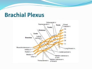
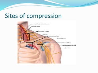
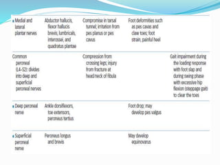
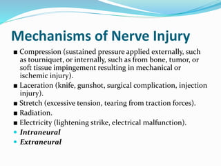
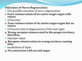
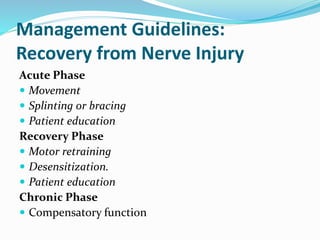
Dr. Maria Idrees; PT
Review of Peripheral
Nerve Structure
Nerve Structure
 Peripheral nerves contain a mix of motor, sensory, and
sympathetic neurons.
■ Alpha motor neurons (somatic efferent fibers): cell bodies located
in anterior column of spinal cord; innervate skeletal muscles
■ Gamma motor neurons (efferent fibers): cell bodies located in
lateral columns of spinal cord; innervate intrafusal muscle fibers
of the muscle spindle
■ Sensory neurons (somatic afferent fibers): cell bodies located in
the dorsal root ganglia; innervate sensory receptors
■ Sympathetic neurons (visceral afferent fibers): cell bodies located
in sympathetic ganglia; innervate sweat glands, blood vessels,
viscera, and glands
Mobility Characteristics of the
Nervous System
H???
Common Sites of Injury to Peripheral
Nerves
 Anywhere along the pathway from the nerve roots to
their termination in the tissues of the trunk and
extremities.
 Inter vertebral foramen
 Symptoms and signs of nerve impairments are sensory
changes or loss and motor weakness in the distribution
of the involved nerve fibers
Nerve Roots
 Nerve roots emerge from the spinal canal and traverse the
foramina of the spine, where they can become impinged as
a result of various pathologies of the spine that reduce the
space in the foramina, such as degenerative disc disease
(DDD), degenerative joint disease (DJD), disc lesions, and
spondylolisthesis.
 With reduced spinal canal or foraminal space (stenosis),
extension, side bending, or rotation to the side of the
stenosis further decreases the space where the nerve root
courses and may cause or perpetuate symptoms
 Nerve roots of the upper quarter include C5 through T1 and
those of the lower quarter L1 through S3.
Brachial Plexus
Sites of compression
Upper plexus injuries (C5, 6): The mechanism involves
shoulder depression and lateral flexion of the neck to the
opposite side. There is loss of abduction and lateral
rotation of the shoulder and weakness in elbow flexion and
forearm supination (waiter’s tip position). Erb’s palsy
occurs with birth injuries when the shoulder is stretched
downward.
 Middle plexus injuries (C7): Rarely seen alone.
 Lower plexus injuries (C8, T1): Usually due to
compression by a cervical rib or stretching the arm
overhead. Klumpke’s paralysis (paralysis of the intrinsics of
the hand) occurs in birth injuries when the baby presents
with its arm overhead.
 Complete or total injury of the plexus: Erb-Klumpke’s
paralysis (Horner’s syndrome)
Myotomes
Lumbosacral Plexus
Mechanisms of Nerve Injury
■ Compression (sustained pressure applied externally, such
as tourniquet, or internally, such as from bone, tumor, or
soft tissue impingement resulting in mechanical or
ischemic injury).
■ Laceration (knife, gunshot, surgical complication, injection
injury).
■ Stretch (excessive tension, tearing from traction forces).
■ Radiation.
■ Electricity (lightening strike, electrical malfunction).
 Intraneural
 Extraneural
Excellent regenerative potential: radial,
musculocutaneous, and femoral nerves
Moderate regenerative potential: median, ulnar, and
tibial nerves
Poor regenerative potential: peroneal nerve
Outcomes of Nerve Regeneration
 Five possible outcomes of nerve regeneration.
1. Exact reinnervation of its native target organ with
return
 of function
2. Exact reinnervation of its native target organ but no
return
 of function due to degeneration of the end organ
3. Wrong receptor reinnervated in the proper territory;
therefore,
 improper input
4. Receptor reinnervation in wrong territory causing
false
 localization of input
5. No connection with an end organ
Management Guidelines:
Recovery from Nerve Injury
Acute Phase
 Movement
 Splinting or bracing
 Patient education
Recovery Phase
 Motor retraining
 Desensitization.
 Patient education
Chronic Phase
 Compensatory function
Suggestions for graded modalities and procedures for
desensitizing:
■ Use multiple types of textures or contact for sensory stimulation,
such as cotton, rough material, sandpaper of various grades, and
Velcro. The textures can be wrapped around dowel rods for
finger manipulation or to stroke along the skin.
■ Place contact particles, such as cotton balls, beans, macaroni,
sand, or other material, with various degrees of roughness in
tubs or cans, so the patient can run the involved hand or foot
through the material. Have the patient begin by manipulating or
placing the extremity in the least irritating texture for 10
minutes. As tolerance improves, progress to the next texture of
slightly more irritating but tolerable stimulus. Maximum
progress occurs when the most irritating texture is tolerated.
■ Use vibration. Pattern of recovery after nerve injury is pain
(hypersensitivity), perception of slow vibration (30 cps), moving
touch, constant touch, rapid vibration (256 cps), and awareness
from proximal to distal.
Suggestions for retraining the brain to recognize a
stimulus
■ Begin by using a moving touch stimulus, such as the eraser
end of a pencil, and stroke over the area. The patient first
watches, then closes his or her eyes, and tries to identify
where touch occurred.
■ Progress from stroking to using constant touch.
■ When the patient is able to localize constant touch,
progress to identification of familiar objects of various
sizes, shapes, and textures.
■ For the hand, use familiar household and personal care
objects, such as keys, eating utensils, blocks, toothbrush,
and safety pins.
■ For the feet, have the patient walk on various surfaces, such
as grass, sand, wood, pebbles, and uneven surfaces.
Neural Testing and Mobilization
Techniques for the Upper Quadrant
 Median Nerve
 Radial Nerve
 Ulner Nerve
Neural Testing and Mobilization
Techniques for the Lower Quadrant
 Sciatica Nerve
 Slump sitting
 Femoral Nerve

Peripheral nerve disorders

  • 1.
  • 2.
    Review of Peripheral NerveStructure Nerve Structure  Peripheral nerves contain a mix of motor, sensory, and sympathetic neurons. ■ Alpha motor neurons (somatic efferent fibers): cell bodies located in anterior column of spinal cord; innervate skeletal muscles ■ Gamma motor neurons (efferent fibers): cell bodies located in lateral columns of spinal cord; innervate intrafusal muscle fibers of the muscle spindle ■ Sensory neurons (somatic afferent fibers): cell bodies located in the dorsal root ganglia; innervate sensory receptors ■ Sympathetic neurons (visceral afferent fibers): cell bodies located in sympathetic ganglia; innervate sweat glands, blood vessels, viscera, and glands
  • 4.
    Mobility Characteristics ofthe Nervous System H???
  • 5.
    Common Sites ofInjury to Peripheral Nerves  Anywhere along the pathway from the nerve roots to their termination in the tissues of the trunk and extremities.  Inter vertebral foramen  Symptoms and signs of nerve impairments are sensory changes or loss and motor weakness in the distribution of the involved nerve fibers
  • 6.
    Nerve Roots  Nerveroots emerge from the spinal canal and traverse the foramina of the spine, where they can become impinged as a result of various pathologies of the spine that reduce the space in the foramina, such as degenerative disc disease (DDD), degenerative joint disease (DJD), disc lesions, and spondylolisthesis.  With reduced spinal canal or foraminal space (stenosis), extension, side bending, or rotation to the side of the stenosis further decreases the space where the nerve root courses and may cause or perpetuate symptoms  Nerve roots of the upper quarter include C5 through T1 and those of the lower quarter L1 through S3.
  • 8.
  • 9.
  • 10.
    Upper plexus injuries(C5, 6): The mechanism involves shoulder depression and lateral flexion of the neck to the opposite side. There is loss of abduction and lateral rotation of the shoulder and weakness in elbow flexion and forearm supination (waiter’s tip position). Erb’s palsy occurs with birth injuries when the shoulder is stretched downward.  Middle plexus injuries (C7): Rarely seen alone.  Lower plexus injuries (C8, T1): Usually due to compression by a cervical rib or stretching the arm overhead. Klumpke’s paralysis (paralysis of the intrinsics of the hand) occurs in birth injuries when the baby presents with its arm overhead.  Complete or total injury of the plexus: Erb-Klumpke’s paralysis (Horner’s syndrome)
  • 12.
  • 15.
  • 18.
    Mechanisms of NerveInjury ■ Compression (sustained pressure applied externally, such as tourniquet, or internally, such as from bone, tumor, or soft tissue impingement resulting in mechanical or ischemic injury). ■ Laceration (knife, gunshot, surgical complication, injection injury). ■ Stretch (excessive tension, tearing from traction forces). ■ Radiation. ■ Electricity (lightening strike, electrical malfunction).  Intraneural  Extraneural
  • 20.
    Excellent regenerative potential:radial, musculocutaneous, and femoral nerves Moderate regenerative potential: median, ulnar, and tibial nerves Poor regenerative potential: peroneal nerve
  • 21.
    Outcomes of NerveRegeneration  Five possible outcomes of nerve regeneration. 1. Exact reinnervation of its native target organ with return  of function 2. Exact reinnervation of its native target organ but no return  of function due to degeneration of the end organ 3. Wrong receptor reinnervated in the proper territory; therefore,  improper input 4. Receptor reinnervation in wrong territory causing false  localization of input 5. No connection with an end organ
  • 22.
    Management Guidelines: Recovery fromNerve Injury Acute Phase  Movement  Splinting or bracing  Patient education Recovery Phase  Motor retraining  Desensitization.  Patient education Chronic Phase  Compensatory function
  • 23.
    Suggestions for gradedmodalities and procedures for desensitizing: ■ Use multiple types of textures or contact for sensory stimulation, such as cotton, rough material, sandpaper of various grades, and Velcro. The textures can be wrapped around dowel rods for finger manipulation or to stroke along the skin. ■ Place contact particles, such as cotton balls, beans, macaroni, sand, or other material, with various degrees of roughness in tubs or cans, so the patient can run the involved hand or foot through the material. Have the patient begin by manipulating or placing the extremity in the least irritating texture for 10 minutes. As tolerance improves, progress to the next texture of slightly more irritating but tolerable stimulus. Maximum progress occurs when the most irritating texture is tolerated. ■ Use vibration. Pattern of recovery after nerve injury is pain (hypersensitivity), perception of slow vibration (30 cps), moving touch, constant touch, rapid vibration (256 cps), and awareness from proximal to distal.
  • 24.
    Suggestions for retrainingthe brain to recognize a stimulus ■ Begin by using a moving touch stimulus, such as the eraser end of a pencil, and stroke over the area. The patient first watches, then closes his or her eyes, and tries to identify where touch occurred. ■ Progress from stroking to using constant touch. ■ When the patient is able to localize constant touch, progress to identification of familiar objects of various sizes, shapes, and textures. ■ For the hand, use familiar household and personal care objects, such as keys, eating utensils, blocks, toothbrush, and safety pins. ■ For the feet, have the patient walk on various surfaces, such as grass, sand, wood, pebbles, and uneven surfaces.
  • 25.
    Neural Testing andMobilization Techniques for the Upper Quadrant  Median Nerve  Radial Nerve  Ulner Nerve
  • 26.
    Neural Testing andMobilization Techniques for the Lower Quadrant  Sciatica Nerve  Slump sitting  Femoral Nerve